Review
. 2021 Oct 14;61(4):1517-1545.
doi: 10.1093/icb/icab078.

Melanosome Biogenesis in the Pigmentation of Mammalian Skin

Affiliations
Review

Melanosome Biogenesis in the Pigmentation of Mammalian Skin

Linh Le et al. Integr Comp Biol. .

Abstract

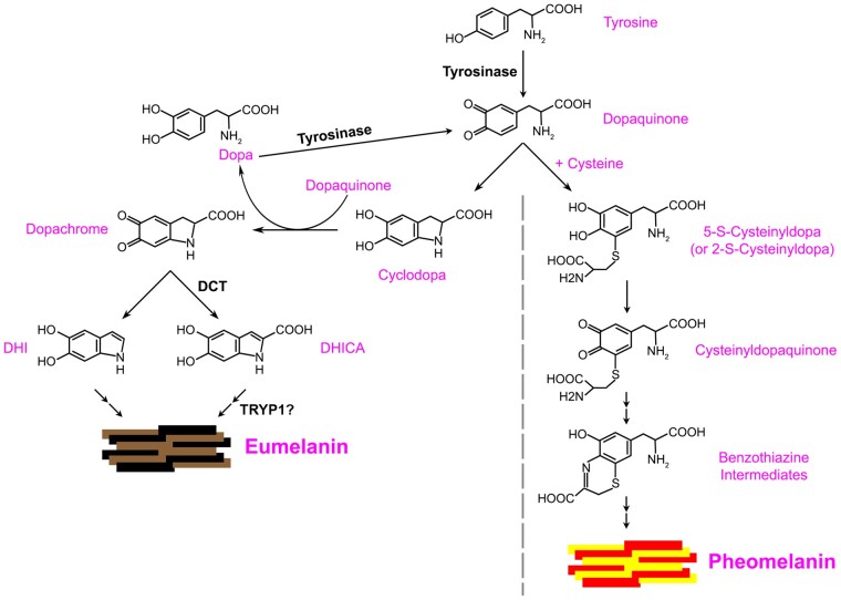
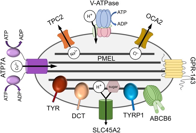
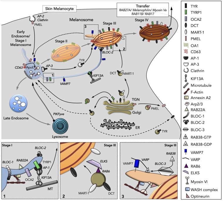
Melanins, the main pigments of the skin and hair in mammals, are synthesized within membrane-bound organelles of melanocytes called melanosomes. Melanosome structure and function are determined by a cohort of resident transmembrane proteins, many of which are expressed only in pigment cells and localize specifically to melanosomes. Defects in the genes that encode melanosome-specific proteins or components of the machinery required for their transport in and out of melanosomes underlie various forms of ocular or oculocutaneous albinism, characterized by hypopigmentation of the hair, skin, and eyes and by visual impairment. We review major components of melanosomes, including the enzymes that catalyze steps in melanin synthesis from tyrosine precursors, solute transporters that allow these enzymes to function, and structural proteins that underlie melanosome shape and melanin deposition. We then review the molecular mechanisms by which these components are biosynthetically delivered to newly forming melanosomes-many of which are shared by other cell types that generate cell type-specific lysosome-related organelles. We also highlight unanswered questions that need to be addressed by future investigation.


Figures

Fig. 1
Fig. 1
Model of eumelanin synthesis. TYR catalyzes the oxidation of tyrosine to generate dopaquinone. In the presence of cysteine, dopaquinone is spontaneously converted to pheomelanin via 5-S-cysteinyldopa or 2-S-cysteinyldopa, cysteinyldopaquinone, and benzothiazine intermediates. In the absence of cysteine, dopaquinone can spontaneously cyclize to cyclodopa, and subsequent spontaneous redox exchange of cyclodopa with dopaquinone gives rise to dopachrome and L-dopa (Cooksey et al. 1997; Ramsden and Riley 2014); L-dopa can then be oxidized by TYR to form more dopaquinone. Dopachrome can spontaneously reorganize to form DHI or can undergo tautomerization by DCT to form DHICA. Both DHI and DHICA undergo oxidation and polymerization to form eumelanins. TYRP1 might support DHICA polymerization either directly or indirectly, or alternatively serve either as a chaperone for TYR or as an antioxidant sink. Adapted from Ito and Wakamatsu (2008).
Fig. 2
Fig. 2
Major melanogenic components of melanosomes. Shown is a schematic of the protein components discussed in the text and their localization within melanosomes of multiple stages (melanosome stage is not indicated). For transporters, the substrate transported is indicated in white circles (and/or the mauve sugar for SLC45A2), and direction of transport is indicated by arrows.
Fig. 3.
Fig. 3.
Working model of intracellular trafficking during melanosome biogenesis. Key molecules indicated in the figure are shown to the right. All melanosomal proteins are synthesized in the ER and transit the Golgi complex and the TGN en route to melanosomes. Transport of cargoes such as TYR, TYRP1, and DCT between compartments from the TGN and beyond are indicated by solid (if known) or dashed (if not yet determined) black arrows. The early endosome/Stage I melanosome progressively matures (gray arrows) to late endosomes and lysosomes or to Stage IV pigmented melanosomes through the sequential delivery of components that originate from the endocytic and exocytic pathways. PMEL is targeted from the TGN to the plasma membrane, from where it is endocytosed PMEL and delivered to Stage I melanosomes. It is then sorted to ILVs, from which it forms elongated amyloid fibrils that distend the organelle to form Stage II melanosomes. This process requires CD63, OA1, Apoliprotein E (ApoE), and other effectors described in the text. Melanin synthesis begins in Stage III due to the delivery of TYR, TYRP1, OCA2, ATP7A, and other cargoes from endosomes/Stage I melanosomes—to which TYRP1 is delivered without passing through the cell surface—and of DCT and MART-1 from the Golgi/TGN. Two transport pathways originate from Stage I melanosomes, a vesicular route requiring AP-3 and a tubular route (box 1) requiring BLOC-1, AP-1, RAB22A, KIF13A, microtubules, and branched actin-associated machineries for tubule formation. The tubules are targeted along microtubules toward maturing Stage III melanosomes in a process requiring BLOC-2, and fuse with these organelles in a VAMP7-dependent manner. A third route (box 2) is mediated by secretory-like vesicles bearing MART-1 and DCT that bud from the Golgi apparatus/TGN in a process requiring RAB6 and are targeted toward maturing Stage III melanosomes in an ELKS-dependent manner. From Stage III melanosomes, some components (e.g., VAMP7 bound to the scaffolding protein VARP) are removed (box 3) via membrane tubules that require BLOC-3, RAB38/RAB32, Myosin VI, OPTN, the WASH complex, and branched actin filaments to promote their formation and release; these tubules might be transported to Stage I melanosomes (dashed arrow). Stage IV melanosomes require the tripartite complex RAB27A, Melanophilin, Myosin Va, and other RAB GTPases to tether to the peripheral actin cytoskeleton prior to transfer to keratinocytes, a poorly understood process that is detailed in the companion paper in this issue (Benito-Martinez et al. 2021). Adapted from Bowman et al. (2019) and Delevoye et al. (2019).

References

    1. Adelmann CH, Traunbauer AK, Chen B, Condon KJ, Chan SH, Kunchok T, Lewis CA, Sabatini DM.. 2020. MFSD12 mediates the import of cysteine into melanosomes and lysosomes. Nature 588:699–704. - PMC - PubMed
    1. Adhikari K, Mendoza-Revilla J, Sohail A, Fuentes-Guajardo M, Lampert J, Chacon-Duque JC, Hurtado M, Villegas V, Granja V, Acuna-Alonzo V, et al.2019. A GWAS in Latin Americans highlights the convergent evolution of lighter skin pigmentation in Eurasia. Nat Commun 10:358. - PMC - PubMed
    1. Altimimi HF, Schnetkamp PP.. 2007. Na+/Ca2+-K+ exchangers (NCKX): functional properties and physiological roles. Channels (Austin) 1:62–9. - PubMed
    1. Ambrosio AL, Boyle JA, Aradi AE, Christian KA, Di Pietro SM.. 2016. TPC2 controls pigmentation by regulating melanosome pH and size. Proc Natl Acad Sci U S A 113:5622–7. - PMC - PubMed
    1. Ancans J, Tobin DJ, Hoogduijn MJ, Smit NP, Wakamatsu K, Thody AJ.. 2001. Melanosomal pH controls rate of melanogenesis, eumelanin/phaeomelanin ratio and melanosome maturation in melanocytes and melanoma cells. Exp Cell Res 268:26–35. - PubMed

LinkOut - more resources