. 2021 May 22:41:558-575.
doi: 10.22203/eCM.v041a36.

Prenatal muscle forces are necessary for vertebral segmentation and disc structure, but not for notochord involution in mice

Affiliations

Prenatal muscle forces are necessary for vertebral segmentation and disc structure, but not for notochord involution in mice

A Levillain et al. Eur Cell Mater. .

Abstract

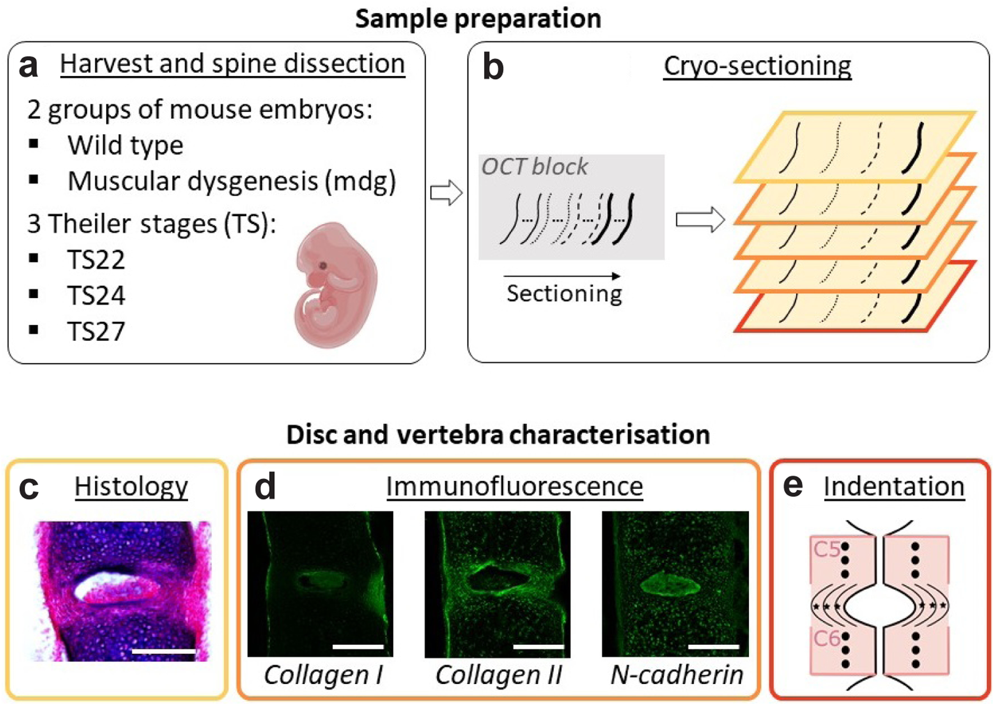
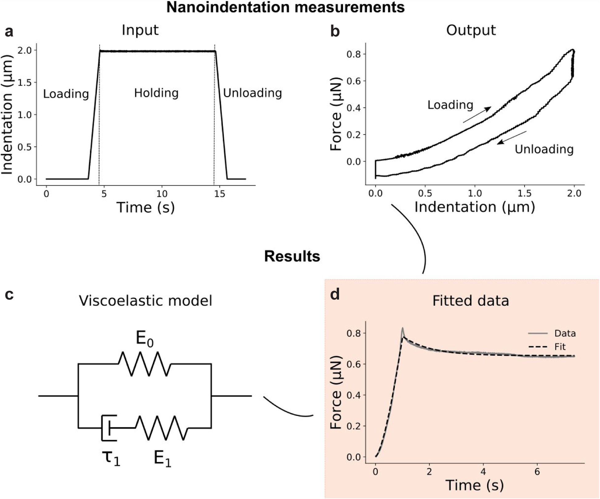
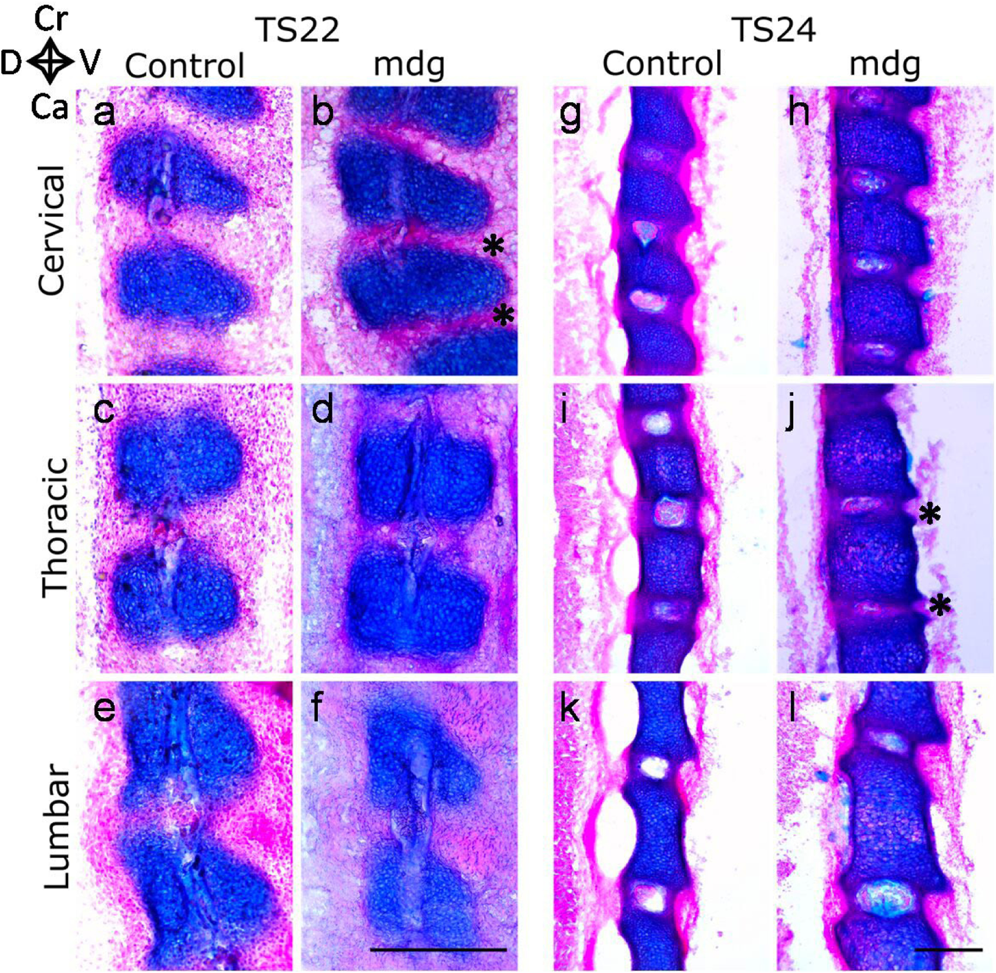
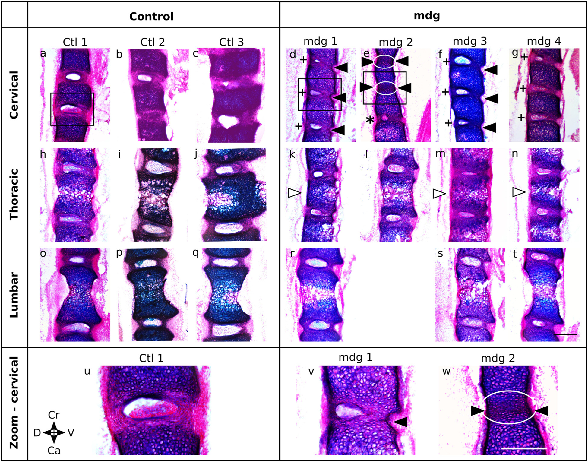
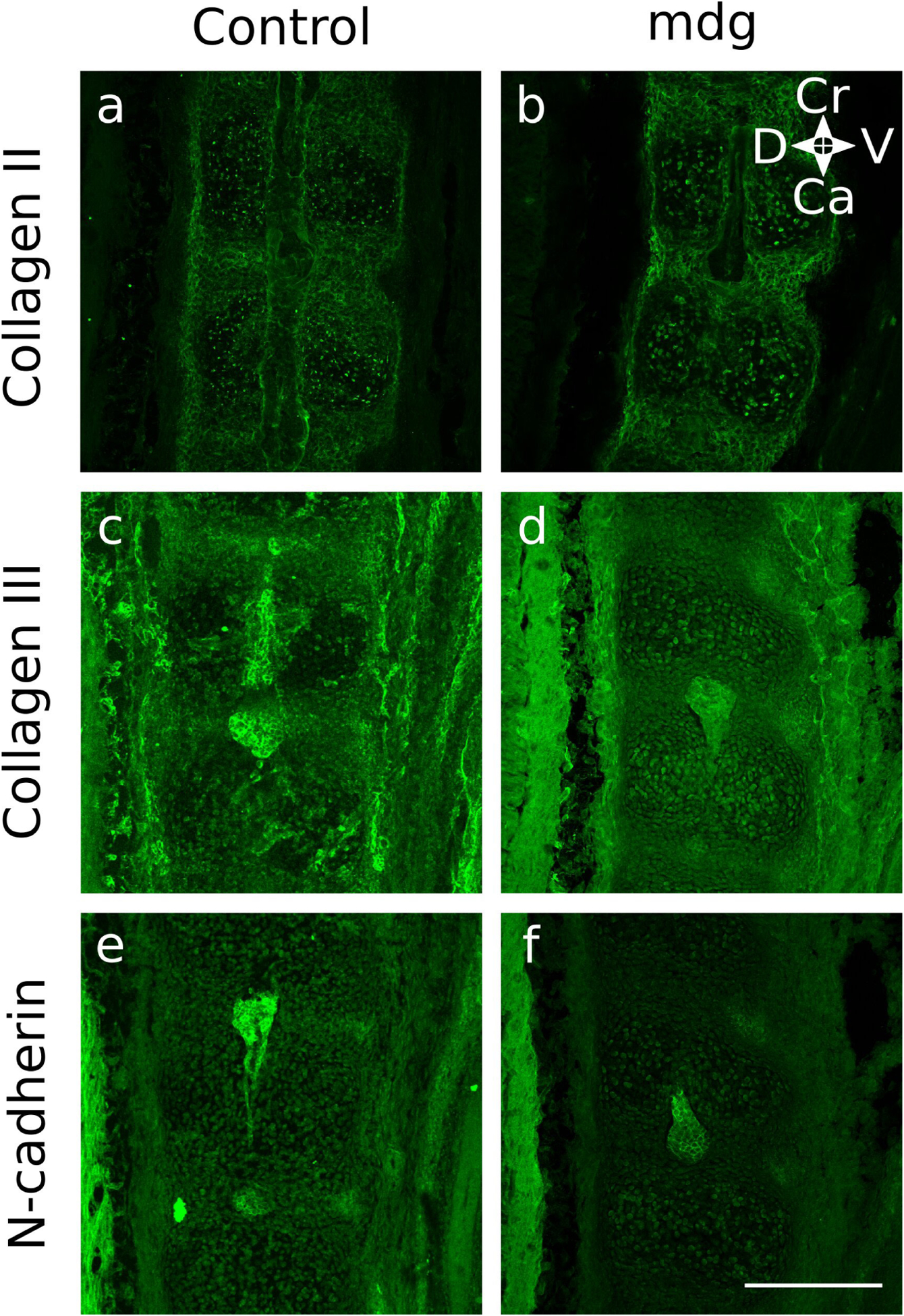
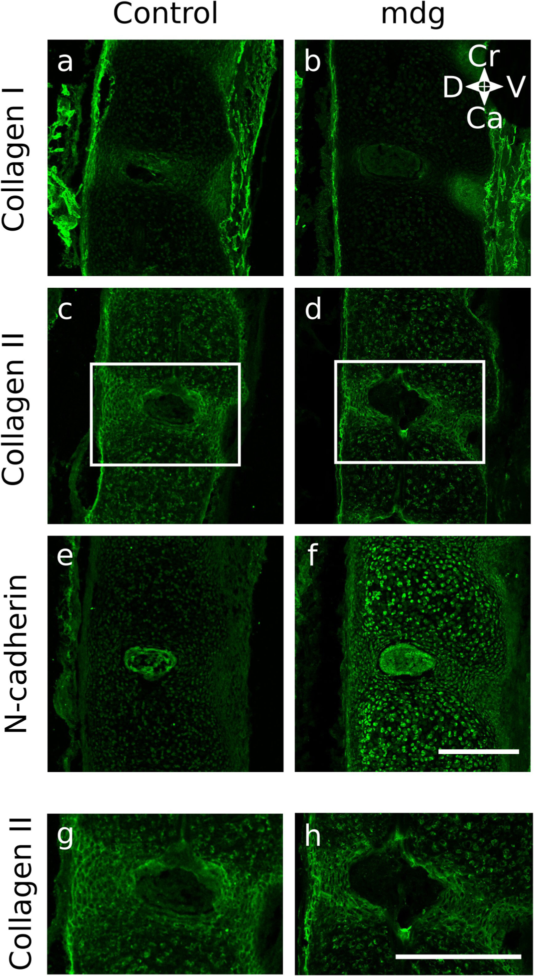
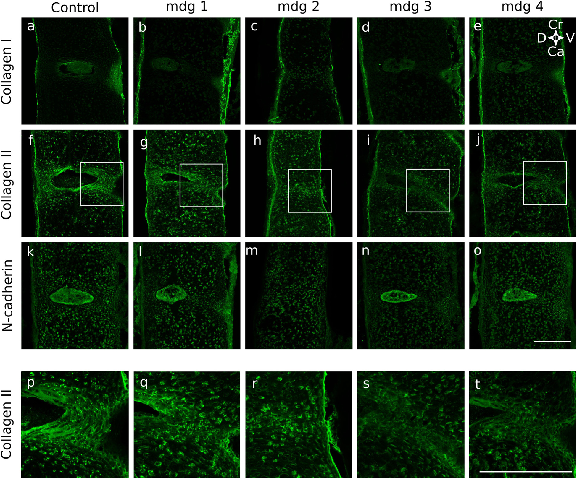
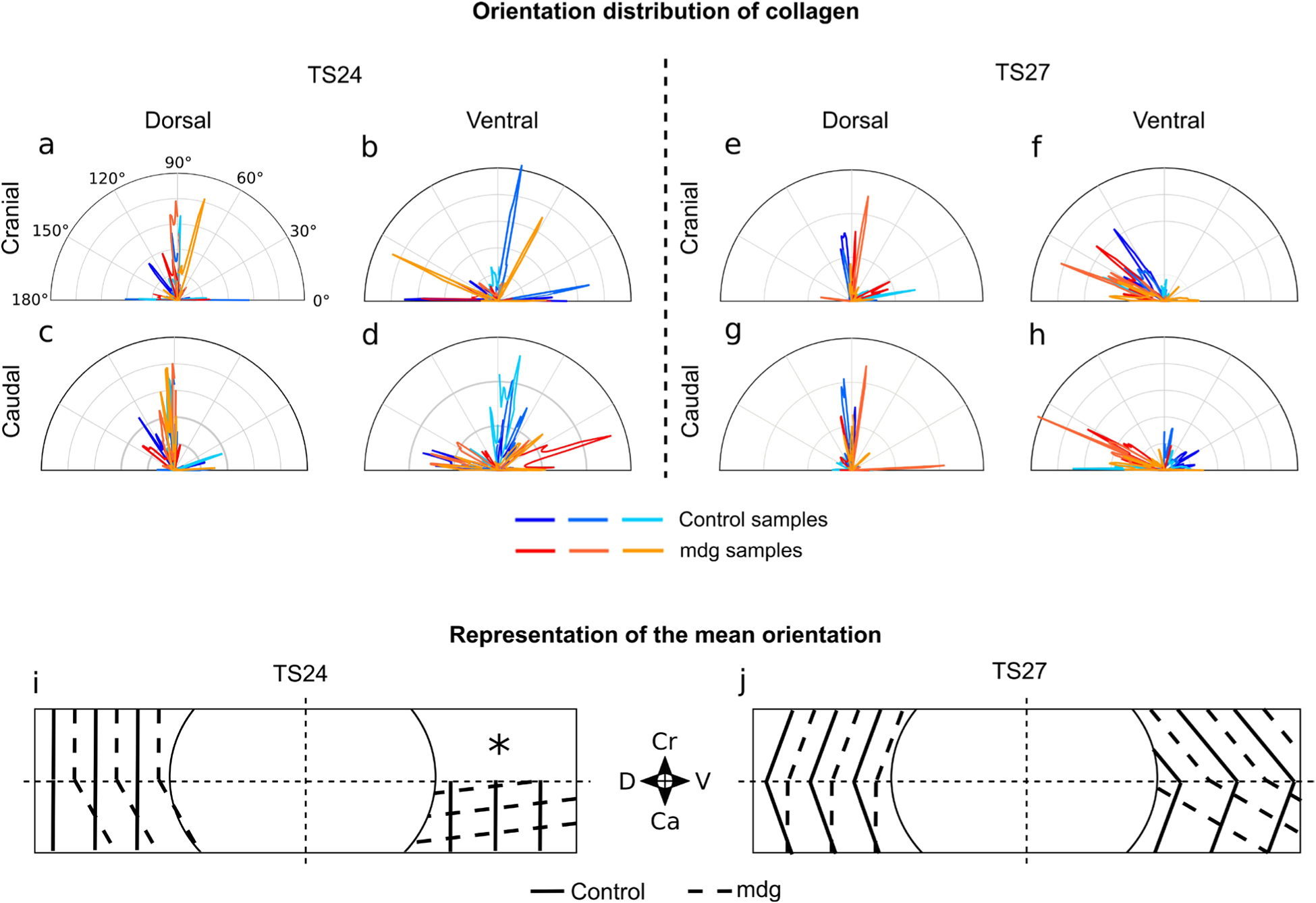
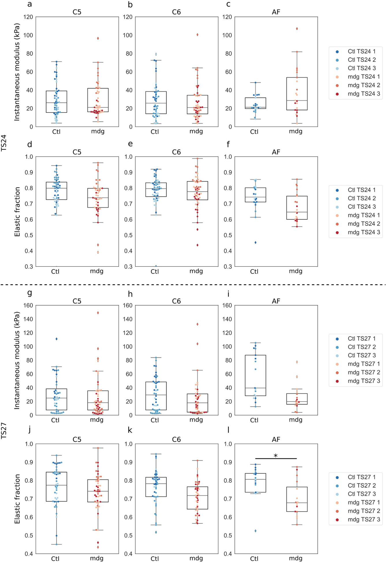
Embryonic muscle forces are necessary for normal vertebral development and spinal curvature, but their involvement in intervertebral disc (IVD) development remains unclear. The aim of the current study was to determine how muscle contractions affect (1) notochord involution and vertebral segmentation, and (2) IVD development including the mechanical properties and morphology, as well as collagen fibre alignment in the annulus fibrosus. Muscular dysgenesis (mdg) mice were harvested at three prenatal stages: at Theiler Stage (TS)22 when notochord involution starts, at TS24 when involution is complete, and at TS27 when the IVD is formed. Vertebral and IVD development were characterised using histology, immunofluorescence, and indentation testing. The results revealed that notochord involution and vertebral segmentation occurred independently of muscle contractions between TS22 and TS24. However, in the absence of muscle contractions, we found vertebral fusion in the cervical region at TS27, along with (i) a displacement of the nucleus pulposus towards the dorsal side, (ii) a disruption of the structural arrangement of collagen in the annulus fibrosus, and (iii) an increase in viscous behaviour of the annulus fibrosus. These findings emphasise the important role of mechanical forces during IVD development, and demonstrate a critical role of muscle loading during development to enable proper annulus fibrosus formation. They further suggest a need for mechanical loading in the creation of fibre-reinforced tissue engineering replacement IVDs as a therapy for IVD degeneration.


Figures

Fig. 1.
Fig. 1.. Methods overview.
(a) Wild type and muscular dysgenesis (mdg) mouse embryos were harvested at Theiler stages (TS) 22, TS24, and TS27. The spine was carefully dissected. (b) 25 μm-thick frozen serial sections were cut and collected on 5 consecutive slides, starting with sections represented by the thin plain line, following by sections represented by the dotted line, dashed line, and thick plain line. In this way, the 5 slides were almost identical. (c) Sections from 1 slide (yellow in b) were stained with alcian blue and picrosirius red to assess vertebral segmentation in the cervical, thoracic, and lumbar regions. (d) Immunofluorescence analyses were performed on sections from 3 slides (orange in b), using collagen I, collagen II, and N-cadherin antibodies. Morphology of each component of the disc was characterised in the cervical region. (e) Mechanical properties of the vertebrae and AF were assessed on 1 section from the last slide (red in b) using instrumented indentation. Approximate locations of indents in the vertebral body (black circle) and AF (black star) of intervertebral discs (sagittal view) are illustrated. Indentation was performed on vertebral bodies (6 to 10 indents per vertebra) and AF located between the cervical C5 and C6 (6 indents per AF). Scale bar = 200 μm.
Fig. 2.
Fig. 2.. Indentation method.
(a) Indentation profile. In the loading phase, the displacement was increased at a constant rate of 2 μm/s until it reached 2 μm. The displacement was then held for 10 s and unloading was carried out at 2 μm/s. (b) Output force-indentation curve showing the loading and unloading phases. (c) Viscoelastic model. The material was represented as a spring in parallel to a Maxwell solid (a spring and dashpot connected in series). (d) The relaxation function was determined by fitting the resultant experimental force-time curve on the loading and holding segments. E0 , E1: elastic moduli associated with each spring; τ1: time constant associated with the dashpot.
Fig. 3.
Fig. 3.. The lack of muscle contractions did not affect vertebral segmentation or notochord involution at TS22 or TS24.
Notochord involution initiated at TS22 (af) and was complete at TS24 in mdg (h,j,l) and control (g,i,k) samples. Vertebral bodies (stained in blue) were clearly separated by the AF in all regions. In mdg samples, reduced intervertebral spaces (*) were observed in the cervical region at TS22 (b) and in the thoracic region at TS24 (j) compared to the controls (a and i, respectively). (af) 25 μm-thick sagittal spine sections stained with alcian blue (cartilage) and picrosirius red (collagen) of representative control and mdg samples at TS22 and (gl) at TS24, in the cervical, thoracic, and lumbar regions. Scale bars: 500 μm. Ca: caudal; Cr: cranial; D: dorsal; V: ventral.
Fig. 4.
Fig. 4.. Absence of muscle contraction resulted in morphological defects of the intervertebral discs at most spinal levels with partial or complete vertebral fusion in the cervical region at TS27, with subtle changes in the thoracic region, and no major changes were in the lumbar region.
Due to variability in the mdg group, all 4 mdg samples are shown and compared with all 3 control samples. In the cervical region, there was vertebral fusion (black arrowhead) in 3 out of 4 mdg samples, to varying degree of severity, from complete vertebral fusion in 1 sample (e,w) to partial vertebral fusion on the ventral side in 2 samples (d,f,v). In 1 particularly severe case, absence of contractile muscles resulted in a disruption of NP formation (e,w), with no NP between C4 and C6 (white ellipse) and a reduced NP between C6 and C7 (*). In the 3 other mdg samples (d,f,g), the NP was decentred towards the dorsal side (+). In the thoracic region, 3 mdg samples displayed wedged vertebral bodies (white arrowhead), while no changes were observed in the lumbar region. (a-t): 25 μm-thick sagittal spine sections stained with alcian blue (cartilage) and picrosirius red (collagen) at TS27, in the cervical (C5–C7 or C4–C7), thoracic (T3–T5), and lumbar (L2–L4) regions of control and mdg spines. No suitable section was obtained for the lumbar region of ‘mdg 2’ due to a staining issue. (uw): Zoomed-in specific regions (black boxes) of (at). Scale bars = 500 μm. Ca: caudal; Cr: cranial; D: dorsal; V: ventral.
Fig. 5.
Fig. 5.. Formation of notochord sheath and initiation of notochord involution at TS22 were not dependent on muscle contractions. The notochord formed normally in the mdg group, and was enveloped in a continuous type II collagen-rich notochord sheath that extended along the craniocaudal axis
(b). Notochord cells expressed N-cadherin and started migrating towards the development site of NP (f). Cervical region of representative TS22 control and mdg spines shown (sagittal view). Due to the size of the samples and the small numbers of sections showing the notochord, different segments of the cervical region were imaged. Scale bar = 200 μm. Ca: caudal; Cr: cranial; D: dorsal; V: ventral.
Fig. 6.
Fig. 6.. Muscle activity was not required for formation of each component of the intervertebral disc at TS24, but ectopic expression of N-cadherin was observed in 2 out of 3 mdg samples.
The NP formed normally in mdg samples (f), indicating completion of notochord involution, and the AF was divided into a collagen II-rich inner part showing a lamellar arrangement (d,h) and a collagen I-rich outer part (f). While N-cadherin was expressed only in the NP of control spines, it was scattered throughout the spine in 2 out of 3 mdg samples (f). (af) Cervical region (C5–C6) of representative control and mdg spines shown (sagittal view). (gh) Zoomed in view of collagen II expression showing the lamellar structure in the inner AF (regions shown from white boxes in cd). Scale bars = 200 μm. Ca: caudal; Cr: cranial; D: dorsal; V: ventral.
Fig. 7.
Fig. 7.. Muscle activity was required for the lamellar arrangement of the inner AF at TS27, and some discs were highly abnormal in the mdg samples.
All mdg samples displayed dense collagen II signal in the inner AF (gj), but the lamellar structure was affected to varying degrees. One mdg sample showed a complete lack of arrangement between cervical C3 and C6 (h,r), while others displayed a partial lack of arrangement on the ventral side (g,i,q,s) or a less prominent structure (j,t) compared to the control (f,p). The outer AF and NP formed normally in mdg specimens and expressed collagen I and N-cadherin, respectively, except in 1 severe case in which both structures were absent between cervical C3 and C6 (c,m). (ao) Collagen I (ae), collagen II (fj), and N-cadherin (ko) in the cervical region (C5–C6) of TS27 control (a,f,k) and mdg spines be, gj, lo). Due to variability in the mdg group, all 4 mdg samples are shown and compared with 1 representative control. (pt) Zoomed in view of collagen II expression in ventral outer AF illustrating loss of lamellar structure in mdg specimens (regions shown from white boxes in fj). Ca: caudal; Cr: cranial; D: dorsal; V: ventral. Scale bars = 200 μm.
Fig. 8.
Fig. 8.. Collagen II orientation in the ventral side of the inner AF was disrupted in mdg samples, while its organisation was not affected on the dorsal side.
On the ventral side of mdg samples, collagen II was orientated in a more oblique direction at TS24 compared to the controls, where it was mostly orientated in the vertical direction (d,i). At TS27, while collagen II in control samples displayed a circumferential pattern with opposite orientations in the cranial and caudal regions, collagen II in mdg samples had the same oblique orientation in both regions, indicating an absence of circumferential organisation (f,h,j). On the dorsal side, collagen II was mainly orientated in the cranio-caudal direction as in controls at both stages (e,g,j). (ah) Circular histograms showing the distribution of collagen II orientation (relative to the dorsoventral axis) of 3 control and 3 mdg samples in 4 regions of the AF C5–C6 at TS24 and TS27. (ij) Schematic of the AF showing the mean orientation of collagen II in the 4 regions examined of control (solid lines) and mdg (dashed lines) groups at TS24 and TS27 (n = 3 per group). *: no predominant orientation. Ca: caudal; Cr: cranial; D: dorsal; V: ventral.
Fig. 9.
Fig. 9.. Elastic fraction of the AF at TS27 lower in mdg samples than in controls, while no effects on the vertebral bodies at TS24 and TS27, or on the AF at TS24, were observed
(a–c). Instantaneous modulus of the cervical vertebrae C5 and C6 and AF C5–C6 of control (blue) and mdg (red) embryos at TS24. (df) Elastic fraction of the cervical vertebrae C5 and C6 and AF C5–C6 of control (blue) and mdg (red) embryos at TS24. (gi) Instantaneous modulus of the cervical vertebrae C5 and C6 and AF C5–C6 of control (blue) and mdg (red) embryos at TS27. (jl) Elastic fraction of the cervical vertebrae C5 and C6 and AF C5–C6 of control (blue) and mdg (red) embryos at TS27. Dots represent individual data points (at least 6 indents per structure and per specimen) and colours represent specimens (n = 3 per group and per stage) * p < 0.05.

Similar articles

Cited by

References

    1. Ahmed S, Nowlan NC (2020) Initiation and emerging complexity of the collagen network during prenatal skeletal development. Eur Cell Mater 39: 136–155. - PubMed
    1. Alkhatib B, Ban GI, Williams S, Serra R (2018) IVD development: nucleus pulposus development and sclerotome specification. Curr Mol Biol Rep 4: 132–141. - PMC - PubMed
    1. Antoniou J, Steffen T, Nelson F, Winterbottom N, Hollander AP, Poole RA, Aebi M, Alini M (1996) The human lumbar intervertebral disc: evidence for changes in the biosynthesis and denaturation of the extracellular matrix with growth, maturation, ageing, and degeneration. J Clin Invest 98: 996–1003. - PMC - PubMed
    1. Aszódi A, Chan D, Hunziker E, Bateman JF, Fässler R (1998) Collagen II is essential for the removal of the notochord and the formation of intervertebral discs. J Cell Biol 143: 1399–1412. - PMC - PubMed
    1. Blecher R, Krief S, Galili T, Biton IE, Stern T, Assaraf E, Levanon D, Appel E, Anekstein Y, Agar G, Groner Y, Zelzer E (2017) The proprioceptive system masterminds spinal alignment: insight into the mechanism of scoliosis. Dev Cell 42: 388–399. - PubMed

Additional References

    1. Flück M, Giraud M-N, Tunç V, Chiquet M (2003) Tensile stress-dependent collagen XII and fibronectin production by fibroblasts requires separate pathways, Biochim Biophys Acta 1593: 239–248. - PubMed
    1. Lindholm ME, Rundqvist H (2016) Skeletal muscle hypoxia-inducible factor-1 and exercise. Exp Physiol 101: 28–32. - PubMed
    1. Sato S, Kimura A, Ozdemir J, Asou Y, Miyazaki M, Jinno T, Ae K, Liu X, Osaki M, Takeuchi Y, Fukumoto S, Kawaguchi H, Haro H, Shinomiya K, Karsenty G, Takeda S (2008) The distinct role of the Runx proteins in chondrocyte differentiation and intervertebral disc degeneration: findings in murine models and in human disease. Arthritis Rheum 58: 2764–2775. - PubMed

Publication types

MeSH terms

LinkOut - more resources