. 2021 Jun 3;108(6):1083-1094.
doi: 10.1016/j.ajhg.2021.04.025. Epub 2021 May 21.

Non-coding region variants upstream of MEF2C cause severe developmental disorder through three distinct loss-of-function mechanisms

Affiliations

Non-coding region variants upstream of MEF2C cause severe developmental disorder through three distinct loss-of-function mechanisms

Caroline F Wright et al. Am J Hum Genet. .

Abstract

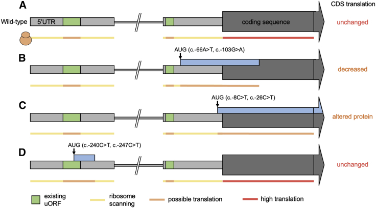
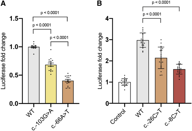
Clinical genetic testing of protein-coding regions identifies a likely causative variant in only around half of developmental disorder (DD) cases. The contribution of regulatory variation in non-coding regions to rare disease, including DD, remains very poorly understood. We screened 9,858 probands from the Deciphering Developmental Disorders (DDD) study for de novo mutations in the 5' untranslated regions (5' UTRs) of genes within which variants have previously been shown to cause DD through a dominant haploinsufficient mechanism. We identified four single-nucleotide variants and two copy-number variants upstream of MEF2C in a total of ten individual probands. We developed multiple bespoke and orthogonal experimental approaches to demonstrate that these variants cause DD through three distinct loss-of-function mechanisms, disrupting transcription, translation, and/or protein function. These non-coding region variants represent 23% of likely diagnoses identified in MEF2C in the DDD cohort, but these would all be missed in standard clinical genetics approaches. Nonetheless, these variants are readily detectable in exome sequence data, with 30.7% of 5' UTR bases across all genes well covered in the DDD dataset. Our analyses show that non-coding variants upstream of genes within which coding variants are known to cause DD are an important cause of severe disease and demonstrate that analyzing 5' UTRs can increase diagnostic yield. We also show how non-coding variants can help inform both the disease-causing mechanism underlying protein-coding variants and dosage tolerance of the gene.

Keywords: developmental disorders, clinical genetic testing, non-coding region variants, 5' UTR variants.


Conflict of interest statement

K.J.K. is a consultant for Vor Biopharma. J.J. and K.R. are employees of GeneDx, Inc. K.R. holds shares in Opko Health, Inc. B.D.Z. is a member of the speakers bureau for Biogen, Neurelis, and Supernus. S.A.C. is co-founder and shareholder of Enleofen Bio Pte Ltd. M.E.H. is co-founder, shareholder, consultant, and non-executive director of Congenica Ltd. All other authors declare no competing interests.

Figures

Figure 1
Figure 1
Schematic of the N terminus of MEF2C Shown are the wild-type structure (A) and the position and effect of uAUG-creating variants identified as de novo in developmental disorder cases (B and C) and in gnomAD population control subjects (D). (A) The two 5′ UTR exons are shown as light gray boxes, separated by an intron shown as a thinner broken gray line. Upstream open reading frames (uORFs) already present in the sequence are shown in green. Variant positions are represented by arrows. New ORFs created by the variants are shown as blue boxes. (B) Two case variants create ORFs that overlap the coding sequence (CDS) out-of-frame (oORF-creating). If translation initiates at the uAUG, the ribosome will not translate the CDS. (C) Two recurrent case variants create uAUGs in-frame with the CDS. If translation initiates at this uAUG, an elongated protein will be translated. (D) Two variants identified in gnomAD create uORFs far upstream of the CDS which would not be predicted to disrupt translation of the normal protein.
Figure 2
Figure 2
uAUG-creating variants decrease translation of MEF2C or transactivation of target genes (A) MEF2C 5′ UTR out-of-frame overlapping ORF (oORF)-creating variants c.−103G>A and c.−66A>T (Figure 1B) reduce downstream luciferase expression relative to wild-type (WT) 5′ UTR in a translation reporter assay. Reduction is stronger for c.−66A>T (moderate uAUG Kozak context) than for c.−103G>A (weak Kozak context). (B) Overexpression of MEF2C with the WT 5′ UTR/CDS induces expression of luciferase from a MEF2C-dependent enhancer-luciferase reporter construct, relative to an empty pcDNA3.1 construct negative control. The MEF2C N-terminus-extending variants c.−26C>T (9 amino acids) and c.−8C>T (3 amino acids; Figure 1C) both reduce transactivation. Bars are colored by Kozak consensus: yellow = weak; orange = moderate; red = strong. Luciferase expression was normalized for transfection efficiency. Error bars represent the standard error of the mean. p values were calculated using 1-way ANOVA followed by Tukey’s post-test.
Figure 3
Figure 3
MEF2C missense variants in individuals with DD are enriched in the N-terminal region and likely disrupt DNA binding (A) The N-terminal region of MEF2C is highly constrained for missense variants in gnomAD (obs/exp = 0.069), with much lower constraint across the rest of the protein (obs/exp = 0.41). This region of high constraint correlates with the location of the majority of de novo missense variants identified in DD-affected individuals (red circles), while gnomAD variants are mostly outside of this N-terminal region (gray circles). (B) The N-terminal portion of the MEF2C dimer (1–92), modeled using structures of the human MEF2A dimer which is 96% identical in sequence to MEF2C, bound directly to its consensus DNA sequence. Side chains of amino acids with pathogenic de novo missense variants from DDD, GeneDx, and ClinVar are shown in yellow, with gnomAD MEF2C missense variants in gray. Most pathogenic missense variants either protrude directly into the DNA or are located in the DNA-binding helix. In particular, the terminal amine (Gly2, top inset) along with Arg3 (bottom inset) act as reader-heads for nucleobase specificity, which is likely disrupted in the N-terminal extension variants (middle inset). All pathogenic and gnomAD variants can be viewed in our interactive protein structure browser (see link in Web Resources). (C and D) Missense variants from DD-affected individuals (DDD, GeneDx, and ClinVar) are significantly more disruptive to the interaction with DNA as measured by ΔΔG values (C) and closer to the bound DNA molecule (D) than MEF2A-D variants in gnomAD (see STAR Methods). p values were calculated using a Wilcoxon rank sum test.
Figure 4
Figure 4
Characteristics and sequencing coverage of the 5′ UTRs of DDG2P genes (A and B) 5′ UTRs of DDG2P haploinsufficient genes (red) are longer (A), and a higher proportion have multiple exons (B) compared to 5′ UTRs of all genes (light gray), and other DDG2P genes (dark gray). Mean lengths for each gene set in (A) are shown as dotted lines. (C) The coverage of 5′ UTRs decays rapidly with distance from the CDS (x axis truncated at 1,000 bps). Note that these figures were calculated using exome sequence data from the DDD study and may vary between different exome capture designs. (D) The position of DNA-binding domains (including homeodomains, zinc-fingers, and specific DNA-binding domains) in DDG2P haploinsufficient genes with respect to the N terminus of the protein; MEF2C is one of three proteins with a DNA-binding domain that starts within 10 bps of the N terminus.

References

    1. Short P.J., McRae J.F., Gallone G., Sifrim A., Won H., Geschwind D.H., Wright C.F., Firth H.V., FitzPatrick D.R., Barrett J.C., Hurles M.E. De novo mutations in regulatory elements in neurodevelopmental disorders. Nature. 2018;555:611–616. - PMC - PubMed
    1. Spielmann M., Mundlos S. Looking beyond the genes: the role of non-coding variants in human disease. Hum. Mol. Genet. 2016;25(R2):R157–R165. - PubMed
    1. An J.-Y., Lin K., Zhu L., Werling D.M., Dong S., Brand H., Wang H.Z., Zhao X., Schwartz G.B., Collins R.L. Genome-wide de novo risk score implicates promoter variation in autism spectrum disorder. Science. 2018;362:362. - PMC - PubMed
    1. Chong J.X., Buckingham K.J., Jhangiani S.N., Boehm C., Sobreira N., Smith J.D., Harrell T.M., McMillin M.J., Wiszniewski W., Gambin T., Centers for Mendelian Genomics The Genetic Basis of Mendelian Phenotypes: Discoveries, Challenges, and Opportunities. Am. J. Hum. Genet. 2015;97:199–215. - PMC - PubMed
    1. Kaplanis J., Samocha K.E., Wiel L., Zhang Z., Arvai K.J., Eberhardt R.Y., Gallone G., Lelieveld S.H., Martin H.C., McRae J.F., Deciphering Developmental Disorders Study Evidence for 28 genetic disorders discovered by combining healthcare and research data. Nature. 2020;586:757–762. - PMC - PubMed

Publication types

Substances