. 2021 Oct:97:13-21.
doi: 10.1016/j.bbi.2021.05.012. Epub 2021 May 19.

Detection of SARS-CoV-2 genome and whole transcriptome sequencing in frontal cortex of COVID-19 patients

Affiliations

Detection of SARS-CoV-2 genome and whole transcriptome sequencing in frontal cortex of COVID-19 patients

Stella Gagliardi et al. Brain Behav Immun. 2021 Oct.

Abstract

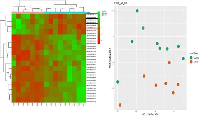
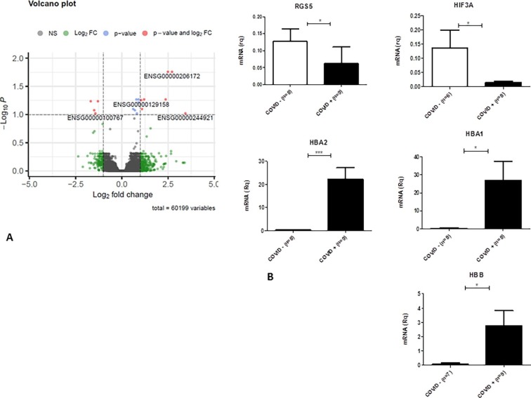
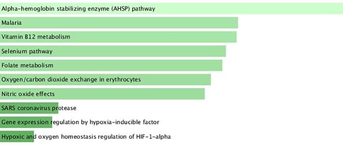
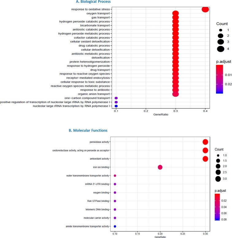
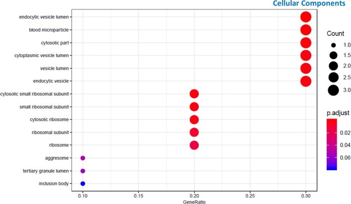
SARS-Cov-2 infection is frequently associated with Nervous System manifestations. However, it is not clear how SARS-CoV-2 can cause neurological dysfunctions and which molecular processes are affected in the brain. In this work, we examined the frontal cortex tissue of patients who died of COVID-19 for the presence of SARS-CoV-2, comparing qRT-PCR with ddPCR. We also investigated the transcriptomic profile of frontal cortex from COVID-19 patients and matched controls by RNA-seq analysis to characterize the transcriptional signature. Our data showed that SARS-CoV-2 could be detected by ddPCR in 8 (88%) of 9 examined samples while by qRT-PCR in one case only (11%). Transcriptomic analysis revealed that 11 genes (10 mRNAs and 1 lncRNA) were differential expressed when frontal cortex of COVID-19 patients were compared to controls. These genes fall into categories including hypoxia, hemoglobin-stabilizing protein, hydrogen peroxide processes. This work demonstrated that the quantity of viral RNA in frontal cortex is minimal and it can be detected only with a very sensitive method (ddPCR). Thus, it is likely that SARS-CoV-2 does not actively infect and replicate in the brain; its topography within encephalic structures remains uncertain. Moreover, COVID-19 may have a role on brain gene expression, since we observed an important downregulation of genes associated to hypoxia inducting factor system (HIF) that may inhibit the capacity of defense system during infection and oxigen deprivation, showing that hypoxia, well known multi organ condition associated to COVID-19, also marked the brain.

Keywords: Frontal cortex; Hemoglobin; Hypoxia; SARS-CoV-2; Transcriptome.


Conflict of interest statement

The authors declare that they have no known competing financial interests or personal relationships that could have appeared to influence the work reported in this paper.

Figures

Fig. 1
Fig. 1
Widespread oedema is clearly evident in supragranular layers of gray matter (A) by Hematoxylin and Eosin (HE) staining. Small vessel disease (SVD) is identified by perivascular space dilatation, features of arteriolosclerosis (B), and myelin loss (C) observed respectively by HE and Luxol Fast Blue stainings. In cases with a low amount of viral RNA perivascular inflammatory infiltrates appear as the most prominent inflammatory feature (D; HE staining); while in cases with dementia and higher amount of viral RNA there are more prominent microglial nodule as shown by both, HE (E) and CD68 antibody (F). In COVID-19 cases affected by Alzheimer’s Disease, 4G8 antibody detects amyloid plaques (G) and, possibly, cerebral amyloid angiopathy (H). In the same cases, AT8 immunoreactivity shows neuritic plaques, tangles and threads in frontal cortex (I), hippocampus (J) and occipital cortex (K). Three COVID-19 cases with dementia are shown in panels A-C and E-K, while panel D shows a COVID-19 case without dementia carrying al low viral RNA load. Images include frontal lobes (A-I), hippocampus (J) and occipital lobe (K). Scale bar: 256 µm (A,G); 340 µm (B,H,I,J,K); 807 µm (C); 122 µm (D,F); 73 µm (E). (For interpretation of the references to color in this figure legend, the reader is referred to the web version of this article.)
Fig. 2
Fig. 2
Expression profiles of differentially expressed genes (DEGs) between COVID-19 patients and healthy controls. In panel (A), Heatmap of log-normalized RNA expression counts for patients and controls are represented, while in panel (B) PCA of DEGs is shown.
Fig. 3
Fig. 3
Volcano Plot and qPCR validation. Volcano plot of differentially expressed genes (DEGs) between COVID-19 patients and healthy controls (A). Gene ID of the most deregulated transcripts are reported, red dots represent DEGs based on p-value and Fold Change. Most DEGs have been validated by qPCR (B). (For interpretation of the references to color in this figure legend, the reader is referred to the web version of this article.)
Fig. 4
Fig. 4
BioPlanet2019 pathway analysis for DEGs in COVID-19 patients compared to healthy controls. In panel A BioPlanet2019 pathways enriched by deregulated genes are shown. The length of the bar represents the significance of that specific gene-set or term. The brighter the color, the more significant that term is. In panel B is showed the cluster gram of DEGs associated to enriched pathways.
Fig. 5
Fig. 5
GO analysis for DEGs in COVID-19 patients compared to healthy controls by AnnotationHub. DEG enriched GO terms for Biological process (A) and Molecular Function (B) are shown.
Supplementary figure 1

References

    1. Ahmad I., Rathore F.A. Neurological manifestations and complications of COVID-19: A literature review. J Clin Neurosci. 2020;77:8–12. doi: 10.1016/j.jocn.2020.05.017. PMID: 32409215; PMCID: PMC7200361. - DOI - PMC - PubMed
    1. Amri F., Ghouili I., Tonon M., Amri M., Masmoudi-Kouki O. Hemoglobin-Improved Protection in Cultured Cerebral Cortical Astroglial Cells: Inhibition of Oxidative Stress and Caspase Activation. Frontiers in Endocrinology. 2017;8:67. doi: 10.3389/fendo.2017.00067. - DOI - PMC - PubMed
    1. K.S. Appelberg S. Gupta svensson akujsärvi, Sara & Ambikan, Anoop & Mikaeloff, Flora & Saccon, Elisa & Végvári, Akos & Benfeitas, Rui & Sperk, Maike & Ståhlberg, Marie & Krishnan, Shuba & Singh, Kamal & Penninger, Josef & Mirazimi, Ali & Neogi, Ujjwal. Dysregulation in Akt/mTOR/HIF-1 signaling identified by proteo-transcriptomics of SARS-CoV-2 infected cells. Emerging Microbes & Infections. 9 2020 1 36 10.1080/22221751.2020.1799723. - PMC - PubMed
    1. Asadi-Pooya A.A., Simani L. Central nervous system manifestations of COVID-19: A systematic review. J Neurol Sci. 2020;15(413) doi: 10.1016/j.jns.2020.116832. PMID: 32299017; PMCID: PMC7151535. - DOI - PMC - PubMed
    1. Baig A.M., Khaleeq A., Ali U., Syeda H. Evidence of the COVID-19 Virus Targeting the CNS: Tissue Distribution, Host-Virus Interaction, and Proposed Neurotropic Mechanisms. ACS Chem Neurosci. 2020;11(7):995–998. doi: 10.1021/acschemneuro.0c00122. PMID: 32167747; PMCID: PMC7094171. - DOI - PubMed

Publication types