

A novel nomogram containing acute radiation esophagitis predicting radiation pneumonitis in thoracic cancer receiving radiotherapy
- PMID: 34022830
- PMCID: PMC8140476
- DOI: 10.1186/s12885-021-08264-y
A novel nomogram containing acute radiation esophagitis predicting radiation pneumonitis in thoracic cancer receiving radiotherapy
Abstract
Background: Radiation-induced pneumonitis (RP) is a non-negligible and sometimes life-threatening complication among patients with thoracic radiation. We initially aimed to ascertain the predictive value of acute radiation-induced esophagitis (SARE, grade ≥ 2) to symptomatic RP (SRP, grade ≥ 2) among thoracic cancer patients receiving radiotherapy. Based on that, we established a novel nomogram model to provide individualized risk assessment for SRP.
Methods: Thoracic cancer patients who were treated with thoracic radiation from Jan 2018 to Jan 2019 in Shandong Cancer Hospital and Institute were enrolled prospectively. All patients were followed up during and after radiotherapy (RT) to observe the development of esophagitis as well as pneumonitis. Variables were analyzed by univariate and multivariate analysis using the logistic regression model, and a nomogram model was established to predict SRP by "R" version 3.6.0.
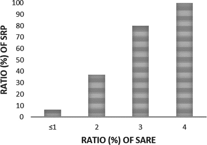
Results: A total of 123 patients were enrolled (64 esophageal cancer, 57 lung cancer and 2 mediastinal cancer) in this study prospectively. RP grades of 0, 1, 2, 3, 4 and 5 occurred in 29, 57, 31, 0, 3 and 3 patients, respectively. SRP appeared in 37 patients (30.1%). In univariate analysis, SARE was shown to be a significant predictive factor for SRP (P < 0.001), with the sensitivity 91.9% and the negative predictive value 93.5%. The incidence of SRP in different grades of ARE were as follows: Grade 0-1: 6.5%; Grade 2: 36.9%; Grade 3: 80.0%; Grade 4: 100%. Besides that, the dosimetric factors considering total lung mean dose, total lung V5, V20, ipsilateral lung mean dose, ipsilateral lung V5, and mean esophagus dose were correlated with SRP (all P < 0.05) by univariate analysis. The incidence of SRP was significantly higher in patients whose symptoms of RP appeared early. SARE, mean esophagus dose and ipsilateral mean lung dose were still significant in multivariate analysis, and they were included to build a predictive nomogram model for SRP.
Conclusions: As an early index that can reflect the tissue's radiosensitivity visually, SARE can be used as a predictor for SRP in patients receiving thoracic radiation. And the nomogram containing SARE may be fully applied in future's clinical work.
Keywords: Acute radiation-induced esophagitis; Nomogram; Radiotherapy; Symptomatic radiation-induced pneumonitis; Thoracic cancer.
Conflict of interest statement
The authors declare that they have no competing interests.
Figures


Similar articles
-
Correlation of dosimetric and clinical factors with the development of esophagitis and radiation pneumonitis in patients with limited-stage small-cell lung carcinoma.Clin Lung Cancer. 2015 May;16(3):216-20. doi: 10.1016/j.cllc.2014.11.008. Epub 2014 Dec 2. Clin Lung Cancer. 2015. PMID: 25532963
-
Predicting Severe Radiation Pneumonitis in Patients With Locally-Advanced Non-Small-Cell Lung Cancer After Thoracic Radiotherapy: Development and Validation of a Nomogram Based on the Clinical, Hematological, and Dose-Volume Histogram Parameters.Clin Lung Cancer. 2025 Jul;26(5):393-406. doi: 10.1016/j.cllc.2025.02.009. Epub 2025 Feb 21. Clin Lung Cancer. 2025. PMID: 40087057
-
Study of the predictors for radiation pneumonitis in patient with non-small cell lung cancer received radiotherapy after pneumonectomy.Cancer Radiother. 2021 Jun;25(4):323-329. doi: 10.1016/j.canrad.2020.11.001. Epub 2021 Jan 11. Cancer Radiother. 2021. PMID: 33446419
-
Is Thoracic Radiotherapy an Absolute Contraindication for Treatment of Lung Cancer Patients With Interstitial Lung Disease? A Systematic Review.Clin Oncol (R Coll Radiol). 2022 Dec;34(12):e493-e504. doi: 10.1016/j.clon.2022.01.043. Epub 2022 Feb 12. Clin Oncol (R Coll Radiol). 2022. PMID: 35168842
-
Acute and Late Pulmonary Effects After Radiation Therapy in Childhood Cancer Survivors: A PENTEC Comprehensive Review.Int J Radiat Oncol Biol Phys. 2024 Jun 1;119(2):533-548. doi: 10.1016/j.ijrobp.2022.01.052. Epub 2022 May 4. Int J Radiat Oncol Biol Phys. 2024. PMID: 35525723 Review.
Cited by
-
Radiation-induced esophagitis and lung injury during esophageal squamous cell cancer therapy is correlated to tumor gene expression phenotype.Toxicol Res (Camb). 2025 May 4;14(3):tfaf062. doi: 10.1093/toxres/tfaf062. eCollection 2025 Jun. Toxicol Res (Camb). 2025. PMID: 40331088
-
Safety and Feasibility of Esophagectomy Following Neoadjuvant Immunotherapy Combined with Chemotherapy for Esophageal Squamous Cell Carcinoma.Front Surg. 2022 May 26;9:851745. doi: 10.3389/fsurg.2022.851745. eCollection 2022. Front Surg. 2022. PMID: 35711710 Free PMC article.
-
Prognostic nomogram for overall survival of elderly esophageal cancer patients receiving neoadjuvant therapy: a population-based analysis.J Gastrointest Oncol. 2024 Dec 31;15(6):2376-2388. doi: 10.21037/jgo-24-392. Epub 2024 Dec 4. J Gastrointest Oncol. 2024. PMID: 39816016 Free PMC article.
-
A simple-to-use nomogram for predicting the risk of radiation pneumonitis in patients with thoracic segment esophageal squamous cell carcinoma.Transl Cancer Res. 2022 Oct;11(10):3754-3766. doi: 10.21037/tcr-22-582. Transl Cancer Res. 2022. PMID: 36388040 Free PMC article.
-
A dynamic nomogram predicting symptomatic pneumonia in patients with lung cancer receiving thoracic radiation.BMC Pulm Med. 2024 Feb 26;24(1):99. doi: 10.1186/s12890-024-02899-w. BMC Pulm Med. 2024. PMID: 38409084 Free PMC article.
References
-
- Seppenwoolde Y, De Jaeger K, Lebesque JV. In regard to Tsujino et al.: predictive value of dose-volume histogram parameters for predicting radiation pneumonitis after concurrent chemoradiation for lung cancer. IJROBP 2003;55:110-115. Int J Radiat Oncol Biol Phys. 2003;56(4):1208–1209. doi: 10.1016/S0360-3016(03)00345-6. - DOI - PubMed
Publication types
MeSH terms
Grants and funding
LinkOut - more resources
Full Text Sources
Other Literature Sources
Research Materials
