. 2021 May 22;19(1):151.
doi: 10.1186/s12951-021-00888-3.

Extracellular vesicles-based pre-targeting strategy enables multi-modal imaging of orthotopic colon cancer and image-guided surgery

Affiliations

Extracellular vesicles-based pre-targeting strategy enables multi-modal imaging of orthotopic colon cancer and image-guided surgery

Boping Jing et al. J Nanobiotechnology. .

Abstract

Backgroud: Colon cancer contributes to high mortality rates as the result of incomplete resection in tumor surgery. Multimodal imaging can provide preoperative evaluation and intraoperative image-guiding. As biocompatible nanocarriers, extracellular vesicles hold great promise for multimodal imaging. In this study, we aim to synthesized an extracellular vesicles-based nanoprobe to visualize colon cancer with positron-emission tomography/computed tomography (PET/CT) and near-infrared fluorescence (NIRF) imaging, and investigated its utility in image-guided surgery of colon cancer in animal models.

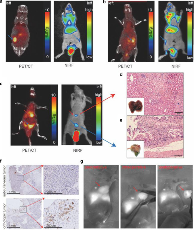
Results: Extracellular vesicles were successfully isolated from adipose-derived stem cells (ADSCs), and their membrane vesicles were observed under TEM. DLS detected that the hydrodynamic diameters of the extracellular vesicles were approximately 140 nm and the zeta potential was - 7.93 ± 0.24 mV. Confocal microscopy showed that extracellular vesicles had a strong binding ability to tumor cells. A click chemistry-based pre-targeting strategy was used to achieve PET imaging in vivo. PET images and the biodistribution results showed that the best pre-targeting time was 20 h, and the best imaging time was 2 h after the injection of 68 Ga-L-NETA-DBCO. The NIRF images showed that the tumor had clear images at all time points after administration of nanoparticles and the Tumor/Muscle ratio peaked at 20 h after injection. Our data also showed that both PET/CT and NIRF imaging clearly visualized the orthotopic colon cancer models, providing preoperative evaluation. Under real-time NIRF imaging, the tumor location and tumor boundary could be clearly observed.

Conclusions: In brief, this novel nanoprobe may be useful for multi-modal imaging of colon cancer and NIRF image-guided surgery. More importantly, this study provides a new possibility for clinical application of extracellular vesicles as nanocarriers.

Keywords: Extracellular vesicles; Image-guided surgery; Multimodal imaging; NIRF; PET/CT.


Conflict of interest statement

The authors declare that there are no conflicts of interest regarding the publication of this paper.

Figures

Scheme 1
Scheme 1
The schematics of multimodal PET and NIRF imaging and real-time NIRF intra-operation based on extracellular vesicles from ADSCs
Fig. 1
Fig. 1
The identification and characteristics of ADSCs-EV. a Membrane vesicles were observed under Transmission Electron Microscope (TEM). b Western blot confirmed the expression of ADSCs-EV markers (CD63, CD9). c The average hydrodynamic diameters of ADSCs-EV and Cy7-EV-N3. b The zeta potential of ADSCs-EV and Cy7-EV-N3. e The changes of hydrodynamic diameters of ADSCs-EV at 4℃ over 8 days. f The changes of zeta potential of ADSCs-EV at 4℃ over 8 days
Fig. 2
Fig. 2
Tumor-binding ability of Cy5-EV-N3 and cell uptakes of Cy7-EV-N3 with different incubation time. a Fluorescence images of HCT116 cells after incubating with Cy5-EV-N3 for different time points (200 ×). b Corresponding quantification of the fluorescent intensity. c Tumor-binding ability was detected by confocal imaging (600 ×). d Uptakes of 68 Ga-L-NETA-DBCO in HCT116 tumor cells (incubated with Cy7-EV-N3 for different time periods) at the indicated time points. Bars represent means ± SD (n = 3). *P < 0.05; **P < 0.01; ***P < 0.001
Fig. 3
Fig. 3
In vivo PET imaging of HCT116 tumor-bearing mice at different pre-targeting time points. a Representative static PET images after 1 h, 2 h and 3 h p.i. of 68 Ga-L-NETA-DBCO. b T/M ratios of different time (1 h, 2 h and 3 h after the injection of 68 Ga-L-NETA-DBCO). c Representative static PET images after 2 h p.i. of 68 Ga-L-NETA-DBCO with different pre-targeting time. d Tumor/muscle ratios of different pre-targeting time
Fig. 4
Fig. 4
Biodistribution analysis at different pre-targeting time points (10 h, 20 h, and 30 h). a Tissues uptakes of HCT116 tumor-bearing mice at 2 h p.i. of 68 Ga-L-NETA-DBCO with different pretargeting time. b Tumor uptakes of different pretargeting time points. ce Tumor/Muscle ratios, Tumor/Liver ratios and Tumor/Spleen ratios of different pretargeting time points. All bars represent as means ± SD (n = 4). *P < 0.05; **P < 0.01; ***P < 0.001
Fig. 5
Fig. 5
NIRF imaging of HCT116 tumor-bearing nude mice and the tissues. a NIRF images of tumor-bearing mice at different time points (1, 5, 10, 20, 30 and 50 h) after the injection of Cy7-EV-N3. b T/M ratios at different time points after Cy7-EV-N3 injection (n = 3, **P < 0.01; ***P < 0.001). c NIRF images of ex vivo tissues at different time points after injection (10, 20 and 30 h). T Tumor, M Muscle, LI Large Intestinal, SI Small Intestinal, K Kidney, SP Spleen, L Liver, ST Stomach
Fig. 6
Fig. 6
Multimodal PET/CT/NIRF images of orthotopic colon cancer and pre-, intra- and postoperative NIRF images. Multimodal PET/CT and NIRF images of the left (a) and right (b) orthotopic colon cancer model. The blue arrows denote colon tumors in situ. c Multimodal PET/CT and NIRF images of orthotopic colon cancer model (blue arrows) with liver metastasis (red arrows). d The visual observation of tumors and HE staining of pathological sections (scale bar: 50 μm). f Immunohistochemistry assay of CD31 in orthotopic and subcutaneous colon cancer. g Representative NIRF images of tumor-bearing mice pre-, intra-, and postoperatively. The red arrows point to the tumor
Fig. 7
Fig. 7
In vivo toxicity evaluation by blood test and histology analysis. ad Liver function makers (ALT, AST and ALP) and kidney function markers (BUN and CRE) after i.v. injection with N3-EV-Cy7 + 68 Ga-L-NETA-DBCO over 1 d and 7 d. e Representative H&E staining images of major organs from the euthanized mice. Bar = 50 μm. All bars represent as means ± SD (n = 4)

Similar articles

Cited by

References

    1. Aliperti LA, Predina JD, Vachani A, Singhal S. Local and systemic recurrence is the Achilles heel of cancer surgery. Ann Surg Oncol. 2011;18:603–607. doi: 10.1245/s10434-010-1442-0. - DOI - PMC - PubMed
    1. Kim MH, Kim SG, Kim DW. A novel dual-modality imaging agent targeting folate receptor of tumor for molecular imaging and fluorescence-guided surgery. Ann Nucl Med. 2019;33:606–616. doi: 10.1007/s12149-019-01369-2. - DOI - PubMed
    1. Teraphongphom N, Chhour P, Eisenbrey JR, Naha PC, Witschey WR, Opasanont B, Jablonowski L, Cormode DP, Wheatley MA. Nanoparticle loaded polymeric microbubbles as contrast agents for multimodal imaging. Langmuir. 2015;31:11858–11867. doi: 10.1021/acs.langmuir.5b03473. - DOI - PMC - PubMed
    1. Lee SY, Jeon SI, Jung S, Chung IJ, Ahn CH. Targeted multimodal imaging modalities. Adv Drug Deliv Rev. 2014;76:60–78. doi: 10.1016/j.addr.2014.07.009. - DOI - PubMed
    1. Li C, Zhang Y, Wang M, Zhang Y, Chen G, Li L, Wu D, Wang Q. In vivo real-time visualization of tissue blood flow and angiogenesis using Ag2S quantum dots in the NIR-II window. Biomaterials. 2014;35:393–400. doi: 10.1016/j.biomaterials.2013.10.010. - DOI - PubMed

Substances

LinkOut - more resources