Review
. 2021 May 22;9(1):96.
doi: 10.1186/s40478-021-01197-4.

Identifying degenerative effects of repetitive head trauma with neuroimaging: a clinically-oriented review

Affiliations
Review

Identifying degenerative effects of repetitive head trauma with neuroimaging: a clinically-oriented review

Breton M Asken et al. Acta Neuropathol Commun. .

Abstract

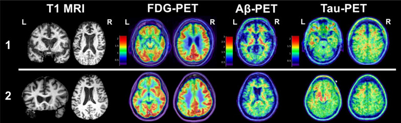
Background and scope of review: Varying severities and frequencies of head trauma may result in dynamic acute and chronic pathophysiologic responses in the brain. Heightened attention to long-term effects of head trauma, particularly repetitive head trauma, has sparked recent efforts to identify neuroimaging biomarkers of underlying disease processes. Imaging modalities like structural magnetic resonance imaging (MRI) and positron emission tomography (PET) are the most clinically applicable given their use in neurodegenerative disease diagnosis and differentiation. In recent years, researchers have targeted repetitive head trauma cohorts in hopes of identifying in vivo biomarkers for underlying biologic changes that might ultimately improve diagnosis of chronic traumatic encephalopathy (CTE) in living persons. These populations most often include collision sport athletes (e.g., American football, boxing) and military veterans with repetitive low-level blast exposure. We provide a clinically-oriented review of neuroimaging data from repetitive head trauma cohorts based on structural MRI, FDG-PET, Aβ-PET, and tau-PET. We supplement the review with two patient reports of neuropathology-confirmed, clinically impaired adults with prior repetitive head trauma who underwent structural MRI, FDG-PET, Aβ-PET, and tau-PET in addition to comprehensive clinical examinations before death.

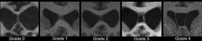
Review conclusions: Group-level comparisons to controls without known head trauma have revealed inconsistent regional volume differences, with possible propensity for medial temporal, limbic, and subcortical (thalamus, corpus callosum) structures. Greater frequency and severity (i.e., length) of cavum septum pellucidum (CSP) is observed in repetitive head trauma cohorts compared to unexposed controls. It remains unclear whether CSP predicts a particular neurodegenerative process, but CSP presence should increase suspicion that clinical impairment is at least partly attributable to the individual's head trauma exposure (regardless of underlying disease). PET imaging similarly has not revealed a prototypical metabolic or molecular pattern associated with repetitive head trauma or predictive of CTE based on the most widely studied radiotracers. Given the range of clinical syndromes and neurodegenerative pathologies observed in a subset of adults with prior repetitive head trauma, structural MRI and PET imaging may still be useful for differential diagnosis (e.g., assessing suspected Alzheimer's disease).

Keywords: Chronic traumatic encephalopathy; Concussion; Magnetic resonance imaging; Neurodegenerative disease; Neuroimaging; Positron emission tomography; Repetitive head trauma; Traumatic brain injury; Traumatic encephalopathy syndrome.


Conflict of interest statement

BMA and GDR declare that they have no competing interests relevant this review.

Figures

Fig. 1
Fig. 1
Enlarged views of representative T1 coronal images for each “grade” of cavum septum pellucidum (CSP). A “Grade 0” septum pellucidum appears crisp without any evidence of cyst or separation (CSP absent). “Grade 1” CSP shows slight interior hypointensity that is not clearly CSF signal intensity (septum unclear/CSP equivocal). Grades 2–4 show clear evidence of CSF signal between separated leaves of the septum pellucidum. The degree of separation between the leaves is used to assign a grade of 2–4: Grade 2 CSP is not wider than the septum, Grade 3 CSP is wider than the septum but less than half the intraventricular width, and Grade 4 CSP is greater than half the intraventricular width. Grading is based on the coronal slice that shows the greatest evidence of separation of the leaves of the septum pellucidum. Figure and caption adapted from Gardner et al. 2016, J Neurotrauma, 33(1):157–61 (permissions pending review and acceptance of manuscript)
Fig. 2
Fig. 2
Representative slices of antemortem structural T1 magnetic resonance imaging (T1 MRI), FDG-PET, Aβ-PET (PIB), and tau-PET (FTP) for 2 clinically impaired adults with prior repetitive head trauma meeting criteria for “Probable CTE” (see text for case descriptions). Patient #1 had a primary neuropathologic diagnosis of CTE (Stage IV) with contributing hippocampal sclerosis and left subiculum microinfarct (no AD pathology observed). Patient #2 had a primary neuropathologic diagnosis of FTLD-tau (corticobasal degeneration) with contributing hippocampal sclerosis and unclassifiable FTLD-TDP-43 inclusions (no CTE or AD pathology observed). For FDG-PET, cooler colors represent areas of decreased glucose uptake (hypometabolism). For PIB-PET, warmer colors represent increased tracer uptake. A positive Aβ-PET scan is represented by increased tracer uptake diffusely throughout the cortex. In both patients, Aβ tracer uptake is restricted to the white matter, which is considered non-specific and represents a negative Aβ-PET scan. For FTP-PET, warmer colors represent areas of increased tracer binding. A “positive” scan for AD neurofibrillary tangles requires contiguous neocortical uptake in the posterolateral temporal, occipital, or parietal/precuneus regions with or without frontal uptake. Neither patient showed a typical AD pattern of FTP tracer uptake, while both showed evidence of nonspecific, scattered frontotemporal uptake and non-specific increased signal in the basal ganglia. Slices were chosen to highlight cavum septum pellucidum (T1 MRI) or representative areas of hypometabolism (FDG-PET) and Aβ/tau tracer uptake. Additional brain slices for PET scans from each case are provided in supplemental material

References

    1. Perry DC, Sturm VE, Peterson MJ, Pieper CF, Bullock T, Boeve BF, et al. Association of traumatic brain injury with subsequent neurological and psychiatric disease: a meta-analysis. J Neurosurg. 2016;124(2):511–526. doi: 10.3171/2015.2.JNS14503. - DOI - PMC - PubMed
    1. Mackay DF, Russell ER, Stewart K, MacLean JA, Pell JP, Stewart W. Neurodegenerative disease mortality among former professional soccer players. N Engl J Med. 2019;381:1801–1808. doi: 10.1056/NEJMoa1908483. - DOI - PMC - PubMed
    1. Gardner RC, Yaffe K. Epidemiology of mild traumatic brain injury and neurodegenerative disease. Mol Cell Neurosci. 2015;66:75–80. doi: 10.1016/j.mcn.2015.03.001. - DOI - PMC - PubMed
    1. Nordström A, Nordström P. Traumatic brain injury and the risk of dementia diagnosis: a nationwide cohort study. PLoS Med. 2018;15(1):e1002496. doi: 10.1371/journal.pmed.1002496. - DOI - PMC - PubMed
    1. Mez J, Daneshvar DH, Kiernan PT, Abdolmohammadi B, Alvarez VE, Huber BR, et al. Clinicopathological evaluation of chronic traumatic encephalopathy in players of American football. JAMA. 2017;318(4):360–370. doi: 10.1001/jama.2017.8334. - DOI - PMC - PubMed

Publication types

MeSH terms