Review
. 2021 Jul 21;49(13):7239-7255.
doi: 10.1093/nar/gkab378.

A comprehensive review of m6A/m6Am RNA methyltransferase structures

Affiliations
Review

A comprehensive review of m6A/m6Am RNA methyltransferase structures

Stephanie Oerum et al. Nucleic Acids Res. .

Abstract

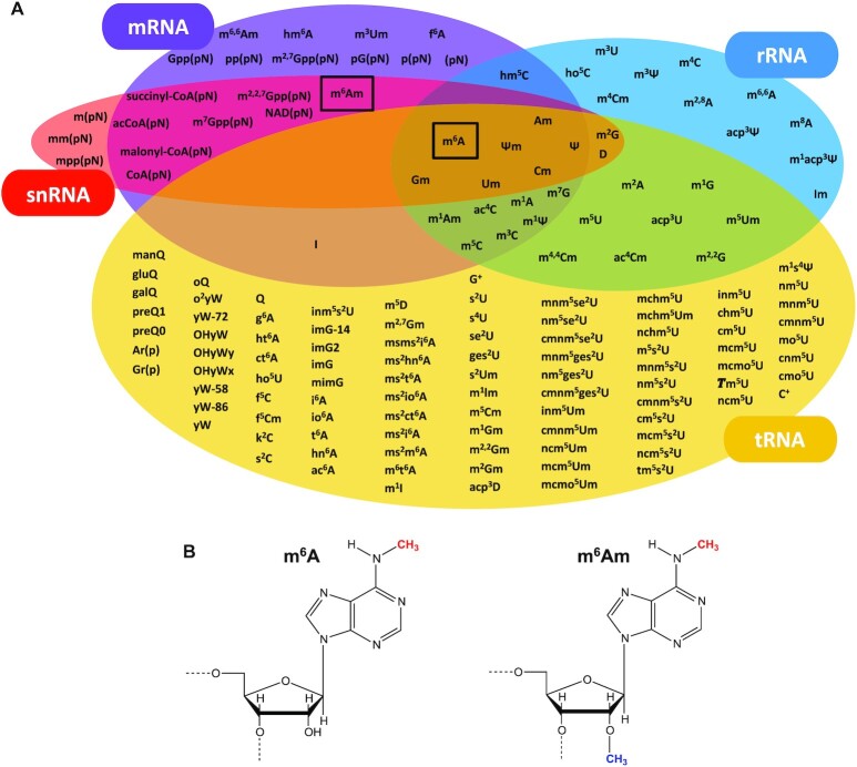
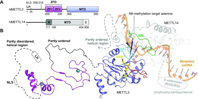
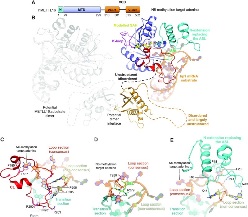
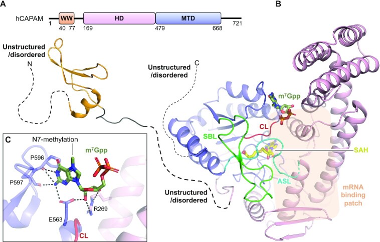
Gene expression is regulated at many levels including co- or post-transcriptionally, where chemical modifications are added to RNA on riboses and bases. Expression control via RNA modifications has been termed 'epitranscriptomics' to keep with the related 'epigenomics' for DNA modification. One such RNA modification is the N6-methylation found on adenosine (m6A) and 2'-O-methyladenosine (m6Am) in most types of RNA. The N6-methylation can affect the fold, stability, degradation and cellular interaction(s) of the modified RNA, implicating it in processes such as splicing, translation, export and decay. The multiple roles played by this modification explains why m6A misregulation is connected to multiple human cancers. The m6A/m6Am writer enzymes are RNA methyltransferases (MTases). Structures are available for functionally characterized m6A RNA MTases from human (m6A mRNA, m6A snRNA, m6A rRNA and m6Am mRNA MTases), zebrafish (m6Am mRNA MTase) and bacteria (m6A rRNA MTase). For each of these MTases, we describe their overall domain organization, the active site architecture and the substrate binding. We identify areas that remain to be investigated, propose yet unexplored routes for structural characterization of MTase:substrate complexes, and highlight common structural elements that should be described for future m6A/m6Am RNA MTase structures.


Figures

Figure 1.
Figure 1.
Post-transcriptional RNA modifications in different types of RNA and m6A/m6Am modifications. (A) Overview of the post-transcriptional modifications on mRNA, rRNA, tRNA and snRNA. m6A is found in all of these RNAs whereas m6Am is so far only identified in mRNA and snRNA. In addition to the outlined RNAs, m6A is found in lncRNA and miRNA. (B) m6A and m6Am nucleosides. The N6-methylation is shown in red and the 2′-O-methylation is shown in blue.
Figure 2.
Figure 2.
Structure of the human METTL3:METTL14 complex. (A) Schematic outline of the human METTL3:METTL14 (hMETTL3:hMETTL14) complex protein domains. Crystallized parts are indicated in boxes. (B) Structure of human METTL3 ZFD (light pink) (PDB 5YZ9), SAM-bound (yellow) MTD (blue) (PDB 5IL1) complexed with METTL14 covering the N-extension (black) and MTD (grey) of human METTL14 (PDB 5IL1). The sections not included in the structures are shows as dotted lines, and their structure/order state is indicated. The catalytic loop (CL, red), the active site loop (ASL, cyan) and the substrate binding loop (SBL, green) are shown for METTL3. Single-stranded DNA (ssDNA) (orange) has been modelled from the DNA-bound m6A DNA MTase EcoP15I (PDB 4ZCF).
Figure 3.
Figure 3.
Structure of human METTL16. (A) Schematic outline of the human METTL16 (hMETTL16) protein domains. Crystallized parts are indicated in boxes. (B) Structure of the human METTL16 VCD lacking the linker region connecting VCR1 and VCR2 shown as a dotted line (light orange) (PDB 6M1U), the MTD (blue) and N-terminal extension (cyan) that replaces the active site loop (ASL), co-crystallized with the substrate hairpin, hp1, from the MAT2A mRNA (dark orange) (PDB 6DU4). SAH (yellow) has been modelled from SAH-bound METTL16 crystallized without substrate (PDB 2H00). The catalytic loop (CL, red) and the substrate binding loop (SBL, green) are shown. A second METTL16 molecule is shown in transparent grey with dimerisation through the VCD. The sections not included in the structures are shows as dotted lines, and their structure/order state is indicated. (C) The interactions between the hMETTL16 catalytic loop (CL, red) with the MAT2A mRNA substrate consensus sequence in the substrate loop (orange), non-consensus sequence in the substrate loop (green), transition section (teal) and stem (dark grey). The enzyme residues that interact with the mRNA are shown in sticks and labelled. The N6-methylated target adenine base is highlighted. Hydrogen bonds are shown in black, dashed lines. (D) As in (C), but for the substrate binding loop (SBL, green). (E) As in (C), but for the N-extension/active site loop (ASL, cyan).
Figure 4.
Figure 4.
Structure of human CAPAM. (A) Schematic outline of the human CAPAM (hCAPAM) protein domains. Crystallized parts are indicated in boxes. (B) Structure of human CAPAM with the WW (orange) (PDB 2JX8), the SAH-bound (yellow) MTD (blue), and the HD (pink) (PDB 6IRW). The active site loop (ASL, cyan) that connects the MTD and HD is partly disordered in human CAPAM and is therefore modelled based on zebrafish CAPAM (PDB 6IRX). m7Gpp (dark green) is modelled from zebrafish CAPAM (PDB 6IS0). The catalytic loop (CL, red) and substrate binding loop (SBL, green) are shown. The mRNA substrate binding patch is indicated with a transparent orange box. The sections not included in the structures are shows as dotted lines, and their structure/order state is indicated. (C) Zoom of the active site showing the build part (m7Gppp) of the m7Gpp-derived ligand, co-crystallized with zebrafish CAPAM (PDB 6IS0). The enzyme residues that interact with the ligand are shown in sticks and labelled. Hydrogen bonds are shown in black, dashed lines, none of which are with the N7-methylation.
Figure 5.
Figure 5.
Structure of the human METTL5:TRMT112 complex. (A) Schematic outline of the human METTL5:TRMT112 (hMETTL5:hTRMT112) complex protein domains. Crystallized parts are indicated in boxes. (B) Structure of the SAM-bound (yellow) full-length, human METTL5 (blue) (PDB 6H2V) bound to human TRMT112 (grey/black) (PDB 6H2U). The active site loop (ASL, cyan), catalytic loop (CL, red) and substrate binding loop (SBL, green) are shown. Substrate 18S rRNA (dark orange) is modelled from positioning of METTL5:TRMT112 on the immature 40S ribosome (PDB 6G53). The immature ribosome structure lacks U1830-A1835 from the rRNA (not build), which is here instead indicated as a dotted line and the target base (A1832) is indicated as an orange dot. (C) Positioning of METTL5:TRMT112 into unoccupied density on the surface of the immature 40S ribosome (PDB 6G53). The unbuild part of the rRNA (U1830-A1835) is shown as a dotted line and the target base (A1832) is indicated as an orange dot.
Figure 6.
Figure 6.
Structure of human ZCCHC4. (A) Schematic outline of the human ZCCHC4 (hZCCHC4) protein domains. Crystallized parts are indicated in boxes. (B) Structure of the SAM-bound (yellow) human ZCCHC4 showing the GRF (grey), C2H2 (pink), MTD (blue) and CCHC (light orange) domains (PDB 6UCA). Zn2+ ions are shown in teal-coloured spheres. The catalytic loop (CL, red), substrate binding loop (SBL, green) and the active site loop (ASL, cyan) connecting C2H2 with the MTD are shown. The rRNA substrate binding patch is indicated with a transparent orange box. The sections not included in the structures are shows as dotted lines, and their structure/order state is indicated. (C) The mature human 80S ribosome with an insert highlighting the ZCCHC4 substrate stem-loop structure from 28S rRNA with target adenine (A4220, dark orange) placed between the stem (teal) and loop (grey) (PDB 6EK0).
Figure 7.
Figure 7.
Structure of E. coli RlmJ. (A) Schematic outline of the E. coli RlmJ (ecRlmJ) protein domains. Crystallized parts are indicated in boxes. (B) Structure of the ecRlmJ SAM-bound (yellow) MTD (blue) with the inserted HSD (pink) (PDB 4BLV), crystallized with a closed tail-conformation of the active site loop (ASL, cyan). The ASL is further shown from apo RlmJ (PDB 4BLU) in the open tail-conformation. The target adenine (dark green) is modelled from RlmJ co-crystallised with a bisubstrate molecule (BA4) (PDB 6QDX, chain B). The catalytic loop (CL, red) and substrate binding loop (SBL, green) are shown. (C) The mature E. coli 70S ribosome with an insert highlighting the RlmJ substrate stem-loop structure from 23S rRNA with target adenine (A2030, dark orange) placed in the loop (grey) followed by a stem (teal) (PDB 7K00).
Figure 8.
Figure 8.
The m6A RNA MTase loops. The active site loop (cyan), catalytic loop (red) and substrate binding loop (green) is shown on each structurally and functionally characterized m6A RNA MTase described herein. For hMETTL16, the active site loop covers the entire N-terminal extension. For comparison, the MTDs (blue) are positioned with similar cofactor (yellow) orientation. hMETTL14, without cofactor, is oriented as hMETTL3. ec: E. coli, h: human.

Similar articles

Cited by

References

    1. Desrosiers R., Friderici K., Rottman F.. Identification of methylated nucleosides in messenger RNA from Novikoff hepatoma cells. Proc. Natl. Acad. Sci. U. S. A. 1974; 71:3971–3975. - PMC - PubMed
    1. Wei C., Gershowitz A., Moss B.. N6, O2′-dimethyladenosine a novel methylated ribonucleoside next to the 5′ terminal of animal cell and virus mRNAs. Nature. 1975; 257:251–253. - PubMed
    1. Zhao B.S., Roundtree I.A., He C.. Post-transcriptional gene regulation by mRNA modifications. Nat. Rev. Mol. Cell Biol. 2016; 18:31–42. - PMC - PubMed
    1. Yue H., Nie X., Yan Z., Weining S.. N6-methyladenosine regulatory machinery in plants: composition, function and evolution. Plant Biotechnol. J. 2019; 17:1194–1208. - PMC - PubMed
    1. Lence T., Soller M., Roignant J.Y.. A fly view on the roles and mechanisms of the m6A mRNA modification and its players. RNA Biol. 2017; 14:1232–1240. - PMC - PubMed

Publication types

LinkOut - more resources