. 2021 Dec;13(2_suppl):1570S-1582S.
doi: 10.1177/19476035211014588. Epub 2021 May 24.

The Natural History of Medial Meniscal Tears in the ACL Deficient and ACL Reconstructed Rat Knee

Affiliations

The Natural History of Medial Meniscal Tears in the ACL Deficient and ACL Reconstructed Rat Knee

Akinori Kaneguchi et al. Cartilage. 2021 Dec.

Abstract

Objective: The process of anterior cruciate ligament (ACL) injury-induced meniscal tear formation is not fully understood. Clinical studies have shown that ACL reconstruction (ACLR) reduces the development of secondary meniscal tears, but it is difficult to gain insight into the protective effects of ACLR from clinical studies alone. Using rat ACL transection (ACLT) and ACLR models, we aimed to reveal (1) the formation process of meniscal tears secondary to ACLT and (2) the protective effects of ACLR on secondary meniscal tears.

Design: ACLT surgery alone or with ACLR was performed on the knees of rats. Histomorphological and histopathological changes were examined in the posteromedial region of the meniscus in intact rats and in rats that received ACLT or ACLR up to 12 weeks postsurgery. In addition, anterior-posterior joint laxity was measured using the universal testing machine to evaluate the effects of ACLT and ACLR on joint laxity.

Results: AAnterior-posterior laxity was significantly increased by ACLT compared to the intact knee. This ACLT-induced joint laxity was partially but significantly reduced by ACLR. Meniscal proliferation and hyaline cartilage-like tissue formation were detected in the medial meniscus at 4 weeks post-ACLT. At 12 weeks post-ACLT, hyaline cartilage-like tissue was replaced by ossicles and meniscal tears were observed. These ACLT-induced abnormalities were attenuated by ACLR.

Conclusions: Our results suggest that ACLT-induced joint laxity induces secondary medial meniscal tears through meniscal proliferation and ossicle formation via endochondral ossification. Joint re-stabilization by ACLR suppresses meniscal proliferation and ossicle formation and consequently prevents secondary meniscal tears.

Keywords: anterior cruciate ligament injury; anterior cruciate ligament reconstruction; joint laxity; medial meniscal proliferation; medial meniscal tear; ossicles.


Conflict of interest statement

Declaration of Conflicting Interests: The author(s) declared no potential conflicts of interest with respect to the research, authorship, and/or publication of this article.

Figures

Figure 1.
Figure 1.
Histomorphometric analysis of the posteromedial meniscus. The height (a), length (b), and area (surrounded by the dotted line) of the posteromedial meniscus were measured.
Figure 2.
Figure 2.
Joint laxity testing. (A) Diagram of the joint laxity testing setup. The femur and tibia were affixed to the universal testing machine via aluminum pipes at 90° knee flexion. Five anterior-posterior loading cycles (target forces ±5 N) were applied to the femur. (B) A representative force-displacement curve. The maximum displacements of the femur during each loading cycle were extracted as anterior and posterior laxities. (C) Measurements were repeated following ACL (anterior cruciate ligament) transection and reconstruction.
Figure 3.
Figure 3.
Representative force-displacement curves showing anterior-posterior joint laxity in the (A) intact knee, (B) anterior cruciate ligament (ACL) transected (ACLT) knee, and (C) ACL reconstructed (ACLR) knee. Black and white arrows indicate the degree of anterior and posterior displacement, respectively. Panel (D) indicates anterior-posterior joint laxity. Compared to the intact knee (A), both anterior and posterior laxities were significantly increased by ACL transection (B, D). ACL reconstruction significantly reduced anterior laxity, but not posterior laxity. (C, D) Values are shown as mean ± standard deviation. *Indicates significant differences compared to the intact (P < 0.05). Indicate significant differences compared to the ACLT (P < 0.05).
Figure 4.
Figure 4.
Representative images of the aldehyde fuchsin–Masson Goldner (AFMG)-stained posterior knee joint showing meniscus size for the (A) pre-surgery group; the anterior cruciate ligament transection (ACLT) group at (B) 1 week, (C) 4 weeks, and (D) 12 weeks after surgery; and the ACL reconstruction (ACLR) group at (E) 1 week, (F) 4 weeks, and (G) 12 weeks after surgery. The (H) height, (I) length, and (J) area of the meniscus are shown. In the pre-surgery group, only the central region of the outer edge of the meniscus attached to the joint capsule (white arrowheads, A). However, the entire outer edge of the meniscus was attached to the joint capsule at 1 week post-surgery in both the ACLT and the ACLR groups (black arrows, B, E). Scale bars = 1 mm. Values are presented as mean ± standard deviation. *Indicates significant differences compared to the pre-surgery group (P < 0.05). Indicates significant differences compared to 1 week post-surgery (P < 0.05). Indicates significant differences compared to 4 weeks post-surgery (P < 0.05). $Indicates significant differences compared to the same side in the ACLT group at the same time point (P < 0.05).
Figure 5.
Figure 5.
Histopathological features in the outer region of the proliferated meniscus. Representative images of the Safranin-O Fast Green–stained outer region of the proliferated meniscus for (A, E) 4 weeks and (C, G) 12 weeks after anterior cruciate ligament transection (ACLT), and (B, F) 4 weeks and (D, H) 12 weeks after ACL reconstruction (ACLR). (E-H) show high magnification images of the boxes in (A-D). Proteoglycan is stained red. In both groups, the outer region was composed of proteoglycan-rich matrix at both 4 and 12 weeks post-surgery (A-H). In both groups, cells in this region included both spindle-shaped fibroblast-like cells and chondrocyte-like cells at 4 weeks post-surgery (E, F) and included mainly chondrocyte-like cells at 12 weeks post-surgery (G, H). Scale bars = 500 µm in A to D and 50 µm in E to H.
Figure 6.
Figure 6.
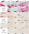
Histopathological features in the inner to middle region of the meniscus. Representative images of the Safranin-O Fast Green–stained sections from the (A) pre-surgery group, and the anterior cruciate ligament transection (ACLT) group at (B) 1 week, (C) 4 weeks, and (D) 12 weeks after surgery, and the ACL reconstruction (ACLR) group at (E) 1 week, (F) 4 weeks, and (G) 12 weeks after surgery. Proteoglycan is stained red. After 4 weeks post-surgery, proteoglycan-expressing hyaline cartilage–like tissue (black arrows), which has lacunas, was found in tibial side in both groups (C, F). Hyaline cartilage–like tissue was replaced by ossicles (white arrows), and tears (black arrowheads) were observed adjacent to the ossicle at 12 weeks post-ACLT (D). Hypocellular regions (white arrowheads) were also detected around tears at 12 weeks post-ACLT (D). In ACLR group, ossicle and tears were not detected (G). Representative images of the type II collagen–stained sections from the (H) pre-surgery group, the ACLT group at (I) 1 week, (J) 4 weeks, and (K) 12 weeks after surgery, and the ACLR group at (L) 1 week, (M) 4 weeks, and (N) 12 weeks after surgery. Staining for type II collagen in extracellular matrix was almost negative in the pre-surgery group (H) and after 1 week post-surgery in both the ACLT and ACLR groups (I, L). At 4 weeks post-surgery, type II collagen, a marker for hyaline cartilage, was detected on the tibial side of the meniscus in both groups (J, M). Expression of type II collagen disappeared in ACLT group at 12 weeks post-surgery (K), but remained in ACLR group (N). Representative images of the type I collagen–stained sections from the (O) pre-surgery group, the ACLT group at (P) 1 week, (Q) 4 weeks, and (R) 12 weeks after surgery, and the ACLR group at (S) 1 week, (T) 4 weeks, and (U) 12 weeks after surgery. Type I collagen, a marker for bone, was expressed in the tibial side of the meniscus at 12 weeks post-ACLT (surrounded by the dotted line, R). Type I collagen expression was not detected in other panels (OQ, SU). Scale bars = 200 µm (100 µm in the inset boxes).
Figure 7.
Figure 7.
Schema illustrating the processes of anterior cruciate ligament (ACL) injury-induced meniscal tears. ACL transection (ACLT) induces joint laxity, which increases mechanical stress (indicated by arrowheads) on the meniscus (A, B). Increased mechanical stress causes meniscal proliferation, and meniscal proliferation further increases mechanical stress (C). Abnormal mechanical stress triggers ossicle formation through endochondral ossification, and eventually, meniscal tears develop adjacent to the ossicle (C, D). Restabilization of the joint by ACL reconstruction (ACLR) attenuates meniscal proliferation via partial reduction of joint laxity (E, F) and prevents ossicle formation and secondary meniscal tears (G).

Similar articles

Cited by

References

    1. Musahl V, Karlsson J. Anterior cruciate ligament tear. N Engl J Med. 2019;380(24):2341-48. - PubMed
    1. Du G, Zhan H, Ding D, Wang S, Wei X, Wei F, et al.. Abnormal mechanical loading induces cartilage degeneration by accelerating meniscus hypertrophy and mineralization after ACL injuries in vivo. Am J Sports Med. 2016;44(3):652-63. - PMC - PubMed
    1. Sonoda M, Harwood FL, Amiel ME, Moriya H, Amiel D. The effects of hyaluronan on the meniscus in the anterior cruciate ligament-deficient knee. J Orthop Sci. 2000;5(2_suppl):157-64. - PubMed
    1. Hagmeijer MH, Hevesi M, Desai VS, Sanders TL, Camp CL, Hewett TE, et al.. Secondary meniscal tears in patients with anterior cruciate ligament injury: relationship among operative management, osteoarthritis, and arthroplasty at 18-year mean follow-up. Am J Sports Med. 2019;47(7):1583-90. - PubMed
    1. Keene GC, Bickerstaff D, Rae PJ, Paterson RS. The natural history of meniscal tears in anterior cruciate ligament insufficiency. Am J Sports Med. 1993;21(5):672-9. - PubMed

Publication types