

White matter changes in psychosis risk relate to development and are not impacted by the transition to psychosis
- PMID: 34024906
- PMCID: PMC8611104
- DOI: 10.1038/s41380-021-01128-8
White matter changes in psychosis risk relate to development and are not impacted by the transition to psychosis
Abstract
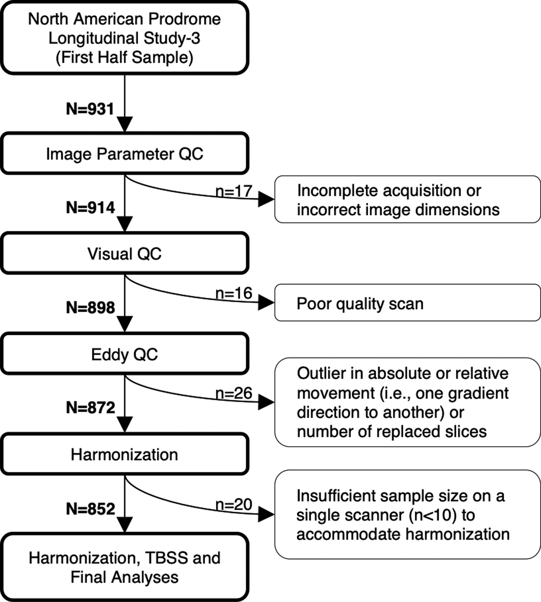
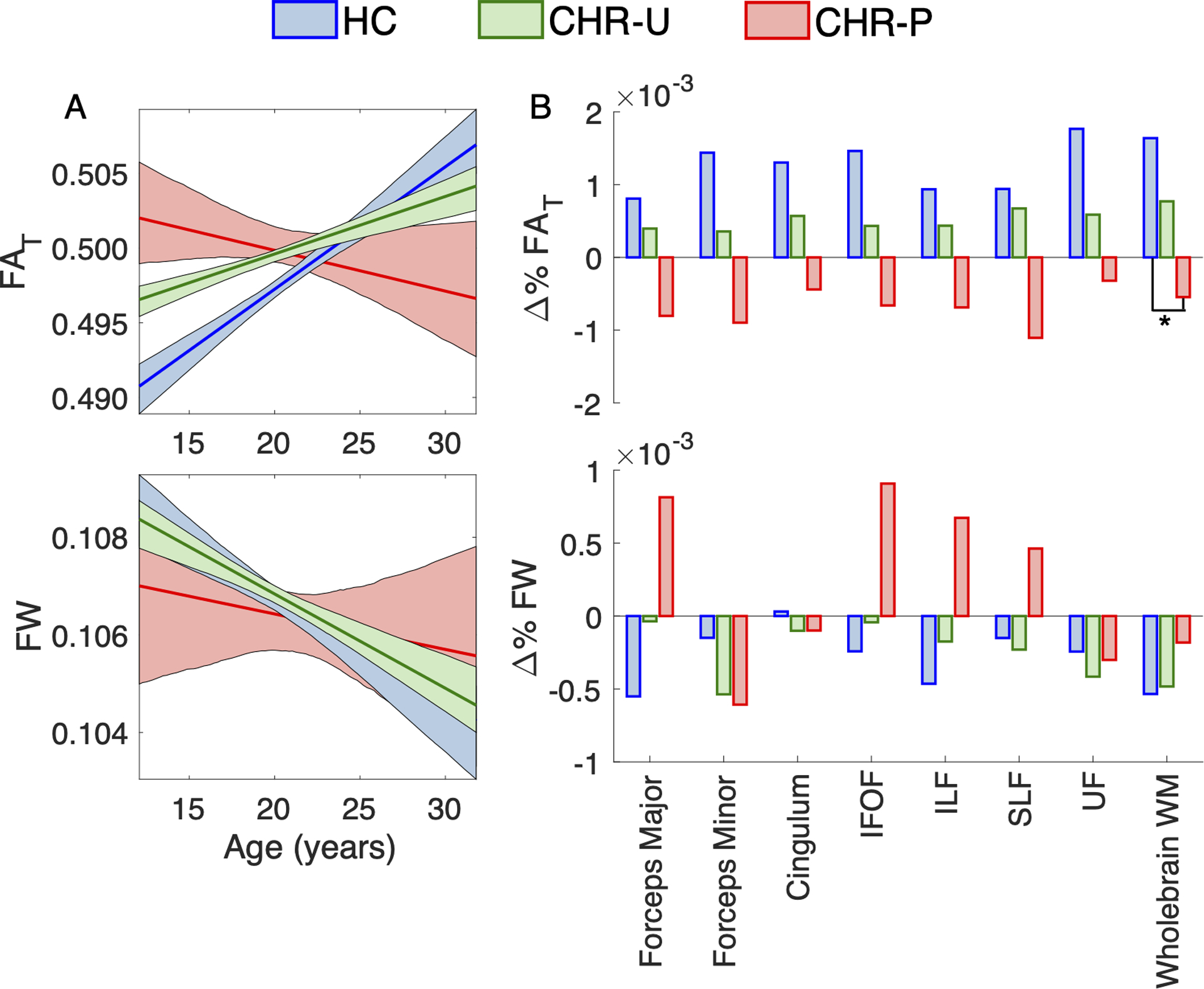
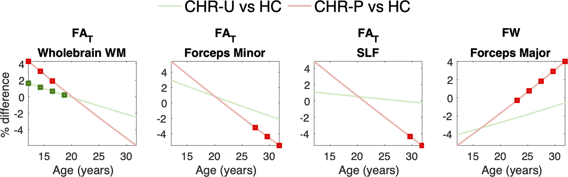
Subtle alterations in white matter microstructure are observed in youth at clinical high risk (CHR) for psychosis. However, the timing of these changes and their relationships to the emergence of psychosis remain unclear. Here, we track the evolution of white matter abnormalities in a large, longitudinal cohort of CHR individuals comprising the North American Prodrome Longitudinal Study (NAPLS-3). Multi-shell diffusion magnetic resonance imaging data were collected across multiple timepoints (1-5 over 1 year) in 286 subjects (aged 12-32 years): 25 CHR individuals who transitioned to psychosis (CHR-P; 61 scans), 205 CHR subjects with unknown transition outcome after the 1-year follow-up period (CHR-U; 596 scans), and 56 healthy controls (195 scans). Linear mixed effects models were fitted to infer the impact of age and illness-onset on variation in the fractional anisotropy of cellular tissue (FAT) and the volume fraction of extracellular free water (FW). Baseline measures of white matter microstructure did not differentiate between HC, CHR-U and CHR-P individuals. However, age trajectories differed between the three groups in line with a developmental effect: CHR-P and CHR-U groups displayed higher FAT in adolescence, and 4% lower FAT by 30 years of age compared to controls. Furthermore, older CHR-P subjects (20+ years) displayed 4% higher FW in the forceps major (p < 0.05). Prospective analysis in CHR-P did not reveal a significant impact of illness onset on regional FAT or FW, suggesting that transition to psychosis is not marked by dramatic change in white matter microstructure. Instead, clinical high risk for psychosis-regardless of transition outcome-is characterized by subtle age-related white matter changes that occur in tandem with development.
© 2021. The Author(s), under exclusive licence to Springer Nature Limited.
Conflict of interest statement
Figures

Similar articles
-
White matter microstructure in individuals at clinical high risk of psychosis: a whole-brain diffusion tensor imaging study.Schizophr Bull. 2014 Jul;40(4):895-903. doi: 10.1093/schbul/sbt079. Epub 2013 Jun 4. Schizophr Bull. 2014. PMID: 23737549 Free PMC article.
-
Sex- and Age-Specific Deviations in Cerebellar Structure and Their Link With Symptom Dimensions and Clinical Outcome in Individuals at Clinical High Risk for Psychosis.Schizophr Bull. 2023 Mar 15;49(2):350-363. doi: 10.1093/schbul/sbac169. Schizophr Bull. 2023. PMID: 36394426 Free PMC article.
-
Cellular and extracellular white matter alterations indicate conversion to psychosis among individuals at clinical high-risk for psychosis.World J Biol Psychiatry. 2021 Mar;22(3):214-227. doi: 10.1080/15622975.2020.1775890. Epub 2020 Jul 9. World J Biol Psychiatry. 2021. PMID: 32643526 Free PMC article.
-
Longitudinal Structural MRI Findings in Individuals at Genetic and Clinical High Risk for Psychosis: A Systematic Review.Front Psychiatry. 2021 Feb 2;12:620401. doi: 10.3389/fpsyt.2021.620401. eCollection 2021. Front Psychiatry. 2021. PMID: 33603688 Free PMC article.
-
The temporal dynamics of transition to psychosis in individuals at clinical high-risk (CHR-P) shows negative prognostic effects of baseline antipsychotic exposure: a meta-analysis.Transl Psychiatry. 2023 Apr 5;13(1):112. doi: 10.1038/s41398-023-02405-6. Transl Psychiatry. 2023. PMID: 37019886 Free PMC article.
Cited by
-
Matrix metalloproteinase 9 (MMP-9) activity, hippocampal extracellular free water, and cognitive deficits are associated with each other in early phase psychosis.Neuropsychopharmacology. 2024 Jun;49(7):1140-1150. doi: 10.1038/s41386-024-01814-5. Epub 2024 Mar 2. Neuropsychopharmacology. 2024. PMID: 38431757 Free PMC article.
-
Characterization of the extracellular free water signal in schizophrenia using multi-site diffusion MRI harmonization.Mol Psychiatry. 2023 May;28(5):2030-2038. doi: 10.1038/s41380-023-02068-1. Epub 2023 Apr 24. Mol Psychiatry. 2023. PMID: 37095352 Free PMC article.
-
Parsing heterogeneity in global and local white matter integrity at different stages across the psychosis continuum.Schizophrenia (Heidelb). 2024 Nov 13;10(1):106. doi: 10.1038/s41537-024-00516-7. Schizophrenia (Heidelb). 2024. PMID: 39537644 Free PMC article.
-
White matter microstructure alterations in early psychosis and schizophrenia.Transl Psychiatry. 2025 May 23;15(1):179. doi: 10.1038/s41398-025-03397-1. Transl Psychiatry. 2025. PMID: 40410167 Free PMC article.
-
Advances in the understanding of the pathophysiology of schizophrenia and bipolar disorder through induced pluripotent stem cell models.J Psychiatry Neurosci. 2024 Mar 15;49(2):E109-E125. doi: 10.1503/jpn.230112. Print 2024 Jan-Feb. J Psychiatry Neurosci. 2024. PMID: 38490647 Free PMC article. Review.
References
-
- Di Biase MA, Pantelis C, Zalesky A. White Matter Pathology in Schizophrenia. Neuroimaging in Schizophrenia. Springer; 2020, pp 71–91.
-
- Saito J, Hori M, Nemoto T, Katagiri N, Shimoji K, Ito S et al. Longitudinal study examining abnormal white matter integrity using a tract‐specific analysis in individuals with a high risk for psychosis. Psychiatry and clinical neurosciences 2017; 71(8): 530–541. - PubMed
-
- Tomyshev AS, Lebedeva IS, Akhadov TA, Omelchenko MA, Rumyantsev AO, Kaleda VG. Alterations in white matter microstructure and cortical thickness in individuals at ultra-high risk of psychosis: A multimodal tractography and surface-based morphometry study. Psychiatry Research: Neuroimaging 2019; 289: 26–36. - PubMed
-
- Peters B, Schmitz N, Dingemans P, Van Amelsvoort T, Linszen D, De Haan L et al. Preliminary evidence for reduced frontal white matter integrity in subjects at ultra-high-risk for psychosis. Schizophrenia research 2009; 111(1): 192–193. - PubMed
Publication types
MeSH terms
Grants and funding
- U01 MH081902/MH/NIMH NIH HHS/United States
- U01 MH076989/MH/NIMH NIH HHS/United States
- R01 MH074794/MH/NIMH NIH HHS/United States
- P41 EB015902/EB/NIBIB NIH HHS/United States
- U01 MH081928/MH/NIMH NIH HHS/United States
- R01 MH102377/MH/NIMH NIH HHS/United States
- U01 MH109977/MH/NIMH NIH HHS/United States
- U01 MH082022/MH/NIMH NIH HHS/United States
- U01 MH081984/MH/NIMH NIH HHS/United States
- UL1 TR001863/TR/NCATS NIH HHS/United States
- T32 GM136651/GM/NIGMS NIH HHS/United States
- R01 MH108574/MH/NIMH NIH HHS/United States
- K01 MH115247/MH/NIMH NIH HHS/United States
- U01 MH081857/MH/NIMH NIH HHS/United States
- U01 MH082004/MH/NIMH NIH HHS/United States
- U01 MH081944/MH/NIMH NIH HHS/United States
LinkOut - more resources
Full Text Sources
Other Literature Sources
Medical
