. 2021 May 24;6(1):186.
doi: 10.1038/s41392-021-00578-4.

FLT3 tyrosine kinase inhibitors synergize with BCL-2 inhibition to eliminate FLT3/ITD acute leukemia cells through BIM activation

Affiliations

FLT3 tyrosine kinase inhibitors synergize with BCL-2 inhibition to eliminate FLT3/ITD acute leukemia cells through BIM activation

Ruiqi Zhu et al. Signal Transduct Target Ther. .

Abstract

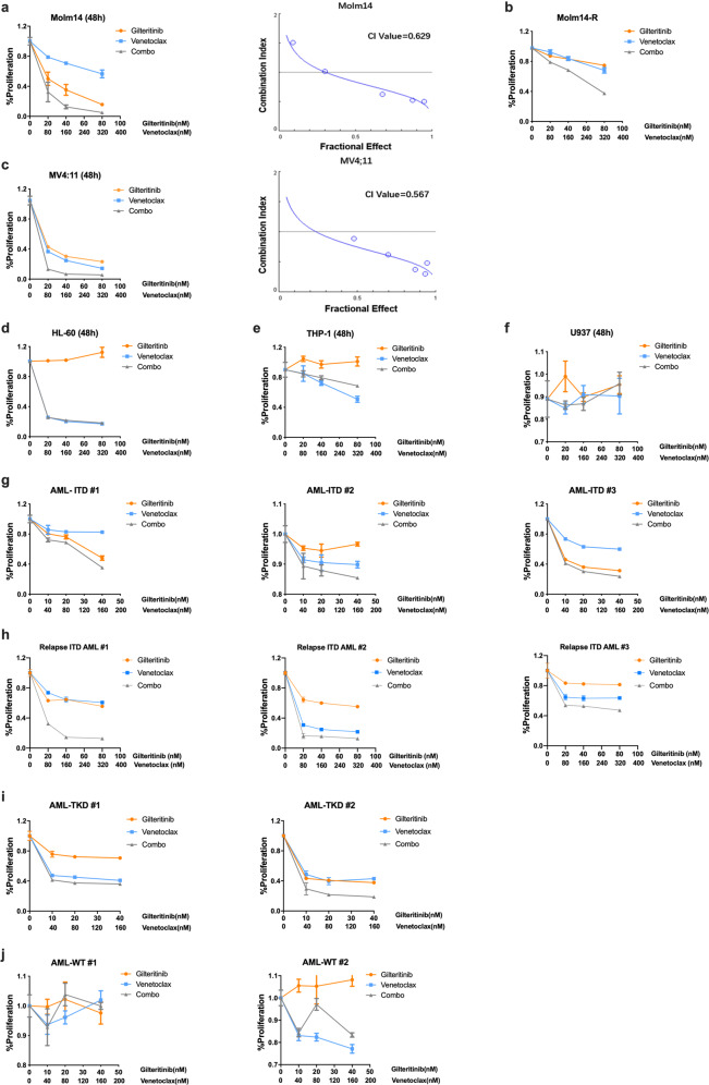
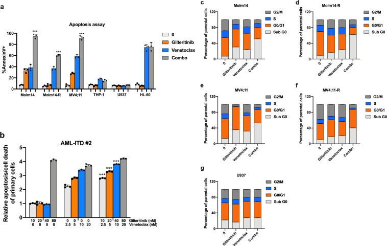
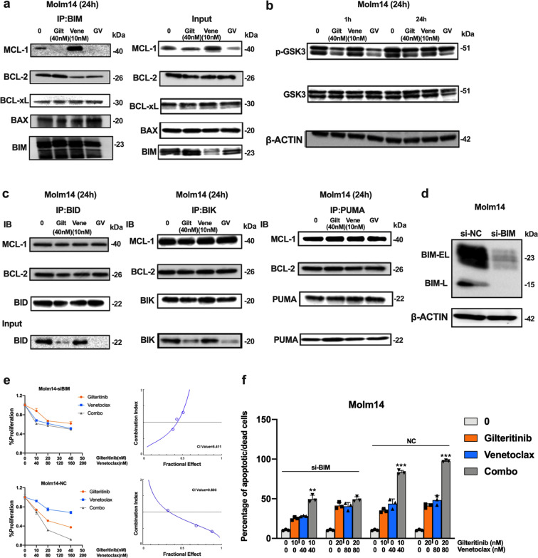
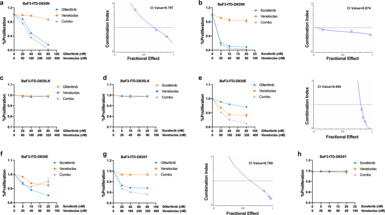
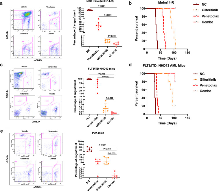
Tyrosine kinase inhibitors (TKIs) targeting FLT3 have shown activity but when used alone have achieved limited success in clinical trials, suggesting the need for combination with other drugs. We investigated the combination of FLT3 TKIs (Gilteritinib or Sorafenib), with Venetoclax, a BCL-2 selective inhibitor (BCL-2i), on FLT3/ITD leukemia cells. The combination of a FLT3 TKI and a BCL-2i synergistically reduced cell proliferation and enhanced apoptosis/cell death in FLT3/ITD cell lines and primary AML samples. Venetoclax also re-sensitized FLT3 TKI-resistant cells to Gilteritinib or Sorafenib treatment, mediated through MAPK pathway inhibition. Gilteritinib treatment alone dissociated BIM from MCL-1 but increased the binding of BIM to BCL-2. Venetoclax treatment enhanced the binding of BIM to MCL-1 but dissociated BIM from BCL-2. Treatment with the drugs together resulted in dissociation of BIM from both BCL-2 and MCL-1, with an increased binding of BIM to the cell death mediator BAX, leading to increased apoptosis. These findings suggest that Venetoclax mitigates the unintended pro-survival effects of FLT3 TKI mainly through the dissociation of BIM and BCL-2 and also decreased BIM expression. This study provides evidence that the addition of BCL-2i enhances the effect of FLT3 TKI therapy in FLT3/ITD AML treatment.


Conflict of interest statement

The authors declare no conflicts of interest. D.S. does serve on the SAB of InSilico Medicine and receives research support and serves as a consultant for an unrelated project from PHarosI&BT co, Ltd. M.L. has received honoraria from Daiichi Sankyo, Novartis, and Agios; has served in a consulting or advisory role for Daiichi Sankyo, Novartis, and Agios; and has received research funding from Astellas and Novartis.

Figures

Fig. 1
Fig. 1
Gilteritinib and Venetoclax synergistically inhibit the proliferation of FLT3/ITD cell lines and primary FLT3/ITD AML samples. 48 h MTT assay of cells treated with the indicated doses of Gilteritinib and Venetoclax: a Molm14, b Molm14 cells resistant to 60 nM Lestaurtinib (Molm14-R), c MV4;11, d HL-60, e THP-1, f U937, g Primary de novo AML patient samples with FLT3/ITD mutation (AML-ITD#1, AML-ITD#2, AML-ITD#3), h Primary AML samples from relapsed patients with FLT3/ITD mutations (Relapse ITD AML #1, Relapse ITD AML #2, Relapse ITD AML #3), i Primary AML patients with FLT3/TKD mutation, or j without FLT3 mutations (AML-WT#1, AML-WT#2)
Fig. 2
Fig. 2
Gilteritinib and Venetoclax induce apoptosis/cell death of FLT3/ITD cells. a Bar graph of 48 h apoptosis/cell death assay of Molm14, Molm14-R, MV4;11, THP-1, HL-60, and U937 cells. (Gilteritinib: 80 nM, Venetoclax: 20 nM, Combo: Gilteritinib 80 nM + Venetoclax 20 nM), b Bar graph of 48 h apoptosis/cell death assay of primary FLT3/ITD AML patient sample #2 treated with the indicated concentration of Gilteritinib and Venetoclax. cg Bar graphs of cell cycle analysis of Molm14, Molm14-R, MV4;11, MV4;11-R, and U937 treated cells
Fig. 3
Fig. 3
Venetoclax sensitizes TKI-resistant FLT3/ITD cells to TKI treatment through inhibiting the MAPK/ERK pathway. Western blot analysis for FLT3 and downstream signaling pathways in Molm14 cells treated for 1 h with the indicated concentrations of a Gilteritinib, b Venetoclax, and c the combination. In all, 24 h Western blot analysis for FLT3 and downstream signaling pathways in Molm14 cells treated with the indicated concentrations of d Gilteritinib, e Venetoclax, and f the combination. 1 h western blot analysis for FLT3 downstream signaling pathways in Molm14-R cells treated with the indicated concentrations of (g) Gilteritinib, (h) Venetoclax, and (i) the combination. In all, 24 h western blot analysis for FLT3 downstream signaling pathways in Molm14 cells treated with the indicated concentrations of (j) Gilteritinib, (k) Venetoclax, and (l) the combination
Fig. 4
Fig. 4
Treatment with Gilteritinib and/or Venetoclax changes expression of some BCL-2 family members. Western blot for BCL-2 family members in Molm14 cells treated for 24 h with the indicated concentrations of a Gilteritinib, b Venetoclax, and c the combination
Fig. 5
Fig. 5
Gilteritinib and Venetoclax combined treatment releases BIM from the pro-survival proteins BCL-2 and MCL-1 to trigger apoptosis/cell death. a Co-immunoprecipitation followed by western blot analysis for BIM binding proteins after 24 h of treatment with Gilteritinib and/or Venetoclax. b Western blot analysis for p-GSK3 and GSK3 expression in Molm14 cells treated for 1 h and 24 h. c Co-immunoprecipitation analysis for BID, BIK, and PUMA binding proteins after 24 h treatment. Input levels of the immunoprecipitated proteins are shown below. d Western blot analysis for BIM protein in negative control (si-NC) and si-BIM group. e 48 h MTT and f 48 h apoptosis analysis for Molm14-si-NC and Molm14-si-BIM cells treated with Gilteritinib and Venetoclax at the indicated concentrations
Fig. 6
Fig. 6
The effects of FLT3 TKI (Gilteritinib or Sorafenib) and/or Venetoclax treatment in BaF3 cells with FLT3 AL point mutations. In all, 48 h MTT assay of a BaF3 D835N, c BaF3 D835L+K, e BaF3 D835E, and g BaF3 D835Y cells treated with indicated doses of Gilteritinib and/or Venetoclax. In all, 48 h MTT assay of b BaF3 D835N, d BaF3 D835L+K, f BaF3 D835E, and h BaF3 D835Y treated with indicated doses of sorafenib and/or Venetoclax
Fig. 7
Fig. 7
Gilteritinib and Venetoclax combine to more effectively eliminate FLT3/ITD cells in vivo. Mice were treated with vehicle, Gilteritinib (15 mg/kg/day), Venetoclax (80 mg/kg/day), or the combination via oral gavage for various length of time. a, b Engraftment in BM and Kaplan–Meier survival curve for Molm14-R transplanted mice; c, d Engraftment in PB and Kaplan–Meier survival curve for FLT3/ITD;NHD13 transplanted mice; e Engraftment of human CD45 + cells in the BM of PDX mice

References

    1. Frohling S, et al. Prognostic significance of activating FLT3 mutations in younger adults (16 to 60 years) with acute myeloid leukemia and normal cytogenetics: a study of the AML Study Group Ulm. Blood. 2002;100:4372–4380. doi: 10.1182/blood-2002-05-1440. - DOI - PubMed
    1. Kottaridis PD, et al. The presence of a FLT3 internal tandem duplication in patients with acute myeloid leukemia (AML) adds important prognostic information to cytogenetic risk group and response to the first cycle of chemotherapy: analysis of 854 patients from the United Kingdom Medical Research Council AML 10 and 12 trials. Blood. 2001;98:1752–1759. doi: 10.1182/blood.V98.6.1752. - DOI - PubMed
    1. Small D. Targeting FLT3 for the treatment of leukemia. Semin. Hematol. 2008;45:S17–S21. doi: 10.1053/j.seminhematol.2008.07.007. - DOI - PMC - PubMed
    1. Ueno Y, et al. Evaluation of gilteritinib in combination with chemotherapy in preclinical models of FLT3-ITD(+) acute myeloid leukemia. Oncotarget. 2019;10:2530–2545. doi: 10.18632/oncotarget.26811. - DOI - PMC - PubMed
    1. Chang E, Ganguly S. The combination of FLT3 and DNA methyltransferase inhibition is synergistically cytotoxic to FLT3/ITD acute myeloid leukemia cells. Leukemia. 2016;30:1025–1032. doi: 10.1038/leu.2015.346. - DOI - PMC - PubMed

Publication types

MeSH terms