

Theaflavins decrease skeletal muscle wasting in disuse atrophy induced by hindlimb suspension in mice
- PMID: 34025025
- PMCID: PMC8129979
- DOI: 10.3164/jcbn.20-68
Theaflavins decrease skeletal muscle wasting in disuse atrophy induced by hindlimb suspension in mice
Abstract
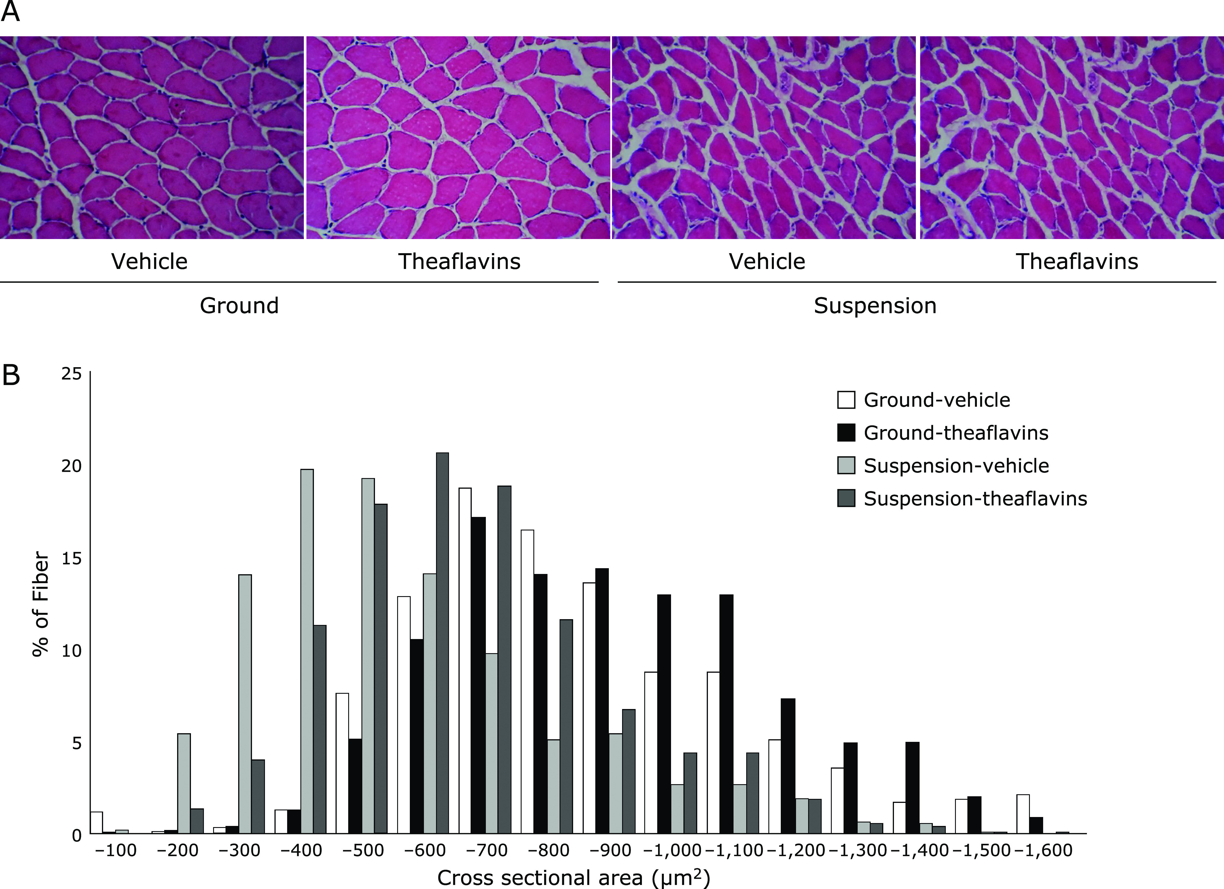
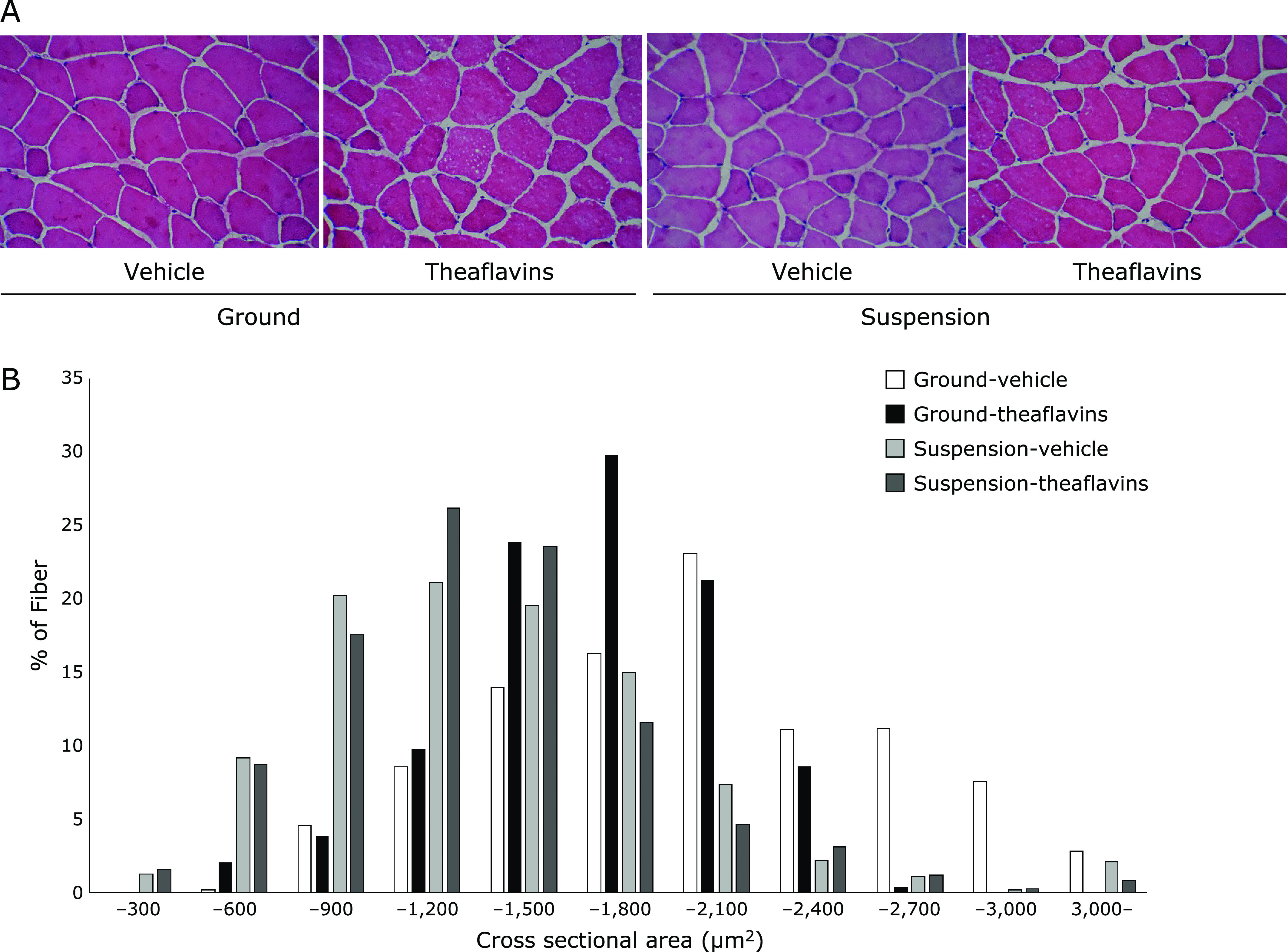
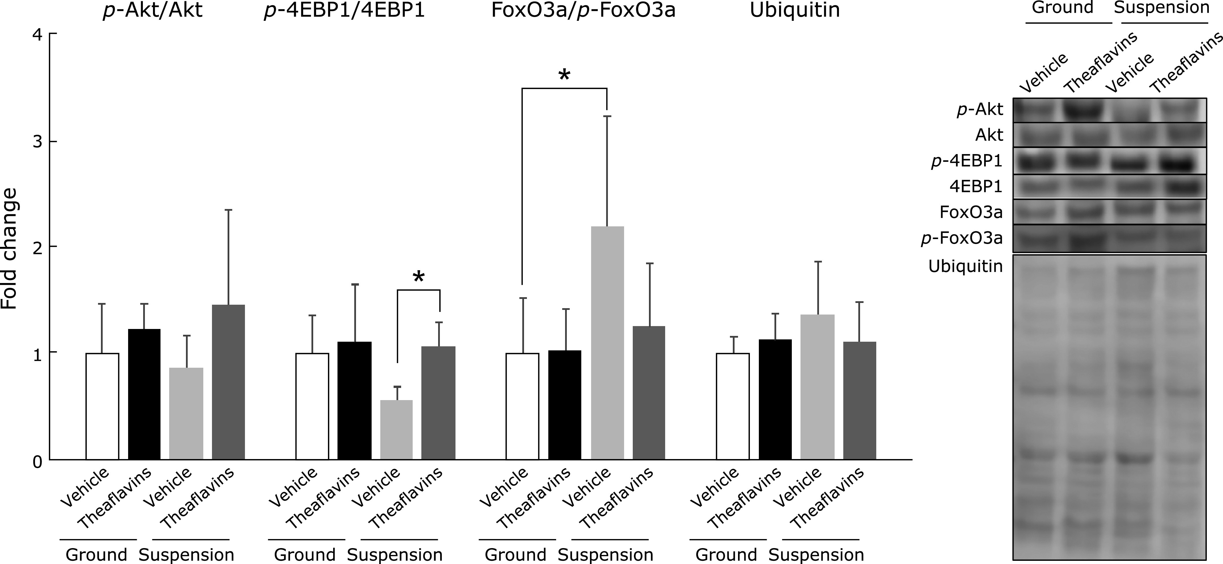
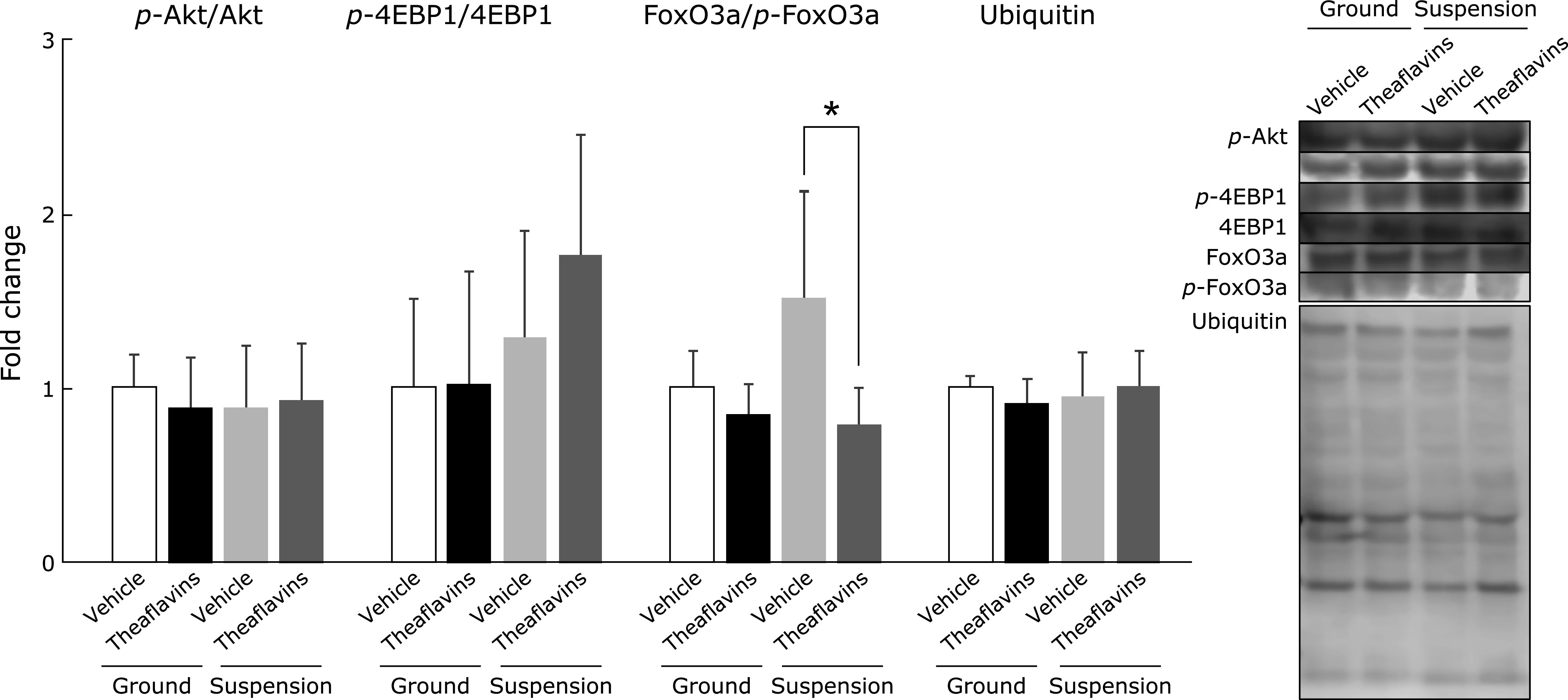
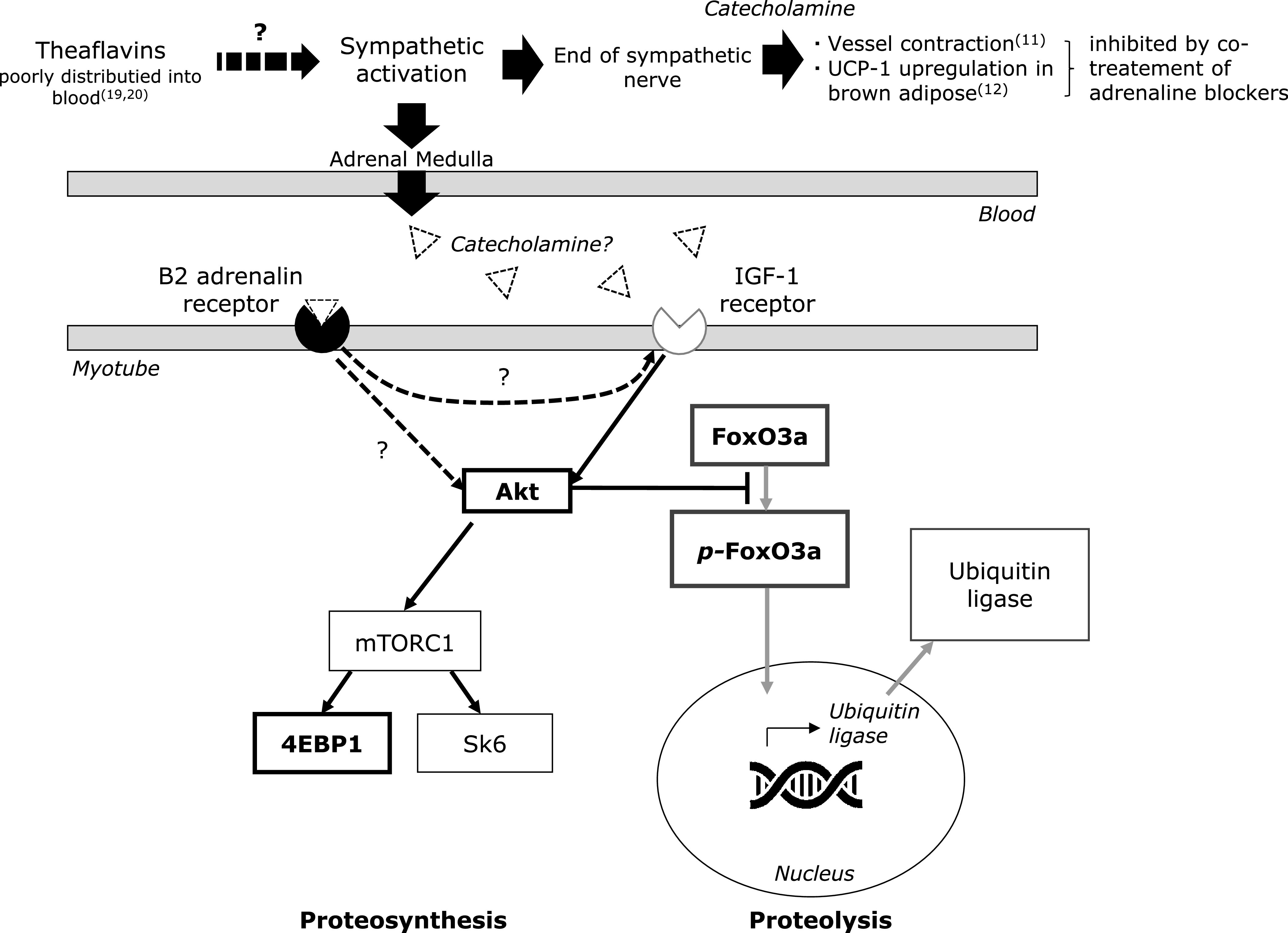
We previously found that a single dose of theaflavins induced skeletal muscle metabolic changes. In this study, we examined the effect of theaflavins on disuse muscle atrophy model mice by hindlimb suspension. Mice were assigned to 4 groups; ground-vehicle, ground-theaflavins, suspension-vehicle, and suspension-theaflavins, dosed with theaflavins (250 mg/kg/day) for 2 weeks. The peak of myotube size of cross sectional area was significantly moved to the smaller side in the suspension-vehicle group compared with the ground-vehicle group, and these shifts were significantly reduced by the treatment with theaflavins in both soleus and extensor digitorum longus. The level of phosphorylated eukaryotic translation initiation factor 4E-binding protein (4EBP)-1, located downstream of the Akt/mTOR pathway, was significantly different between suspension-vehicle and suspension-theaflavins in soleus. The ratio of forkhead box O (FoxO) 3a to phosphorylated FoxO3a significantly increased in soleus or tended to rise in extensor digitorum longus of suspension-vehicle group compared with ground-vehicle. In contrast, these changes were not observed in suspension-theaflavins group. These results suggested that theaflavins inhibited the progress of disuse muscle atrophy through modulation of protein metabolism.
Keywords: FoxO3a; hindlimb suspension; skeletal muscle atrophy; theaflavins.
Copyright © 2021 JCBNCopyright © 2021 JCBN.
Conflict of interest statement
This study was funded by Yaizu Suisankagaku Ind. Co. Ltd. AY and TU are employees of Yaizu Suisankagaku Ind. Co. Ltd. and provide research source. KS, NH, YF, TF, RA, and NO declare no competing interests.
Figures
Similar articles
-
Cinnamtannin A2, (-)-epicatechin tetramer, attenuates skeletal muscle wasting in disuse atrophy model mice induced by hindlimb suspension.J Clin Biochem Nutr. 2023 Sep;73(2):124-130. doi: 10.3164/jcbn.23-12. Epub 2023 Jul 25. J Clin Biochem Nutr. 2023. PMID: 37700845 Free PMC article.
-
Flavan 3-ol delays the progression of disuse atrophy induced by hindlimb suspension in mice.Exp Gerontol. 2017 Nov;98:120-123. doi: 10.1016/j.exger.2017.07.010. Epub 2017 Aug 12. Exp Gerontol. 2017. PMID: 28807824
-
Balanced Diet-Fed Fat-1 Transgenic Mice Exhibit Lower Hindlimb Suspension-Induced Soleus Muscle Atrophy.Nutrients. 2017 Oct 6;9(10):1100. doi: 10.3390/nu9101100. Nutrients. 2017. PMID: 28984836 Free PMC article.
-
Glucocorticoid-induced skeletal muscle atrophy.Int J Biochem Cell Biol. 2013 Oct;45(10):2163-72. doi: 10.1016/j.biocel.2013.05.036. Epub 2013 Jun 24. Int J Biochem Cell Biol. 2013. PMID: 23806868 Review.
-
Rat hindlimb unloading: Soleus and Extensor Digitorum Longus histochemistry, mitochondrial DNA content and mitochondrial DNA deletions.Biosci Rep. 2002 Feb;22(1):115-25. doi: 10.1023/a:1016069208073. Biosci Rep. 2002. PMID: 12418554 Review.
Cited by
-
Cinnamtannin A2, (-)-epicatechin tetramer, attenuates skeletal muscle wasting in disuse atrophy model mice induced by hindlimb suspension.J Clin Biochem Nutr. 2023 Sep;73(2):124-130. doi: 10.3164/jcbn.23-12. Epub 2023 Jul 25. J Clin Biochem Nutr. 2023. PMID: 37700845 Free PMC article.
-
Miso, fermented soybean paste, suppresses high-fat/high-sucrose diet-induced muscle atrophy in mice.J Clin Biochem Nutr. 2024 Jan;74(1):63-69. doi: 10.3164/jcbn.23-36. Epub 2023 Jul 20. J Clin Biochem Nutr. 2024. PMID: 38292116 Free PMC article.
-
A semi-automated observation approach to quantify mouse skeletal muscle differentiation using immunohistochemistry.Physiol Rep. 2025 Apr;13(7):e70330. doi: 10.14814/phy2.70330. Physiol Rep. 2025. PMID: 40223406 Free PMC article.
-
Sarcopenia, a condition shared by various diseases: can we alleviate or delay the progression?Intern Emerg Med. 2023 Oct;18(7):1887-1895. doi: 10.1007/s11739-023-03339-z. Epub 2023 Jul 25. Intern Emerg Med. 2023. PMID: 37490203 Free PMC article. Review.
-
Theaflavin mitigates acute gouty peritonitis and septic organ injury in mice by suppressing NLRP3 inflammasome assembly.Acta Pharmacol Sin. 2023 Oct;44(10):2019-2036. doi: 10.1038/s41401-023-01105-7. Epub 2023 May 23. Acta Pharmacol Sin. 2023. PMID: 37221235 Free PMC article.
References
-
- Magne H, Savary-Auzeloux I, Rémond D, Dardevet D. Nutritional strategies to counteract muscle atrophy caused by disuse and to improve recovery. Nutr Res Rev 2013; 26: 149–165. - PubMed
LinkOut - more resources
Full Text Sources
Research Materials
Miscellaneous