. 2021 May 7:12:664955.
doi: 10.3389/fmicb.2021.664955. eCollection 2021.

Heat Shock Protein A6, a Novel HSP70, Is Induced During Enterovirus A71 Infection to Facilitate Internal Ribosomal Entry Site-Mediated Translation

Affiliations

Heat Shock Protein A6, a Novel HSP70, Is Induced During Enterovirus A71 Infection to Facilitate Internal Ribosomal Entry Site-Mediated Translation

Yu-Siang Su et al. Front Microbiol. .

Abstract

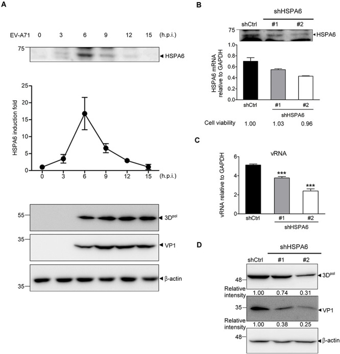
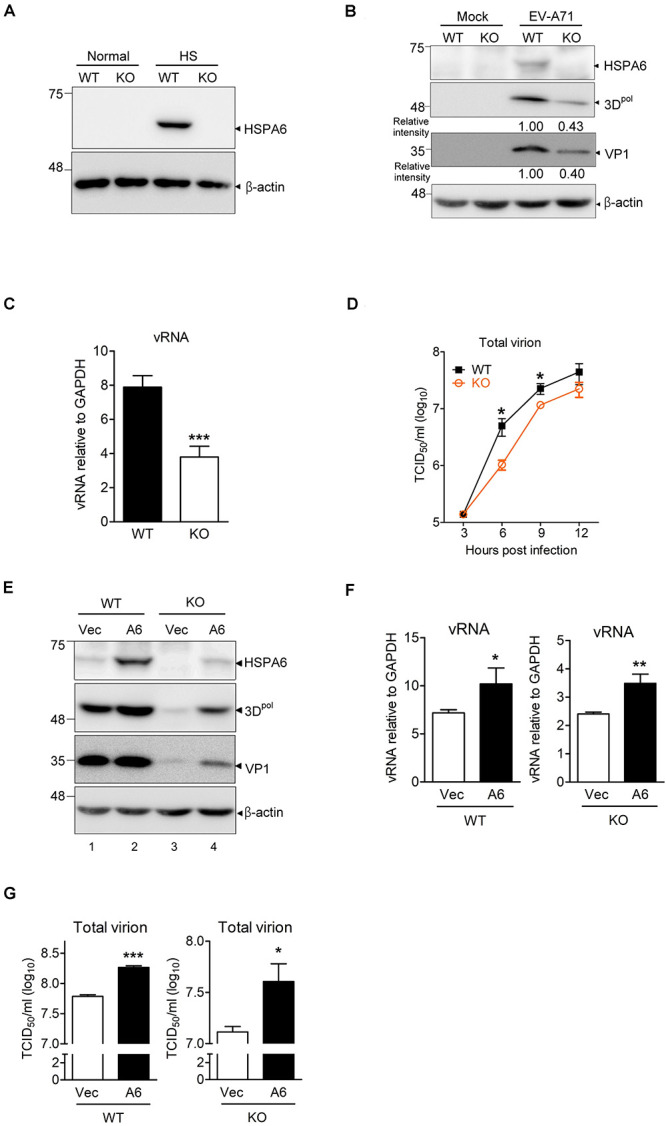
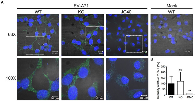
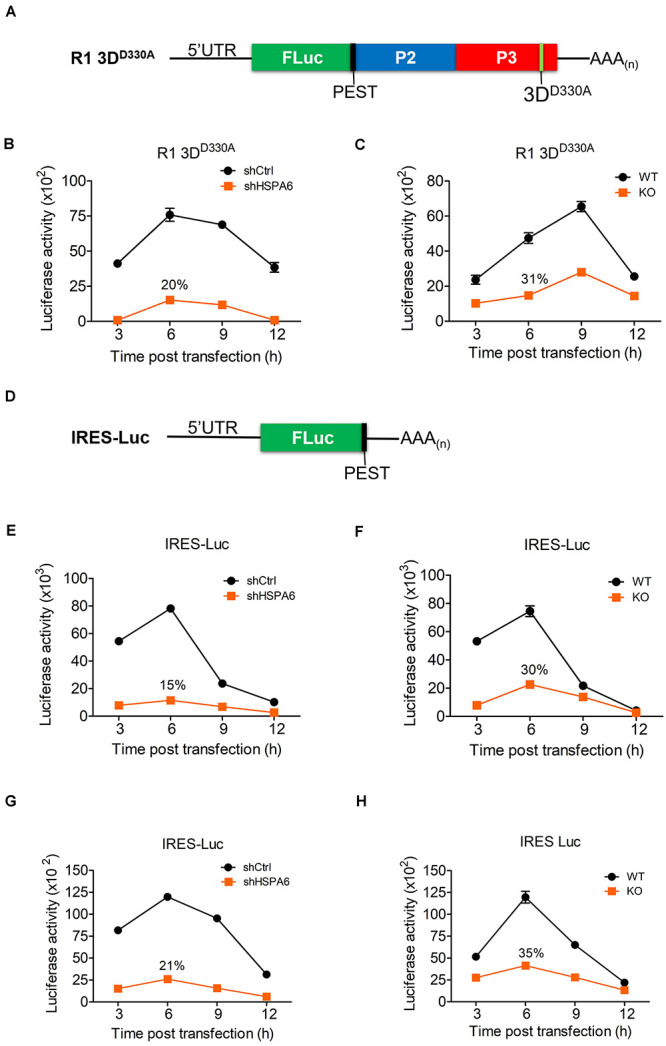
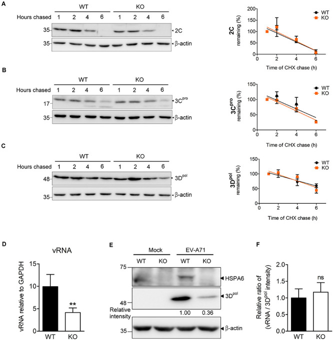
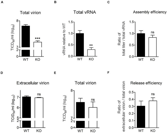
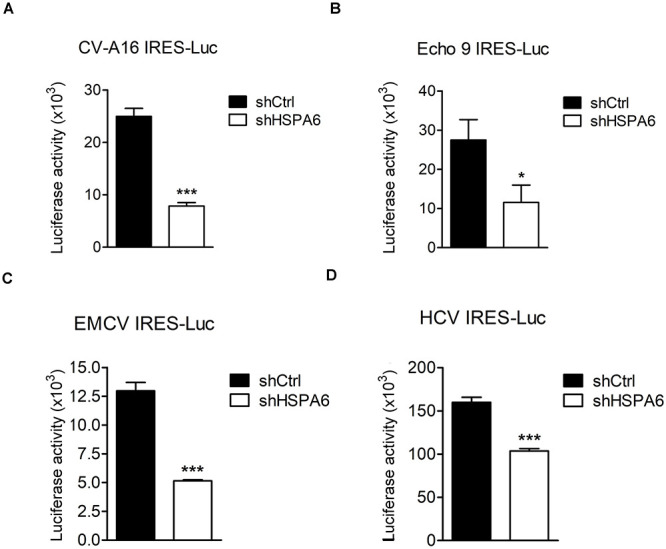
Enterovirus A71 (EV-A71) is a human pathogen causing hand, foot, and mouth disease (HFMD) in children. Its infection can lead to severe neurological diseases or even death in some cases. While being produced in a large quantity during infection, viral proteins often require the assistance from cellular chaperones for proper folding. In this study, we found that heat shock protein A6 (HSPA6), whose function in viral life cycle is scarcely studied, was induced and functioned as a positive regulator for EV-A71 infection. Depletion of HSPA6 led to the reductions of EV-A71 viral proteins, viral RNA and virions as a result of the downregulation of internal ribosomal entry site (IRES)-mediated translation. Unlike other HSP70 isoforms such as HSPA1, HSPA8, and HSPA9, which regulate all phases of the EV-A71 life, HSPA6 was required for the IRES-mediated translation only. Unexpectedly, the importance of HSPA6 in the IRES activity could be observed in the absence of viral proteins, suggesting that HSPA6 facilitated IRES activity through cellular factor(s) instead of viral proteins. Intriguingly, the knockdown of HSPA6 also caused the reduction of luciferase activity driven by the IRES from coxsackievirus A16, echovirus 9, encephalomyocarditis virus, or hepatitis C virus, supporting that HSPA6 may assist the function of a cellular protein generally required for viral IRES activities.

Keywords: HSPA6; enterovirus A71 (EV-A71); induced HSP70; internal ribosomal entry site; viral IRES.


Conflict of interest statement

The authors declare that the research was conducted in the absence of any commercial or financial relationships that could be construed as a potential conflict of interest.

Figures

FIGURE 1
FIGURE 1
HSPA6 is induced to support EV-A71 replication cycle. (A) RD cells were infected with EV-A71 (MOI = 5) and harvested for the protein expression of HSPA6 and viral 3Dpol at the time points indicated. HSPA6 protein expression was quantitated from three independent experiments and presented as induction fold over 0 time point. (B–D) RD cells were infected with lentivirus expressing control shRNA (shCtrl) or shRNA specific to HSPA6 (shHSPA6 clone #1 and clone #2). 48 h later, the cells were infected with EV-A71 (MOI = 5) and harvested at 6 hpi for analyzing the productions of (B) HSPA6 mRNA, (C) cell-associated vRNA, and (D) viral proteins 3Dpol and VP1. Cell viability of shHSPA6-treated cells was also indicated in (B). HSPA6 mRNA and vRNA were measured by RT-qPCR and normalized with GAPDH mRNA. Protein production was examined by Western blot analysis using specific antibody indicated. RT-qPCR results were presented as means ± standard deviation (SD) (n = 3). One-way ANOVA followed by Dunnett’s post hoc analysis was performed to compare the differences between the shCtrl group and the shHSPA6 groups in (C). ***P < 0.001.
FIGURE 2
FIGURE 2
HSPA6 is a positive regulator for EV-A71 cycle. HSPA6 KO RD cells were generated using the CRISPR/Cas9 approach. (A) HSPA6 protein production was examined in WT RD and HSPA6 KO RD cells cultured in normal condition (37°C) or heat shock condition (42°C) for 2 h. (B–D) The WT RD and HSPA6 KO RD cells were infected with EV-A71 (MOI = 5). Cell lysates were prepared for the analyses of (B) HSPA6 and viral proteins 3Dpol and VP1, and (C) cell-associated vRNA at 6 hpi. (D) Total virion were prepared from culture supernatant and cell lysates collected at 3, 6, 9, and 12 hpi and then titrated. (E–G) WT RD and HSPA6 KO RD cells were transfected with vector control (Vec) or Flag-HSPA6 expressing plasmid (A6) (0.2 μg/6 × 105 cells). 24 h post transfection, cells were infected with EV-A71 (MOI = 5) and harvested for the analyses of (E) viral proteins 3Dpol and VP1, (F) cell-associated vRNA at 6 hpi, and (G) total viral titer at 12 hpi. Protein production was measured by Western analysis using specific antibody indicated. HSPA6 mRNA and vRNA were measured by RT-qPCR and normalized with GAPDH mRNA. Viral titer was determined by a 50% tissue culture infective dose (TCID50) assay. Data were presented as means ± SD (n = 3) and compared with unpaired Student’s t-test. ***P < 0.001, **P < 0.01, and *P < 0.05.
FIGURE 3
FIGURE 3
HSPA6 is not needed for EV-A71 internalization. WT RD or HSPA6 KO RD cells, pretreated with cycloheximide (CHX, 100 μg/ml) to prevent viral RNA translation, were infected with EV-A71 (MOI = 300). JG40 (5 μM), an EV-A71 inhibitor, was used as a control. One hour later, the cells were fixed, permeabilized, and incubated with anti-VP0/VP2 primary antibody, followed by Alexa Fluor 488 labeled secondary antibody. (A) Cell images captured by confocal fluorescent microscopy at 63× and 100× were shown (green: VP0/VP1; blue: nuclei). (B) The integrated green fluorescence intensities, averaged from 10–15 fields (20–30 cells/field), were compared between signals from WT RD and HSPA6 KO RD cells. The results, presented as means ± SD of one set of images, are shown here. Similar results were obtained from at least two other sets of experiments. Unpaired Student’s t-test was performed to compare the differences between WT and HSPA6 KO cells. ns, not significant.
FIGURE 4
FIGURE 4
HSPA6 upregulates EV-A71 IRES-meditated translation. (A) Schematic representation shows the mutant replicon R1 3DD330A. This replicon has a D330A mutation in the 3Dpol region a PEST (Pro, Glu, Ser, and Thr) sequence, which caused rapid degradation of the luciferase protein. The in vitro transcribed RNA of R1 3DD330A was transfected into knockdown control (shCtrl) RD, shHSP6A RD, WT RD, and HSPA6 KO RD cells. Cells were harvested at 3, 6, 9, and 12 h post transfection for luciferase assay. Luciferase activity of reporter R1 3DD330A was compared between (B) shCtrl RD and shHSPA6 RD cells and between (C) WT RD and HSPA6 KO RD cells. (D) Schematic representation shows the reporter construct IRES-Luc, which contains no additional EV-A71 genes. (E,F) The in vitro transcribed RNA of IRES-Luc was transfected into RD cells 6 h post EV-A71 infection (MOI = 5). Cells were harvested at 3, 6, 9, and 12 h post transfection for luciferase assay. Luciferase activity was compared between (E) shCtrl RD and shHSP6A RD cells, and (F) WT RD and HSPA6 KO RD cells. (G,H) The in vitro transcribed RNA of IRES-Luc was transfected into RD cells without EV-A71 infection. Cells were harvested at 3, 6, 9, and 12 h post transfection for the luciferase activity assay. Luciferase activity was compared between (G) shCtrl RD RD and shHSPA6 RD cells and between (H) WT RD and HSPA6 KO RD cells. All measurements were normalized with RNA transfection efficiency. The data were presented as means ± SD (n = 3) and analyzed with unpaired Student’s t-test. ***P < 0.001, **P < 0.01, and *P < 0.05.
FIGURE 5
FIGURE 5
HSPA6 depletion does not affect the protein stabilities of viral 2C, 3Cpro, and 3Dpol proteins, nor does it reduce EV-A71 replication efficiency. WT RD and HSPA6 KO RD cells were infected with EV-A71 (MOI = 5) and then treated with CHX (100 μg/ml) at 9 hpi to stop translation. The remaining viral proteins (A) 2C, (B) 3Cpro, and (C) 3Dpol at 1, 2, 4, and 6 h after CHX treatment were examined by Western blot analysis (left panels). At each time point, the intensity for band specific to each protein from the Western analysis was quantified by ImageJ software. The value of each band was normalized with β-actin and plotted for the slope of linear regression curve. The band intensity from 1 h post CHX chase was arbitrarily set as 100% (right panels). (D–F) WT RD and HSPA6 KO RD cells were infected with EV-A71 (MOI = 5) and harvested for the analyses of (D) cell-associated vRNA and (E) viral protein 3Dpol at 6 hpi. (F) A normalization of cell-associated vRNA with viral polymerase 3Dpol expression (vRNA/3Dpol) was calculated to represent the replication efficiency in WT RD and HSPA6 KO RD cells. The data were presented as means ± SD (n = 3) and analyzed by unpaired Student’s t-test. ns, not significant.
FIGURE 6
FIGURE 6
HSPA6 depletion does not reduce virion assembly and release efficiency of EV-A71. (A–C) WT RD or HSPA6 KO RD cells were infected with EV-A71 (MOI = 5). At 9 hpi, (A) total virion and (B) total vRNA were prepared from the culture supernatant together with cell lysates for TCID50 assay and RT-qPCR, respectively. (C) The assembly efficiencies, represented by the ratio of titer of total virion over total vRNA, are shown. (D–F) WT RD or HSPA6 KO RD cells were infected with EV-A71 (MOI = 5). At 12 hpi, (D) the extracellular virion from supernatant and (E) total virion were harvested and titrated using TCID50 assay. (F) The release efficiencies, represented by the ratio of extracellular virion over total virion, are shown. The results were presented as means ± SD (n = 3) and analyzed by unpaired Student’s t-test. ***P < 0.001, **P < 0.01. ns, not significant.
FIGURE 7
FIGURE 7
HSPA6 facilitates IRES activities from other viruses. HeLa and Huh7 cells were first infected with shCtrl or shHSPA6 expressing lentiviruses, followed by puromycin selection for 48 h to generate HSPA6-knockdown cells. The in vitro transcribed RNA of (A) CV-A16 IRES-Luc, (B) Echo 9 IRES-Luc, (C) EMCV IRES-Luc, or (D) HCV IRES-Luc was transfected into HeLa cells, whereas the RNA of HCV IRES-Luc was transfected into Huh7 cells. Luciferase activities expressed in WT RD cells and HSPA6-knockdown cells were compared at 6 h post transfection. All the values of luciferase activity were normalized with the RNA transfection efficiency. The data were presented as means ± SD (n = 3), and analyzed with unpaired student’s t-test. ***P < 0.001, and *P < 0.05.

Similar articles

Cited by

References

    1. Baggen J., Thibaut H. J., Strating J. R. P. M., Van Kuppeveld F. J. M. (2018). The life cycle of non-polio enteroviruses and how to target it. Nat. Rev. Microbiol. 16 368–381. 10.1038/s41579-018-0005-4 - DOI - PubMed
    1. Basavappa R., Syed R., Flore O., Icenogle J. P., Filman D. J., Hogle J. M. (1994). Role and mechanism of the maturation cleavage of VP0 in poliovirus assembly: structure of the empty capsid assembly intermediate at 2.9 A resolution. Protein Sci. 3 1651–1669. 10.1002/pro.5560031005 - DOI - PMC - PubMed
    1. Bird S. W., Maynard N. D., Covert M. W., Kirkegaard K. (2014). Nonlytic viral spread enhanced by autophagy components. Proc. Natl. Acad Sci. U.S.A. 111 13081–13086. 10.1073/pnas.1401437111 - DOI - PMC - PubMed
    1. Bolhassani A., Agi E. (2019). Heat shock proteins in infection. Clin. Chim. Acta 498 90–100. - PubMed
    1. Chen Y. H., Du W., Hagemeijer M. C., Takvorian P. M., Pau C., Cali A., et al. (2015). Phosphatidylserine vesicles enable efficient en bloc transmission of enteroviruses. Cell 160 619–630. 10.1016/j.cell.2015.01.032 - DOI - PMC - PubMed

LinkOut - more resources