. 2021 May 6:12:600017.
doi: 10.3389/fimmu.2021.600017. eCollection 2021.

Epidermal Growth Factor Modulates Palmitic Acid-Induced Inflammatory and Lipid Signaling Pathways in SZ95 Sebocytes

Affiliations

Epidermal Growth Factor Modulates Palmitic Acid-Induced Inflammatory and Lipid Signaling Pathways in SZ95 Sebocytes

Dániel Törőcsik et al. Front Immunol. .

Abstract

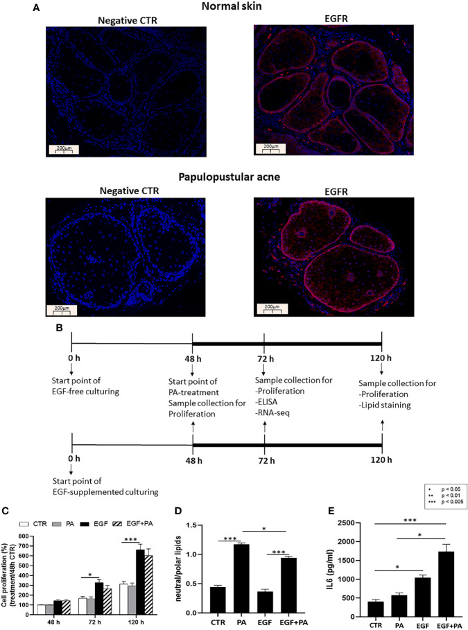
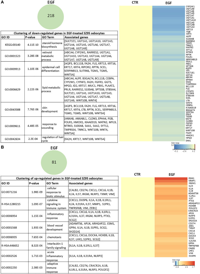
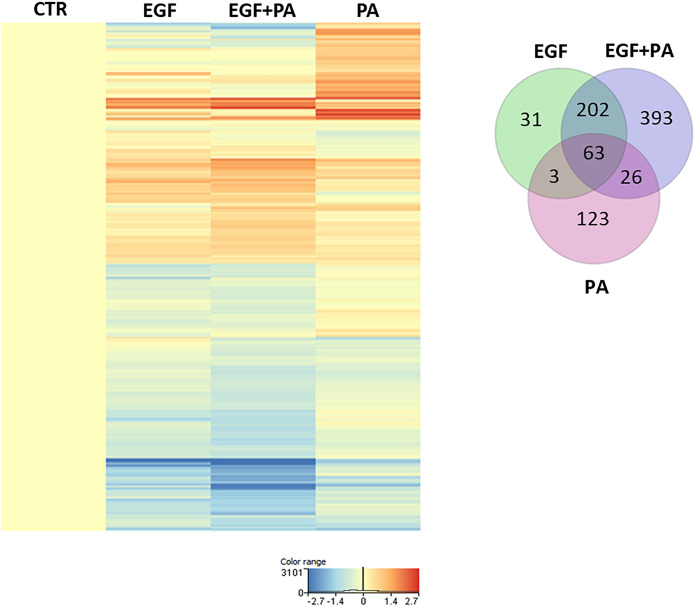
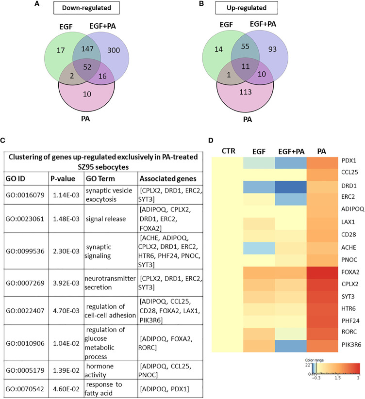
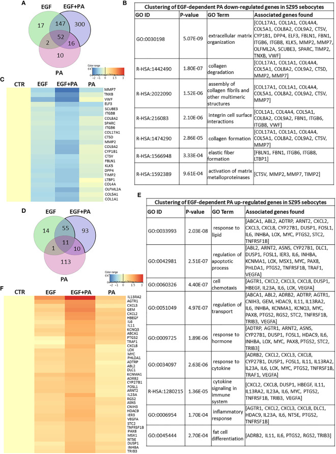
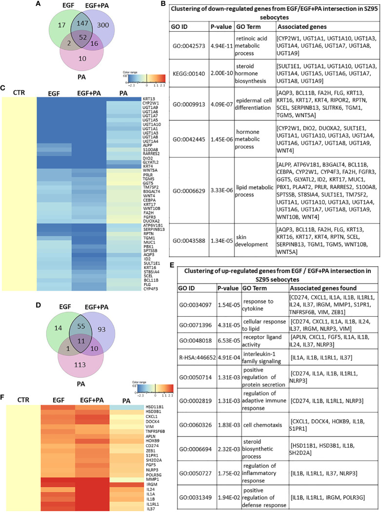
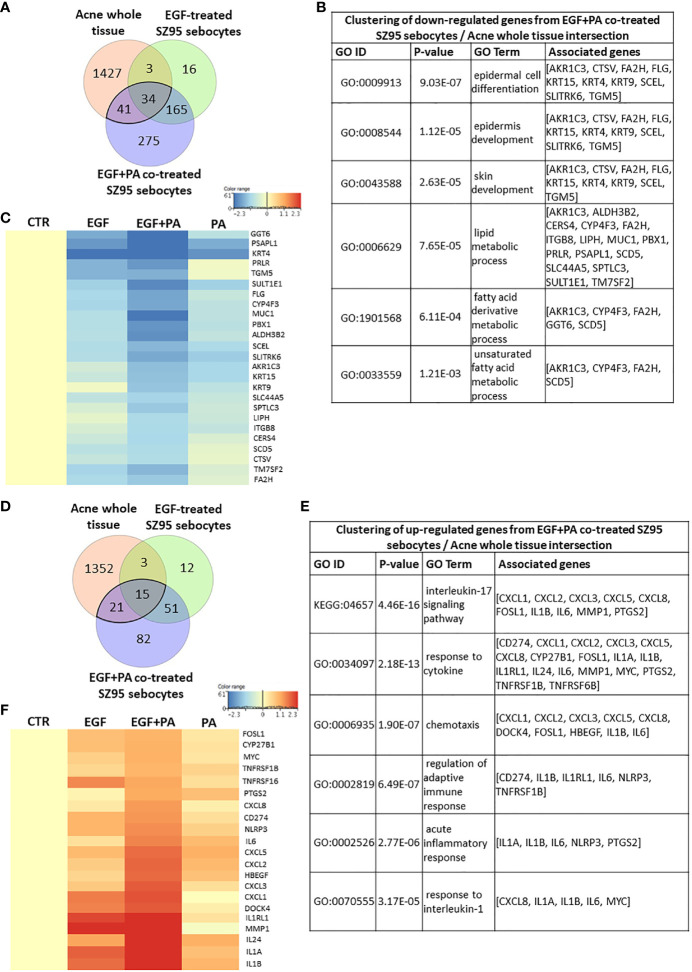
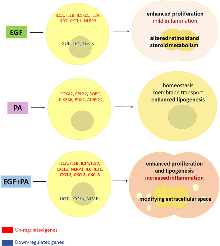
Epidermal growth factor (EGF) acts as a paracrine and autocrine mediator of cell proliferation and differentiation in various types of epithelial cells, such as sebocytes, which produce the lipid-rich sebum to moisturize the skin. However, sebum lipids via direct contact and by penetrating through the epidermis may have regulatory roles on epidermal and dermal cells as well. As EGF receptor (EGFR) is expressed throughout the proliferating and the lipid-producing layers of sebaceous glands (SGs) in healthy and acne-involved skin, we investigated the effect of EGF on SZ95 sebocytes and how it may alter the changes induced by palmitic acid (PA), a major sebum component with bioactive roles. We found that EGF is not only a potent stimulator of sebocyte proliferation, but also induces the secretion of interleukin (IL)6 and down-regulates the expression of genes involved in steroid and retinoid metabolism. Importantly, when applied in combination with PA, the PA-induced lipid accumulation was decreased and the cells secreted increased IL6 levels. Functional clustering of the differentially regulated genes in SZ95 sebocytes treated with EGF, PA or co-treated with EGF+PA further confirmed that EGF may be a potent inducer of hyperproliferative/inflammatory pathways (IL1 signaling), an effect being more pronounced in the presence of PA. However, while a group of inflammatory genes was up-regulated significantly in EGF+PA co-treated sebocytes, PA treatment in the absence of EGF, regulated genes only related to cell homeostasis. Meta-analysis of the gene expression profiles of whole acne tissue samples and EGF- and EGF+PA -treated SZ95 sebocytes showed that the EGF+PA co-activation of sebocytes may also have implications in disease. Altogether, our results reveal that PA-induced lipid accumulation and inflammation can be modulated by EGF in sebocytes, which also highlights the need for system biological approaches to better understand sebaceous (immuno)biology.

Keywords: EGF; acne; palmitic acid; sebocytes; sebum.


Conflict of interest statement

The authors declare that the research was conducted in the absence of any commercial or financial relationships that could be construed as a potential conflict of interest.

Figures

Figure 1
Figure 1
EGFR is expressed in sebaceous glands of healthy and acne involved skin and EGF modulates proliferation, lipid production and inflammation in SZ95 sebocytes. (A) FFPE tissue samples were stained with an anti-EGFR rabbit polyclonal antibody as described in Materials and Methods. Note that similar staining intensities are observed in normal and in papulopustular acne skin samples, localizing to both the proliferating as well as the lipid producing layers of sebaceous glands. Negative controls were processed without using primary antibody. Slides were mounted with Vectashield mounting medium with DAPI. Scale bar = 200 µm. (B) SZ95 sebocytes were cultured in the presence or absence of EGF. At 48 hours 150 µmol/L PA or vehicle was added to the medium and SZ95 sebocytes were cultured for further 24 and 72 hours (marked as 72 h and 120 h). Samples were collected, processed, and measured as described in Materials and Methods at the relevant timepoints. (C) Sulforhodamine B assay was used as described in Materials and Methods to detect the rate of proliferating sebocytes cultured in the presence or absence of EGF for 48, 72 and 120 hours and treated with 150 µmol/L PA, at 48 hours timepoint. EGF significantly induced cell proliferation at 72 and 120 hours, whereas proliferation was tendentiously decreased after co-treatment with PA. Samples from three subcultures of SZ95 sebocytes were used for the experiments and three independent measurements were performed. SRB data were normalized to the absorbance of the 48 hours CTR samples which was taken for 100%. Two-way ANOVA and Tukey post-hoc tests were used in the analysis of SRB data. All data are presented as mean ± SEM. *p < 0.5, ***p < 0.005. (D) AdipoRed staining used to detect polar and neutral lipids as described in Materials and Methods in SZ95 sebocytes cultured in the presence or absence of EGF and treated with 150 µmol/L PA for 72 hours showed that EGF decreased PA-induced lipogenesis. Amount of neutral lipids are normalized with polar lipids, which correlate with cell numbers. Results are expressed as average of three independent measurements using samples from three subcultures of SZ95 sebocytes. Kruskal-Wallis test and Dunn post-hoc tests were used in the analysis of data. All data are presented as mean ± SEM. *p < 0.05, ***p < 0.005. (E) Supernatants from SZ95 sebocytes cultured in the presence or absence of EGF and treated with 150 µmol/L PA for 24 hours were used to determine the levels of IL6 by ELISA as described in Materials and Methods. Results are expressed as average of protein concentrations (pg/mL) of five independent measurements using samples from three subcultures of SZ95 sebocytes. Kruskal-Wallis test and Dunn post-hoc tests were used in the analysis of ELISA data. All data are presented as mean ± SEM. *p < 0.05, ***p < 0.005.
Figure 2
Figure 2
EGF-induced transcriptional changes in SZ95 sebocytes. SZ95 sebocytes were cultured in the presence or absence of EGF. Samples for RNA-seq measurements were harvested at 72 hours of culturing, processed and measured as described in Materials and Methods. (A) The 218 transcripts that were significantly down-regulated in SZ95 sebocytes cultured in the presence of EGF (EGF) when compared to cells cultured in the absence of EGF (CTR) were clustered into the groups of steroid hormone biosynthesis, lipid metabolic process, skin development and response to wounding. Heat map display of the down-regulated transcripts contributing to the clusters. Differentially expressed genes were normalized to control and results are expressed as average mean of three samples from subcultures of SZ95 sebocytes. Color intensities reflect the ratios of signal intensities. (B) The 81 transcripts that were significantly up-regulated in SZ95 sebocytes cultured in the presence of EGF (EGF) when compared to cells cultured in the absence of EGF (CTR), were clustered into the groups of IL1 signaling, blood vessel development and inflammatory responses. Heat map display of the up-regulated transcripts contributing to the clusters. Differentially expressed genes were normalized to control and results are expressed as average mean of three samples from subcultures of SZ95 sebocytes. Color intensities reflect the ratios of signal intensities as shown.
Figure 3
Figure 3
Gene expression profile of EGF-treated, PA-treated and EGF+PA-co-treated sebocytes. Differentially expressed genes shown as a heat map in SZ95 sebocytes treated with PA for 24 hours in the absence (PA) or presence of EGF (EGF+PA), and in SZ95 sebocytes cultured in the presence of EGF (EGF) when compared to cells cultured in the absence of EGF (CTR) as observed by our RNA-seq analysis. Differentially expressed genes were normalized to control and results are expressed as average mean of three samples from subcultures of SZ95 sebocytes. Color intensities reflect the ratios of signal intensities as shown. Venn diagram visualizes the number of genes which are significantly regulated between treatment conditions (EGF (green), EGF+PA (purple) and PA (pink) treated SZ95 sebocytes) 24 hours after PA treatment when compared to SZ95 sebocytes cultured in the absence of EGF (CTR) as observed by our RNA-seq analysis.
Figure 4
Figure 4
Transcriptional changes induced by PA exclusively when SZ95 sebocytes are cultured without EGF. (A) Venn diagram visualizing the number of genes which are significantly down-regulated between treatment conditions (EGF (green), EGF+PA (purple) and PA (pink) treated SZ95 sebocytes) 24 hours after PA treatment when compared to SZ95 sebocytes cultured in the absence of EGF (CTR) as observed by our RNA-seq analysis. Note that the 10 transcripts which were down-regulated only in the PA treated sebocytes could not be grouped into any functional cluster. (B) Venn diagram visualizing the number of transcripts which are significantly up-regulated between treatment conditions (EGF (green), EGF+PA (purple) and PA (pink) treated SZ95 sebocytes) 24 hours after PA treatment when compared to SZ95 sebocytes cultured in the absence of EGF (CTR) as observed by our RNA-seq analysis, revealed that 113 transcripts were specific for the PA treatment. (C) The 113 transcripts that were up-regulated only in PA (PA vs. CTR) but not in EGF+PA co-treated SZ95 sebocytes were functionally categorized into groups related to cell homeostasis. (D) Heat map display of the transcripts significantly up-regulated only in PA treated (PA), but not in EGF+PA co-treated (EGF+PA) SZ95 sebocytes that contributed to the clusters. Differentially expressed genes were normalized to control and results are expressed as average mean of three samples from subcultures of SZ95 sebocytes. Color intensities reflect the ratios of signal intensities as shown.
Figure 5
Figure 5
Transcriptional changes induced by PA exclusively when SZ95 sebocytes are cultured in the presence of EGF. (A) Venn diagram visualizing that 300 transcripts were significantly down-regulated only in the condition when both EGF and PA was present in the culturing medium of the SZ95 sebocytes as observed by our RNA-seq analysis. (B) The 300 transcripts that were down-regulated only in SZ95 sebocytes cultured in the presence of both EGF and PA were functionally categorized into groups of extracellular matrix organization, collagen and elastic fiber formation. (C) Heat map display of the ones out of the 300 transcripts with significant down-regulation only in the EGF+PA co-treated sebocytes which were forming clusters. Differentially expressed genes were normalized to control and results are expressed as average mean of three samples from subcultures of SZ95 sebocytes. Color intensities reflect the ratios of signal intensities as shown. (D) Venn diagram visualizing that 93 transcripts were significantly up-regulated only when both EGF and PA was present in the culturing medium of the SZ95 sebocytes as observed by our RNA-seq analysis. (E) Functional clustering of the 93 transcripts showing significant up-regulation only in SZ95 sebocytes cultured in the presence of both EGF and PA confirmed that PA together with EGF had a complex regulation on genes involved in both lipid metabolism and inflammation. (F) Heat map display of the cluster forming 93 differentially expressed transcripts that showed a significant up-regulation only in the EGF+PA co-treated SZ95 sebocytes. Differentially expressed genes were normalized to control and results are expressed as average mean of three samples from subcultures of SZ95 sebocytes. Color intensities reflect the ratios of signal intensities as shown. Note that many of the transcripts had a tendency of up-regulation by EGF and/or by PA treatment, but the co-treatment was necessary to reach the level of significance.
Figure 6
Figure 6
Combined actions of PA and EGF on the gene expression profile of SZ95 sebocytes. (A) Venn diagram showing the 199 transcripts which were significantly down-regulated when EGF or EGF+PA was used in combination to treat SZ95 sebocytes, as observed by our RNA-seq analysis. (B) Functional clustering of the 199 transcripts showing significant down-regulation in the EGF and the EGF+PA co-treated SZ95 sebocytes revealed that the most significant changes were related to genes involved in hormone and lipid metabolic processes of sebocytes. (C) Heat map display of the transcripts which were clustered out of the 199 transcripts with significant down-regulation both in the EGF and the EGF+PA co-treated SZ95 sebocytes. Differentially expressed genes were normalized to control and results are expressed as average mean of three samples from subcultures of SZ95 sebocytes. Color intensities reflect the ratios of signal intensities. (D) Venn diagram showing the 66 transcripts which were significantly up-regulated when EGF or EGF+PA was used in combination to treat SZ95 sebocytes, as observed by our RNA-seq analysis. (E) Functional clustering of the 66 genes displayed in the Venn diagram defined that EGF alone and in combination with PA regulated immune features of SZ95 sebocytes, notably IL1 signaling. (F) Heat map display of the genes which were clustered out of the 66 differentially expressed transcripts. Differentially expressed genes were normalized to control and results are expressed as average mean of three samples from subcultures of SZ95 sebocytes. Color intensities reflect the ratios of signal intensities. Note the interaction between EGF and PA to regulate the expression of the genes which were already significantly regulated either by EGF or by PA.
Figure 7
Figure 7
Meta-analysis using gene expression profiles from acne samples, EGF-treated and EGF+PA- co-treated SZ95 sebocytes. (A) Venn diagram visualizing the number of transcripts that were significantly down-regulated in acne whole tissue samples from the available gene expression profiles of Kelhala HL et al. (NCBI GEO accession number: GSE5379) (56) and in EGF and EGF+PA co-treated SZ95 sebocytes. (B) Functional clustering of the 75 (41 + 34) transcripts that were significantly down-regulated both in the EGF+PA co-treated SZ95 sebocytes and in acne whole tissue samples (56) revealed that the most significant changes were related to genes involved in skin development and fatty acid metabolic process. (C) Heat map display of the genes which were clustered out of the 75 significantly down-regulated transcripts. Differentially expressed genes were normalized to control and results are expressed as average mean of three samples from subcultures of SZ95 sebocytes. Color intensities reflect the ratios of signal intensities as shown. (D) Venn diagram visualizing the number of transcripts that were significantly up-regulated in acne whole tissue samples from the available gene expression profiles of Kelhala et al. (56) and in EGF and EGF+PA co-treated SZ95 sebocytes. (E) Functional clustering of the 36 transcripts that were significantly up-regulated both in the EGF and EGF+PA co-treated SZ95 sebocytes and in acne whole tissue samples (56) revealed that EGF in combination with PA may contribute to the (patho)physiologically relevant IL1 and IL17 signaling in SZ95 sebocytes. (F) Fold change value based hierarchical clustering of the cluster forming genes, showing their expression levels in the PA, EGF and EGF+PA co-treated SZ95 sebocytes, that are significantly up-regulated in the EGF and EGF+PA co-treated SZ95 sebocytes when compared to untreated cells at 24 hours and in the available gene expression profiles of acne whole tissue samples (56). Differentially expressed genes were normalized to control and results are expressed as average mean of three samples from subcultures of SZ95 sebocytes. Color intensities reflect the ratios of signal intensities as shown. Note that the majority of the identified genes, showing a prominent expression in SZ95 sebocytes treated with PA in combination with EGF (such as CXCL8, IL1A, IL1B, IL6, NLRP3 and PTGS2), are pivotal in the pathogenesis of acne.
Figure 8
Figure 8
Interaction between EGF and PA to modulate cell proliferation, lipogenesis and inflammatory responses in sebocytes. Based on our results, EGF induces proliferation and inflammatory pathways in sebocytes while down regulates the expression of genes involved in steroid and retinoid metabolism. PA treatment regulates lipogenesis and induces genes related to cell homeostasis in human sebocytes. When both EGF and PA is present, sebocytes exert an increased proliferation, lipogenesis and a prominent up-regulation of inflammatory genes, while genes involved in the extracellular matrix formation are down-regulated, which may have (patho)physiological relevance in shaping the inflammatory dermal environment.

Similar articles

Cited by

References

    1. Kligman AM. The Uses of Sebum. Br J Dermatol (1963) 75:307–19. 10.1111/j.1365-2133.1963.tb13567.x - DOI - PubMed
    1. Ottaviani M, Camera E, Picardo M. Lipid Mediators in Acne. Mediators Inflammation (2010) 2010:858176. 10.1155/2010/858176 - DOI - PMC - PubMed
    1. Smith KR, Thiboutot DM. Thematic Review Series: Skin Lipids. Sebaceous Gland Lipids: Friend or Foe? J Lipid Res (2008) 49:271–81. 10.1194/jlr.R700015-JLR200 - DOI - PubMed
    1. Pappas A, Anthonavage M, Gordon JS. Metabolic Fate and Selective Utilization of Major Fatty Acids in Human Sebaceous Gland. J Invest Dermatol (2002) 118:164–71. 10.1046/j.0022-202x.2001.01612.x - DOI - PubMed
    1. Wille JJ, Kydonieus A. Palmitoleic Acid Isomer (C16:1delta6) in Human Skin Sebum is Effective Against Gram-Positive Bacteria. Skin Pharmacol Appl Skin Physiol (2003) 16:176–87. 10.1159/000069757 - DOI - PubMed

Publication types

MeSH terms