

Robotic in situ 3D bio-printing technology for repairing large segmental bone defects
- PMID: 34026288
- PMCID: PMC8132211
- DOI: 10.1016/j.jare.2020.11.011
Robotic in situ 3D bio-printing technology for repairing large segmental bone defects
Abstract
Introduction: The traditional clinical treatment of long segmental bone defects usually requires multiple operations and depends on donor availability. The 3D bio-printing technology constitutes a great potential therapeutic tool for such an injury. However, in situ 3D bio-printing remains a major challenge.
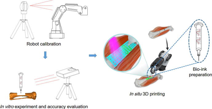
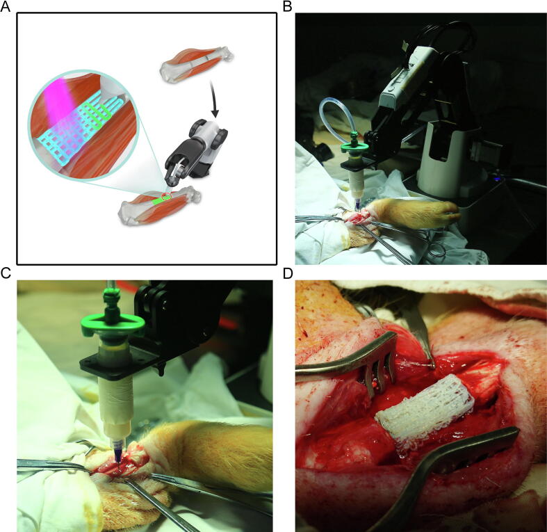
Objectives: In this study, we report the repair of long segmental bone defects by in situ 3D bio-printing using a robotic manipulator 3D printer in a swine model.
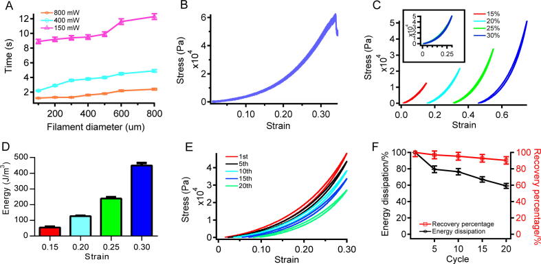
Methods: We systematically optimized bio-ink gelation under physiological conditions to achieve desirable mechanical properties suitable for bone regeneration, and a D-H kinematic model was used to improve printing accuracy to 0.5 mm.
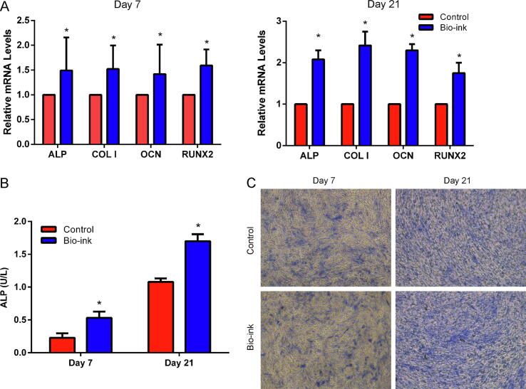
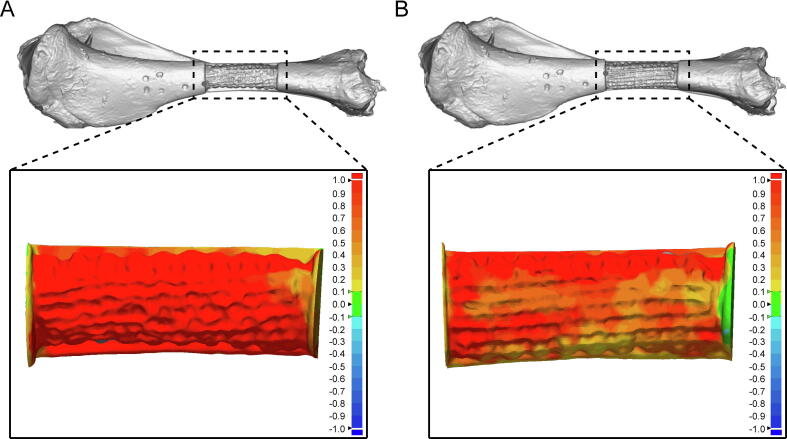
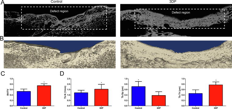
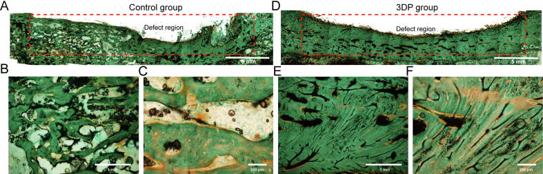
Results: These technical improvements allowed the repair of long segmental defects generated on the right tibia of pigs using 3D bio-printing within 12 min. The 3D bio-printing group showed improved treatment effects after 3 months.
Conclusion: These findings indicated that robotic in situ 3D bio-printing is promising for direct clinical application.
Keywords: 3D bio-printing; In situ; Regenerative medicine; Robotic; Tissue engineering.
© 2020 The Authors. Published by Elsevier B.V. on behalf of Cairo University.
Conflict of interest statement
The authors declare that they have no known competing financial interests or personal relationships that could have appeared to influence the work reported in this paper.
Figures
References
-
- Nauth A., Mckee M.D., Einhorn T.A., Watson J.T., Li R., Schemitsch E.H. Managing bone defects. J Orthop Trauma. 2011;25:462–466. - PubMed
-
- Cyril M., Brian Thomas B., Wade S. Management of segmental bone defects. J Am Acad Orthop Surgeons. 2015;23:143–153. - PubMed
-
- Loeffler B.J., Kellam J.F., Sims S.H., Bosse M.J. Prospective observational study of donor-site morbidity following anterior iliac crest bone-grafting in orthopaedic trauma reconstruction patients. J Bone Joint Surgery-Am. 2012;94A:1649–1654. - PubMed
-
- Seol Y.J., Kang H.W., Lee S.J., Atala A., Yoo J.J. Bioprinting technology and its applications. Eur J Cardio-thoracic Surgery: Off J Eur Assoc Cardio-thoracic Surg. 2014;46:342–348. - PubMed
Publication types
MeSH terms
Substances
LinkOut - more resources
Full Text Sources
Other Literature Sources
