. 2020 Nov 4:30:103-112.
doi: 10.1016/j.jare.2020.10.006. eCollection 2021 May.

Rescuing key native traits in cultured dermal papilla cells for human hair regeneration

Affiliations

Rescuing key native traits in cultured dermal papilla cells for human hair regeneration

Carla M Abreu et al. J Adv Res. .

Abstract

Introduction: The dermal papilla (DP) represents the major regulatory entity within the hair follicle (HF), inducing hair formation and growth through reciprocal interactions with epithelial cells. However, human DP cells rapidly lose their hair inductive ability when cultured in an epithelium-deficient environment.

Objectives: To determine if the conditioned medium collected from interfollicular keratinocytes (KCs-CM) is capable of improving DP cell native properties and inductive phenotype.

Methods: DP cells were cultured with KCs-CM both in 2D and 3D culture conditions (spheroids). Further, the hair-inductive capacity of DP cells precultured with KCs-CM was tested in a hair reconstitution assay, after co-grafting with human keratinocytes in nude mice.

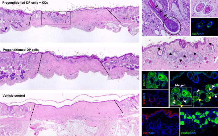
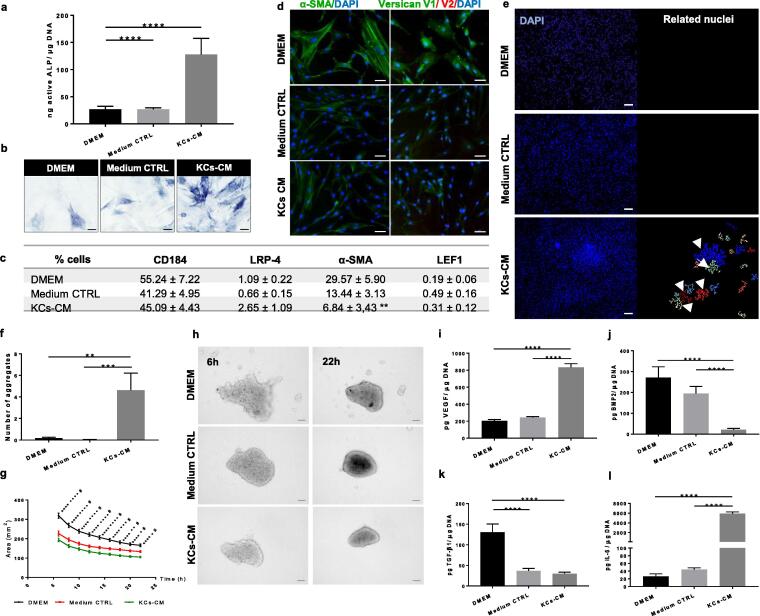
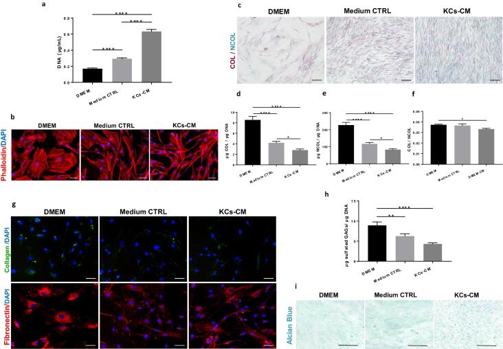
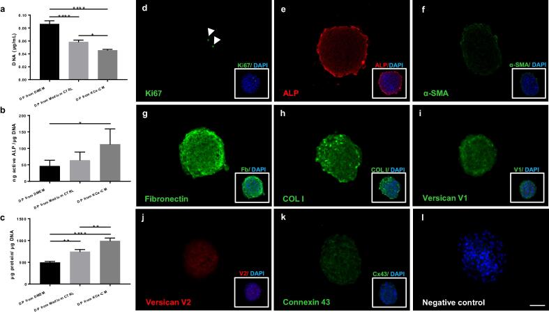
Results: We demonstrate that KCs-CM contributes to restore the inductivity of cultured human DP cells in a more effective mode than the conventional 3D-cultures. This is supported by the higher active alkaline phosphatase (ALP) levels in DP cells, the improved self-aggregative capacity and the reduced expression of α-SMA and the V1-isoform of versican. Moreover, DP cells cultured with KCs-CM displayed a secretome profile (VEGF, BMP2, TGF- β1, IL-6) that matches the one observed during anagen. KCs-CM also enhanced DP cell proliferation, while preventing cells to undergo morphological changes characteristic of high passage cells. In opposition, the amount of collagenous and non-collagenous proteins deposited by DP cells was lower in the presence of KCs-CM. The improvement in ALP activity was maintained in 3D spheroidal cultures, even after KCs-CM retrieval, being superior to the effect of the gold-standard culture conditions. Moreover, DP cells cultured with KCs-CM and grafted with human keratinocytes supported the formation of HF- and sebaceous gland-like structures in mice.

Conclusion: The proposed strategy encourages future cell-based strategies for HF regeneration not only in the context of hair-associated disorders, but also in the management of wounds to aid in restoring critical skin regulatory appendages.

Keywords: Dermal papilla cells; Hair follicle; Hair follicle regeneration; Hair inductivity; Keratinocyte-conditioned medium.


Conflict of interest statement

The authors declare that they have no known competing financial interests or personal relationships that could have appeared to influence the work reported in this paper.

Figures

None
Graphical abstract
Fig. 1
Fig. 1
KCs-CM improves DP cell inductive phenotype. (a) Amount of active ALP and (b) respective images of ALP-active DP cells showing an improvement of the DP cell inductive-related phenotype after culture with KCs-CM (n = 6). (c) Percentage of DP cells expressing CD184, LRP-4, α-SMA (n = 7) and LEF1 determined by flow cytometry (n = 5, **p < 0.01 vs DMEM). (d) Expression of α-SMA and the V1-isoform of versican in DP cells confirming a decrease after culture with KCs-CM. (e) Representative DAPI-stained nuclei images used to perform image analysis and (f) quantify the number of DP cell aggregates formed after 5 days in culture showing that DP cells treated with KCs-CM have improved capacity to self-aggregate (n = 3) (g) Variation of the area occupied by the DP cells up to 22 h of culture as obtained by the (h) analysis of time-lapse images showing that DP cells cultured in KCs-CM form more compact spheroids and faster than controls (n = 6). (i-k) Amount of growth factors and (l) cytokine secreted by DP cells showing the difference of their secretome with the culture conditions. Data shown are mean ± s.e.m. Differences are indicated by * KCs-CM vs DMEM, # KCs-CM vs Medium CTRL and Medium CTRL vs DMEM. *p < 0.05; **p < 0.01; ***p < 0.00; ****p < 0.0001. Scale bars are 50 µm for (b,d), 100 µm for (h) and 200 µm for (e).
Fig. 2
Fig. 2
KCs-CM beneficially affect DP cells proliferation but decreases matrix deposition ability. (a) DNA levels demonstrating that KCs-CM increases DP cell numbers (n = 9). (b) Phaloidin-TRITC stained F-actin cytoskeleton showing that CM-treatment prevents an enlarged morphology in DP cells. (c) Representative images of collagenous (COL, red) and non-collagenous protein deposition by DP cells (NCOL, green) and the (d-e) respective quantification showing a reduced secretion by CM-treated DP cells. The same tendency was observed for (f) COL/NCOL proteins ratio by (n = 6). (g) Expression of collagen type-I and fibronectin showing a dimmed signal in the condition where DP cells were treated with KCs-CM. (h) Amount of sulfated GAGs showing a reduced production by DP cells cultured with KCs-CM (n = 4) as well as different (i) deposition patern of carboxylated and sulfated GAGs in relation to controls. Data shown are mean ± s.e.m. *p < 0.05; **p < 0.01; ****p < 0.0001. Scale bars are 50 µm for (b,g), 100 µm for (c) and 200 µm for (i).
Fig. 3
Fig. 3
Preconditioning with KCs-CM further enhances DP signature recovery in 3D culture conditions. DP spheroids derived from cells previously cultured with KCs-CM had a significantly (a) lower DNA content, (b) higher amount of active ALP and (c) higher protein content (n = 3). Immunolabelling of DP spheroids preconditioned with KCs-CM showing (d) the proliferation-associated marker Ki67 (arrowheads) and the expression of (e) ALP, (f) α-SMA, (g) fibronectin, (h) collagen type I, (i) V1-isoform of versican, (j) V2-isoform of versican and the (k) gap junction protein connexin 43. Negative control is depicted on (l) and DAPI was used as the nuclear counterstaining (merged images, boxes). Data shown are mean ± s.e.m. *p < 0.05; **p < 0.01; ****p < 0.0001. Scale bars = 50 μm.
Fig. 4
Fig. 4
DP cells preconditioning promotes hair induction in mice. (a) H&E staining of the wound sites showing the formation of structures morphologically resembling HFs and SGs 6 weeks after grafting CM-precultured DP cells and KCs in immune-deficient mice. (b) Hair bulb formation (dashed circle) and hair shaft elongation (arrows) were observed in one of the seven co-grafted animals. (c) DP cells grafted alone led to the formation of less complex and less abundant structures which (d) were not observed in controls (dotted lines limit the wound area). (e) High magnification image of the recreated structures of the experimental condition demonstrating their complexity, including the presence of a differentiated core morphologically alike to hair shafts (arrowheads). (f) The folliculoid structures featured distinct epithelial layers, including (g) one layer expressing the IRS marker K25, (h) a layer co-expressing both K15 and K14 or layers only positive for K14. Arrowheads indicate K14 and K15 co-expression in HF-like (white) and SG-like structure (yellow). (i)K10 expression was limited to the skin epithelium, whereas (j) the sebocyte marker FABP4 was expressed in the structures resembling SG. Scale bars are 500 µm for (a-c), 100 µm for (d) and 50 µm for (e-j).

References

    1. Yang C.-C., Cotsarelis G. Review of hair follicle dermal cells. J. Dermatol. Sci. 2010;57:2–11. doi: 10.1016/j.jdermsci.2009.11.005. - DOI - PMC - PubMed
    1. Driskell R.R., Clavel C., Rendl M., Watt F.M. Hair follicle dermal papilla cells at a glance. J. Cell Sci. 2011;124:1179–1182. doi: 10.1242/jcs.082446. - DOI - PMC - PubMed
    1. Ohyama M., Zheng Y., Paus R., Stenn K.S. The mesenchymal component of hair follicle neogenesis: background, methods and molecular characterization. Exp. Dermatol. 2010;19:89–99. doi: 10.1111/j.1600-0625.2009.00935.x. - DOI - PubMed
    1. Higgins C.A., Chen J.C., Cerise J.E., Jahoda C.A.B., Christiano A.M. Microenvironmental reprogramming by three-dimensional culture enables dermal papilla cells to induce de novo human hair-follicle growth. Proc. Natl. Acad. Sci. 2013;110:19679–19688. doi: 10.1073/pnas.1309970110. - DOI - PMC - PubMed
    1. Higgins C.A., Richardson G.D., Ferdinando D., Westgate G.E., Jahoda C.A.B. Modelling the hair follicle dermal papilla using spheroid cell cultures. Exp. Dermatol. 2010;19:546–548. doi: 10.1111/j.1600-0625.2009.01007.x. - DOI - PubMed

Publication types