. 2021 Mar 15;8(10):2001879.
doi: 10.1002/advs.202001879. eCollection 2021 May.

CD38-Targeted Theranostics of Lymphoma with 89Zr/177Lu-Labeled Daratumumab

Affiliations

CD38-Targeted Theranostics of Lymphoma with 89Zr/177Lu-Labeled Daratumumab

Lei Kang et al. Adv Sci (Weinh). .

Abstract

Lymphoma is a heterogeneous disease with varying clinical manifestations and outcomes. Many subtypes of lymphoma, such as Burkitt's lymphoma and diffuse large B cell lymphoma, are highly aggressive with dismal prognosis even after conventional chemotherapy and radiotherapy. As such, exploring specific biomarkers for lymphoma is of high clinical significance. Herein, a potential marker, CD38, is investigated for differentiating lymphoma. A CD38-targeting monoclonal antibody (mAb, daratumumab) is then radiolabeled with Zr-89 and Lu-177 for theranostic applications. As the diagnostic component, the Zr-89-labeled mAb is highly specific in delineating CD38-positive lymphoma via positron emission tomography (PET) imaging, while the Lu-177-labeled mAb serves well as the therapeutic component to suppress tumor growth after a one-time administration. These results strongly suggest that CD38 is a lymphoma-specific marker and prove that 89Zr/177Lu-labeled daratumumab facilitates immunoPET imaging and radioimmunotherapy of lymphoma in preclinical models. Further clinical evaluation and translation of this CD38-targeted theranostics may be of significant help in lymphoma patient stratification and management.

Keywords: CD38; Lu‐177; daratumumab; lymphoma; positron emission tomography (PET); radioimmunotherapy; theranostics.


Conflict of interest statement

Weibo Cai is scientific advisor, stockholder, and grantee of Focus‐X Therapeutics, Inc. The other authors declare no conflict of interest.

Figures

Figure 1
Figure 1
Theranostic design of radiolabeled daratumumab and in vitro evaluation. a) Scheme of the theranostic role of 89Zr‐ and 177Lu‐labeled daratumumab. b) Western blot showed that Daudi cells had the highest CD38 expression relative to β‐tubulin, whereas Ly10 cells had the lowest level. c) Flow cytometry proved the cellular binding affinity of daratumumab. d) Flow cytometry displayed that FITC‐labeled Df‐ or DTPA‐conjugated daratumumab had a similar cellular binding affinity. e) Receptor binding assay showed that 89Zr‐ and 177Lu‐labeled daratumumab had high binding ability in Daudi cells (n = 3). f) SDS‐PAGE showed that 177Lu–dara kept stability after being incubated in PBS and 10% FBS for 16 days. g) TLC presented that the radiochemical purities were higher than 90% over 16 days.
Figure 2
Figure 2
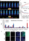
ImmunoPET imaging, biodistribution, and immunofluorescent staining results. a) The maximum intensity projection (MIP) PET images showed that 89Zr–dara exhibited an increasing and persistent tumor uptake in CD38‐positive Daudi models but near‐background uptake in CD38‐negative Ly10 models (n = 4). b) Quantitative data obtained via ROI analysis showed that the tumor uptake in Daudi models was significantly higher than in Ly10 models (p < 0.05). The blood uptake gradually decreased in both tumor models without significant difference. c) Ex vivo biodistributions at 120 h p.i. verified the imaging results. d) Immunofluorescent staining confirmed the strong intensity of CD38 signal in Daudi tumor tissue and limited signal in Ly10 tumor tissue.
Figure 3
Figure 3
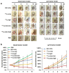
Tumor size monitoring. a) Representative tumor region photos were displayed for different groups in Daudi and Ly10 tumor models for 10 days. b) The standardized tumor volume for different groups in Daudi and Ly10 tumor models (n = 5–6).
Figure 4
Figure 4
18F‐FDG PET imaging for evaluation of the therapeutic efficacy. a) Representative 18F‐FDG PET images were performed for different groups in Daudi and Ly10 models at 8 days (circles indicate tumor region, arrows point to the necrotic region). b) Quantitative data of tumor uptake were obtained after drawing region of interest on the tumor. ** represents p < 0.05 (n = 4).
Figure 5
Figure 5
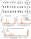
Verification of distribution of 177Lu‐labeled probes. a) Planar radiography was acquired for radioactive probes and showed significantly high tumor accumulation of 177Lu‐dara‐high, compared with 177Lu and 177Lu–IgG for Daudi model (n = 4). b) The biodistribution results at 10 days verified the high tumor uptake of 177Lu‐dara‐high in Daudi model, whereas no significant difference among four groups. 177Lu was mainly found in the liver and spleen, n = 4. c) The biodistribution of 177Lu‐dara‐high in Daudi model at 1, 4, 7, and 10 days showed a high and persistent tumor uptake up to 10 days p.i. of 17.2 ± 3.7 %ID g−1. Blood displayed a descendent uptake correlating with uptake in other normal organs.
Figure 6
Figure 6
H&E and TUNEL staining of tumor tissues. a) H&E staining showed that the Daudi tumor tissues treated with 177Lu‐dara had necrosis inside (yellow box), whereas the Ly10 tumor tissues showed uniformly small lymphoma cells. b) TUNEL staining displayed that there was large scale of brown staining area inside the Daudi tumor treated with 177Lu–dara, suggesting apoptosis, but few staining in other groups.
Figure 7
Figure 7
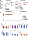
Biosafety evaluation. a) Standardized body weight of CB/17 SCID mice for Daudi and Ly10 tumor models were measured for 10 days and showed the decrease in all 177Lu‐related groups, suggesting potential toxicity (n = 4). b) The body weights of normal BALB/c mice were evaluated further after being treated similarly up to 26 d. The results showed no decrease or significant difference among different groups (n = 4, p > 0.05). c) Hematological analysis was performed for 177Lu‐dara‐high group in Daudi tumor model at 1, 4, 7, and 10 days. WBC, LYM, and MON cell counts dropped in the first 4 days but recovered gradually, indicating no sustained blood toxicity or inflammation after treatment.

References

    1. Shanbhag S., Ambinder R. F., Ca‐Cancer J. Clin. 2018, 68, 116. - PMC - PubMed
    1. Siegel R. L., Miller K. D., Jemal A., Ca‐Cancer J. Clin. 2018, 68, 7. - PubMed
    1. Saltarella I., Desantis V., Melaccio A., Solimando A. G., Lamanuzzi A., Ria R., Storlazzi C. T., Mariggio M. A., Vacca A., Frassanito M. A., Cells 2020, 9, 167. - PMC - PubMed
    1. Green D. J., Orgun N. N., Jones J. C., Hylarides M. D., Pagel J. M., Hamlin D. K., Wilbur D. S., Lin Y., Fisher D. R., Kenoyer A. L., Frayo S. L., Gopal A. K., Orozco J. J., Gooley T. A., Wood B. L., Bensinger W. I., Press O. W., Cancer Res. 2014, 74, 1179. - PMC - PubMed
    1. Bailly C., Clery P. F., Faivre‐Chauvet A., Bourgeois M., Guerard F., Haddad F., Barbet J., Cherel M., Kraeber‐Bodere F., Carlier T., Bodet‐Milin C., Int. J. Mol. Sci. 2016, 18, 57. - PMC - PubMed

Publication types

MeSH terms