. 2021 May 7:9:658877.
doi: 10.3389/fchem.2021.658877. eCollection 2021.

The High-Spin Heme b L Mutant Exposes Dominant Reaction Leading to the Formation of the Semiquinone Spin-Coupled to the [2Fe-2S]+ Cluster at the Qo Site of Rhodobacter capsulatus Cytochrome bc 1

Affiliations

The High-Spin Heme b L Mutant Exposes Dominant Reaction Leading to the Formation of the Semiquinone Spin-Coupled to the [2Fe-2S]+ Cluster at the Qo Site of Rhodobacter capsulatus Cytochrome bc 1

Marcin Sarewicz et al. Front Chem. .

Abstract

Cytochrome bc 1 (mitochondrial complex III) catalyzes electron transfer from quinols to cytochrome c and couples this reaction with proton translocation across lipid membrane; thus, it contributes to the generation of protonmotive force used for the synthesis of ATP. The energetic efficiency of the enzyme relies on a bifurcation reaction taking place at the Qo site which upon oxidation of ubiquinol directs one electron to the Rieske 2Fe2S cluster and the other to heme b L. The molecular mechanism of this reaction remains unclear. A semiquinone spin-coupled to the reduced 2Fe2S cluster (SQo-2Fe2S) was identified as a state associated with the operation of the Qo site. To get insights into the mechanism of the formation of this state, we first constructed a mutant in which one of the histidine ligands of the iron ion of heme b L Rhodobacter capsulatus cytochrome bc 1 was replaced by asparagine (H198N). This converted the low-spin, low-potential heme into the high-spin, high-potential species which is unable to support enzymatic turnover. We performed a comparative analysis of redox titrations of antimycin-supplemented bacterial photosynthetic membranes containing native enzyme and the mutant. The titrations revealed that H198N failed to generate detectable amounts of SQo-2Fe2S under neither equilibrium (in dark) nor nonequilibrium (in light), whereas the native enzyme generated clearly detectable SQo-2Fe2S in light. This provided further support for the mechanism in which the back electron transfer from heme b L to a ubiquinone bound at the Qo site is mainly responsible for the formation of semiquinone trapped in the SQo-2Fe2S state in R. capusulatus cytochrome bc 1.

Keywords: cytochrome bc1 (complex III); electron paramagnetic resonance; electron transfer; quinol oxidation; semiquinone.


Conflict of interest statement

The authors declare that the research was conducted in the absence of any commercial or financial relationships that could be construed as a potential conflict of interest.

Figures
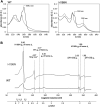
FIGURE 1
FIGURE 1
Comparison of spectra for isolated WT and H198N mutants of Cytbc 1. (A) Optical spectra of WT (left) and H198N mutant (right) measured for ascorbate (gray) and dithionite-reduced (black) Cytbc 1. Maximum at ∼560 nm originates from absorption of hemes b. Additional absorption band at ∼590 nm is likely to originate from heme b L with changed ligand in H198N mutant. (B) X-band EPR spectra of air-oxidized WT (black) and H198N mutant (gray) measured at 10 K. In the range between ∼70 and ∼160 mT the contribution from high-spin iron centers is detected. In the range between ∼160 and 200 mT, the g z transitions of HALS hemes b transitions are detected, while the reduced 2Fe2S contributes to transitions at the magnetic field above ∼320 mT.
FIGURE 2
FIGURE 2
Sensitivity of the EPR spectra of the 2Fe2S cluster to changes in redox state of the Q pool for WT and the H198N mutant at pH 8. (A) The spectra measured for chromatophores poised at + 200 mV (Q pool oxidized) in dark. (B) The spectra measured for chromatophores poised at + 20 mV (Q pool half reduced). The vertical lines indicate the positions of g y and g x transitions of the 2Fe2S cluster, which shifts to higher and lower g values, respectively, upon the reduction of the Q pool. The spectra of the mutant were magnified ∼5 times to normalize the g y signals to the same level as in WT.
FIGURE 3
FIGURE 3
X-band EPR spectra measured for illuminated chromatophores containing WT Cytbc 1 at pH 8 and E h = +77 mV. Gray and black spectra were obtained for samples containing 40 and 20 μM, respectively. Dashed vertical lines show g y = 1.89 and g x = 1.80 transitions of 2Fe2S cluster. The maximum at 1.95 originates from SQo-2Fe2S.
FIGURE 4
FIGURE 4
Comparison of efficiency of light activation of RC in chromatophores containing WT and H198N mutant of Cytbc 1. (A) Amplitude of the normalized g x transition of the 2Fe2S cluster of WT Cytbc 1 which is proportional to the amount of UQ in the Q pool in dark (closed circles) and in illuminated samples (open circles). The experiential data points were simulated using an appropriate Nernst equation with n = 2. (B) Amplitude of the g y transition of the 2Fe2S cluster measured for the illuminated chromatophores containing H198N mutant of Cytbc 1 (open circles) and chromatophores in dark (closed circles).
FIGURE 5
FIGURE 5
Comparison of SQo-2Fe2S generation in chromatophores containing WT (A) and H198N mutant (B) of Cytbc 1. Amplitude of SQo-2Fe2S was measured for illuminated samples (closed circles) and titrated in dark (open circles). Optical transparencies were the same for A and B, and the concentrations of cytochrome c 1 were 20 and 10 μM, respectively. The solid line in A represents the fit of f SQo-2Fe2S = f A/{1 + exp [0.039n (E hE 1)] + exp [0.039n (E 2E h)]} function described in detail by Sarewicz et al. (2018). The fit yielded E 1 = +92 ± 8 mV and E 2 = +40 ± 7 mV for n = 2 and f A = 1.7 ± 0.4. The small bar denoted marked as SD shows the typical uncertainty of the amplitude reads and includes the baseline and the noise of the EPR spectrum. It does not include uncertainty related to changes in efficiency of SQo-2Fe2S generation dominated by variations in light activation and/or changes in the freezing time.
FIGURE 6
FIGURE 6
Simplified scheme of two possible reactions of SQo-2Fe2S formation. (A) The semiforward mechanism of UQH2 oxidation by the 2Fe2S cluster at the time when heme b L is unable to accept electron (because of being already reduced) from resulting SQo, leading to the creation of the SQo-2Fe2S state. (B) The semireverse mechanism assumes that the reduced heme b L donates electrons to UQ at the time when the reduced 2Fe2S is close to the Qo site. This leads to the creation of the SQo-2Fe2S state. According to the mechanism shown in A, the redox-active heme b L is not necessary, while the mechanism shown in B requires heme b L for the generation of the SQo-2Fe2S state.

References

    1. Al-Attar S., de Vries S. (2013). Energy Transduction by Respiratory Metallo-Enzymes: From Molecular Mechanism to Cell Physiology. Coord. Chem. Rev. 257, 64–80. 10.1016/j.ccr.2012.05.022 - DOI
    1. Atta-Asafo-Adjei E., Daldal F. (1991). Size of the Amino Acid Side Chain at Position 158 of Cytochrome B Is Critical for an Active Cytochrome Bc1 Complex and for Photosynthetic Growth of Rhodobacter Capsulatus. Proc. Natl. Acad. Sci. 88, 492–496. 10.1073/pnas.88.2.492 - DOI - PMC - PubMed
    1. Baniulis D., Yamashita E., Zhang H., Hasan S. S., Cramer W. A. (2008). Structure-Function of the Cytochrome b6f Complex. Photochem. Photobiol. 84, 1349–1358. 10.1111/j.1751-1097.2008.00444.x - DOI - PubMed
    1. Berry E. A., Guergova-Kuras M., Huang L.-S., Crofts A. R. (2000). Structure and Function of CytochromebcComplexes. Annu. Rev. Biochem. 69, 1005–1075. 10.1146/annurev.biochem.69.1.1005 - DOI - PubMed
    1. Berry E. A., Huang L.-S., Saechao L. K., Pon N. G., Valkova-Valchanova M., Daldal F. (2004). X-Ray Structure of Rhodobacter Capsulatus Cytochrome Bc1: Comparison with its Mitochondrial and Chloroplast Counterparts. Photosynth. Res. 81, 251–275. 10.1023/b:pres.0000036888.18223.0e - DOI - PubMed

LinkOut - more resources