Review
. 2021 May 7:9:664820.
doi: 10.3389/fchem.2021.664820. eCollection 2021.

Single-Entity Detection With TEM-Fabricated Nanopores

Affiliations
Review

Single-Entity Detection With TEM-Fabricated Nanopores

Hongcheng Yang et al. Front Chem. .

Abstract

Nanopore-based single-entity detection shows immense potential in sensing and sequencing technologies. Solid-state nanopores permit unprecedented detail while preserving mechanical robustness, reusability, adjustable pore size, and stability in different physical and chemical environments. The transmission electron microscope (TEM) has evolved into a powerful tool for fabricating and characterizing nanometer-sized pores within a solid-state ultrathin membrane. By detecting differences in the ionic current signals due to single-entity translocation through the nanopore, solid-state nanopores can enable gene sequencing and single molecule/nanoparticle detection with high sensitivity, improved acquisition speed, and low cost. Here we briefly discuss the recent progress in the modification and characterization of TEM-fabricated nanopores. Moreover, we highlight some key applications of these nanopores in nucleic acids, protein, and nanoparticle detection. Additionally, we discuss the future of computer simulations in DNA and protein sequencing strategies. We also attempt to identify the challenges and discuss the future development of nanopore-detection technology aiming to promote the next-generation sequencing technology.

Keywords: TEM fabrication; electron-beam drilling; sequencing; single entity detection; solid-state nanopores.


Conflict of interest statement

The authors declare that the research was conducted in the absence of any commercial or financial relationships that could be construed as a potential conflict of interest.

Figures
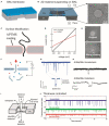
Figure 1
Figure 1
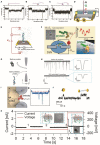
(A,B) Schematic illustration of SiNx and MoS2 membrane suspended on a SiNx supporting membrane. (A) Reprinted from Wang et al. (2020) with permission from the Royal Society of Chemistry. (B) Reprinted from Liu et al. (2014) with permission from the American Chemical Society. (C) Schematic showing DNA threading through an APTMS-coated SiNx nanopore. (D) I–V curves of coated and uncoated pores. Reprinted from Anderson et al. (2013) with permission from the American Chemical Society. (E) TEM images showing the original and silane-coated nanopores. Reprinted from Wanunu and Meller (2007) with permission from the American Chemical Society. (F) Image showing a nanopore that separates two chambers containing electrolyte solutions and small molecules are passed through the nanopore driven by the applied potential. (G) An amplifier showing the detection of the partial current blockade due to the passage of molecules through the nanopore. The translocation event of an individual molecule is usually characterized through dwell time and the amplitude of the current blockade. Reprinted from Fragasso et al. (2020) with permission from the American Chemical Society. (H) Example showing the DNA (48 kbp) translocation events through uncoated and DOPA-coated nanopore. Reprinted from Karmi et al. (2020) with permission from the American Chemical Society. (I) Diagram of STEM-thinning method. The electron beams are collected using electron energy-loss spectroscopy (EELS) and HAADF signal detectors after interaction with the film. Reprinted from Rodriguez-Manzo et al. (2015) with permission from the American Chemical Society. (J) The translocation signals were enhanced in thinner membranes with the translocation of 40 nt ssDNA through nanopore, providing the higher SNR for nanopore sensing. Reprinted from Lee et al. (2014) with permission from Springer Nature. APTMS, 3-(aminopropyl)trimethoxysilane; TEM, transmission electron microscope; DOPA, amino l-3,4-dihydroxyphenylalanine; STEM, scanning transmission electron microscopy; EELS, ssDNA, single-stranded DNA; SNR, signal-to-noise ratio; HAADF, high-angle annular dark-field.
Figure 2
Figure 2
Characteristic translocation current blockade signals for representative structures of TDN bonded to linear DNA molecules. (A) One end bonded to a 7-bp TDN, (B) the middle part bonded to a 13-bp TDN, and (C) a 13-bp and a 7-bp TDN bonded to the middle and end, respectively. Insets are cartoon images of these structures. Reprinted from Zhao et al. (2019) with permission from the Royal Society of Chemistry. (D) DNA sequencing setup showing transversal current rectification during translocation of ssDNA through a nanopore with N-terminated CNT electrodes. Reprinted from Djurišić et al. (2020) with permission from the American Chemical Society. (E) Schematic showing the detection of dsDNA in electrolyte using transverse current and ionic current measurements. Reprinted from Xiong et al. (2020) with permission from the American Chemical Society. (F) Schematic showing the translocation of a model protein (peptides) and lysine residues through MoS2 nanopores with polylysine tags. The right panel shows the relationship between the translocation sequence of events and ionic conductance. Reprinted from Nicolai et al. (2019) with permission from the American Chemical Society. (G) Illustration of translocation events of native β-amylase protein and their sodium dodecyl sulfate-unfolded treatment structures through nanopore. (H) Current signals generated by the translocation of protein molecules; wherein the right panel displays their characteristic events. Reprinted from Restrepo-Perez et al. (2017) with permission from the Royal Society of Chemistry. (I) Illustration representing the translocation of a dsDNA molecule with a bound antibody (left panel) and corresponding spike responses. Reprinted from Plesa et al. (2015a) with permission from the American Chemical Society. (J) Current traces of AuNP translocation through the pore. Reprinted from Karmi et al. (2021) with permission from the American Chemical Society. (K) Schematic of the AuNP growth in nanopore; (left panel inset) the voltage difference triggers the reaction and drives the reagents into nanopore (right panel inset) the formation of a AuNP further blocks the pore and prevents the mixing of reagents. Current or voltage vs. time traces representing a particle growth procedure into three steps. Zero current indicates the NP formation. The time delay (td) before formation of AuNP is represented with vertical [after step (3)] dashed. Inserts are the TEM images of nanopore before/after the AuNP growth. The scale bar is 5 nm. Reprinted from Venta et al. (2013) with permission from the American Chemical Society. TDN, tetrahedral DNA nanostructure; ssDNA, single-stranded DNA; CNT, carbon nanotube; dsDNA, double-stranded DNA; AuNP, gold nanoparticle; NP, nanoparticle; TEM, transmission electron microscope.

Similar articles

Cited by

References

    1. Acharya S., Jiang A., Kuo C., Nazarian R., Li K., Ma A., et al. . (2020). Improved measurement of proteins using a solid-state nanopore coupled with a hydrogel. ACS Sens. 5, 370–376. 10.1021/acssensors.9b01928 - DOI - PubMed
    1. Albrecht T. (2019). Single-molecule analysis with solid-state nanopores. Annu. Rev. Anal. Chem. 12, 371–387. 10.1146/annurev-anchem-061417-125903 - DOI - PubMed
    1. Alibakhshi M. A., Halman J. R., Wilson J., Aksimentiev A., Afonin K. A., Wanunu M. (2017). Picomolar fingerprinting of nucleic acid nanoparticles using solid-state nanopores. ACS Nano 11, 9701–9710. 10.1021/acsnano.7b04923 - DOI - PMC - PubMed
    1. Amarasinghe S. L., Su S., Dong X., Zappia L., Ritchie M. E., Gouil Q. (2020). Opportunities and challenges in long-read sequencing data analysis. Genome Biol. 21:30. 10.1186/s13059-020-1935-5 - DOI - PMC - PubMed
    1. Anderson B. N., Muthukumar M., Meller A. (2013). pH tuning of DNA translocation time through organically functionalized nanopores. ACS Nano 7, 1408–1414. 10.1021/nn3051677 - DOI - PMC - PubMed

LinkOut - more resources