. 2020 Dec 30;6(2):e10209.
doi: 10.1002/btm2.10209. eCollection 2021 May.

Mitochondrial transplantation therapy inhibit carbon tetrachloride-induced liver injury through scavenging free radicals and protecting hepatocytes

Affiliations

Mitochondrial transplantation therapy inhibit carbon tetrachloride-induced liver injury through scavenging free radicals and protecting hepatocytes

Zizhen Zhao et al. Bioeng Transl Med. .

Abstract

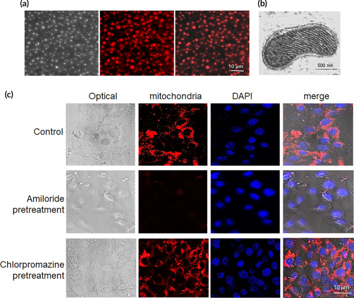
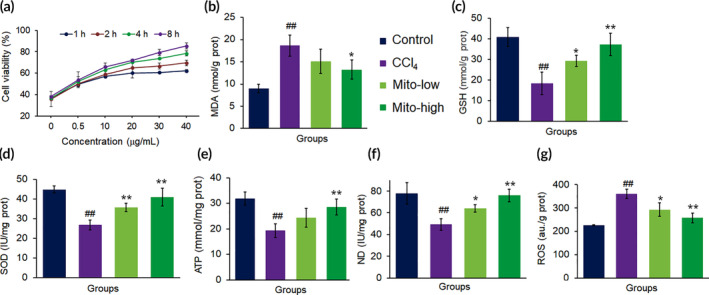
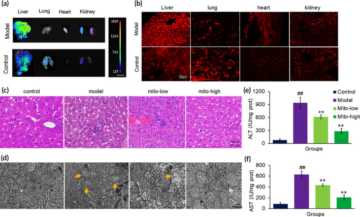
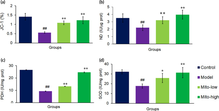
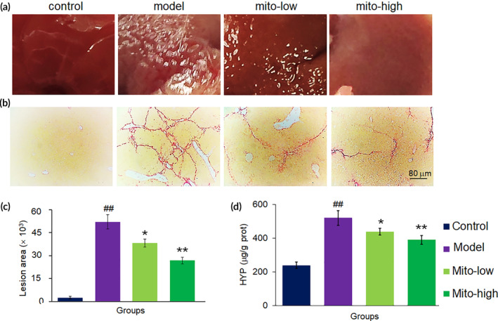
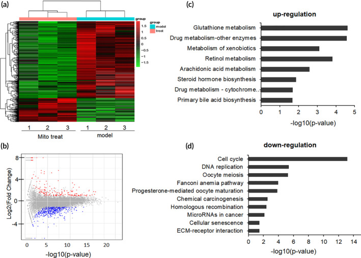
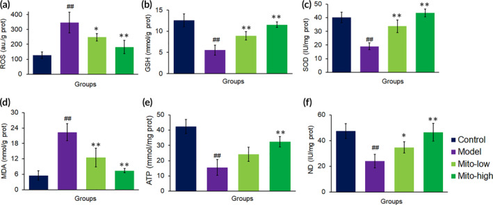
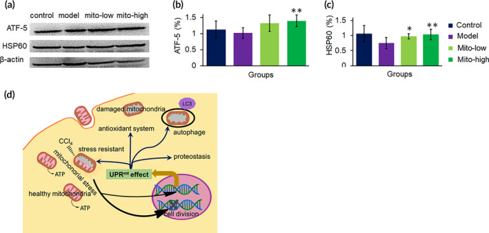
Carbon tetrachloride (CCl4)-induced liver injury is predominantly caused by free radicals, in which mitochondrial function of hepatocytes is impaired, accompanying with the production of ROS and decreased ATP energy supply in animals intoxicated with CCl4. Here we explored a novel therapeutic approach, mitochondrial transplantation therapy, for treating the liver injury. The results showed that mitochondria entered hepatocytes through macropinocytosis pathway, and thereby cell viability was recovered in a concentration-dependent manner. Mitochondrial therapy could increase ATP supply and reduce free radical damage. In liver injury model of mice, mitochondrial therapy significantly improved liver function and prevented tissue fibrogenesis. Transcriptomic data revealed that mitochondrial unfold protein response (UPRmt), a protective transcriptional response of mitochondria-to-nuclear retrograde signaling, would be triggered after mitochondrial administration. Then the anti-oxidant genes were up-regulated to scavenge free radicals. The mitochondrial function was rehabilitated through the transcriptional activation of respiratory chain enzyme and mitophage-associated genes. The protective response re-balanced the cellular homeostasis, and eventually enhanced stress resistance that is linked to cell survival. The efficacy of mitochondrial transplantation therapy in the animals would suggest a novel approach for treating liver injury caused by toxins.

Keywords: UPRmt; energy supply; free radical; mitochondrial therapy.


Conflict of interest statement

The authors declare that there is no conflict of interest that could be perceived as prejudicing the impartiality of the research reported.

Figures

FIGURE 1
FIGURE 1
Mitochondria arrived into hepatocyte through macropinocytosis pathway. (a) Isolated mitochondria. Mitochondria were stained by Mitotracker CMXRos, a mitochondrial specific dye. The dye conjugates thiols of mitochondrial proteins with covalent bonding. (b) Mitochondria morphology under TEM. (c) Cell internalization of mitochondria was prevented by macropinocytosis inhibitor. The cells were respectively added 8 × 104 mitochondria, and then were observed under TEM at 4 h after addition
FIGURE 2
FIGURE 2
The mitochondria rescued hepatocytes damaged by CCl4. (a) Mitochondria increased cell viability in concentration‐ and time‐dependent manners. (b) MDA content decreased, whereas (c) GSH, (d) SOD, (e) ATP, and (f) ND increased in cell homogenates after the mitochondria were introduced into the cell media for incubation 8 h. Mito‐low, low concentration mitochondria (20 μg/ml); Mito‐high, high concentration mitochondria (40 μg/ml). Data were expressed as mean ± SD. ## p < 0.01 comparison with the normal control; *p < 0.05, **p < 0.01 compared with the CCl4 group. The values were averaged for six independent experiments
FIGURE 3
FIGURE 3
Mitochondrial improved mouse liver function. (a) Tissue imaging with in vivo imaging after 4 h injection of mitochondria (0.4 mg/kg body weight). (b) Tissue sections under confocal microscopy after mitochondrial administration for 4 h. Mitochondria were labeled by Mitotracker CMXRos. (c) Liver sections stained with HE from mice treated with saline, CCl4, CCl4 + mito‐low (low dosage of mitochondria; 0.2 mg/kg body weight), and CCl4 + mito‐high (high dosage of mitochondria; 0.4 mg/kg body weight). (d) Mitochondrial morphology of livers under TEM. (e) Serum ALT activity. (f) Serum AST activity. Data were expressed as mean ± SD (n = 10 mice for each group). ## p < 0.01 compared with the normal control; **p < 0.01 compared with the CCl4 group
FIGURE 4
FIGURE 4
Biochemical measurement of mitochondrial activities in mouse livers after mitochondrial treatment. (a) JC‐1 assay. Also, activities of mitochondrial ND (b) PDH (c) and SDH (d) were respectively measured after mitochondria were administered into the mice. Data were expressed as mean ± SD of the mean (n = 10 mice for each group). ## p < 0.01 compared with the control; *p < 0.05, **p < 0.01 comparison with the CCl4 group
FIGURE 5
FIGURE 5
Mitochondria prevented liver fibrosis caused by CCl4. (a) Mouse liver surface of each group. (b) Liver sections stained by Sirius red. (c) Quantitative determination of fibrotic area. (d) Hydroxyproline level of liver tissue in each group. Data were expressed as mean ± SD (n = 10 mice for each group). ## p < 0.01 compared with the control; *p < 0.05, **p < 0.01 compared with the CCl4 group
FIGURE 6
FIGURE 6
Enriched KEGG pathways after mitochondrial therapy. (a) Heatmap for proteins and enzymes were showed in mouse liver tissues of CCl4‐induced injury and mitochondrial treatment group (mitochondrial dosage 0.4 mg/kg body weight), values were represented by log10 (fold changes). (b) A summary of the numbers of up‐ and down‐regulated DEGs for each GO subcategory. (c) The up‐regulated eight significantly enriched KEGG pathways and (d) the down‐regulated 10 KEGG pathways. The pathways were generalized according to p < 0.05
FIGURE 7
FIGURE 7
Mitochondria diminished ROS level and increased ATP supply in mouse liver tissues. The redox system were measured, including (a) ROS, (b) GSH, (c), SOD and (d) MDA. Moreover, the levels of (e) ATP content and (f) ND activity were determined, respectively. Each group contained eight mice (n = 8 for each group). ## p < 0.01 compared with the control; *p < 0.05, **p < 0.01 compared with the CCl4 group
FIGURE 8
FIGURE 8
Representative proteins in transcriptomic analysis were further examined by WB in mouse liver tissues. (a) WB bonds of parkin, LC3B, cyclin A and B. (b and c) Respective ratio of the gray value of autophagy proteins, parkin and LC3B, to β‐actin. (d) A representative picture showed numbers of autophagosome appeared after mitochondrial treatment. The sections were observed under TEM. (e and f) Ratios of the gray value of cyclin A or B, to β‐actin. (g) Ugt level. N = 6 for each group. ## p < 0.01 compared with the control; *p < 0.05, **p < 0.01 compared with the CCl4 group
FIGURE 9
FIGURE 9
Molecular signal pathways involved in mitochondrial therapy on CCl4 induced liver injury. (a) WB bonds of ATF5 and HSP60. (b and c) Respective ratio of the gray value of ATF‐5 and HSP60 to β‐actin. (d) The mechanism of mitochondrial therapy. Mitochondria stress was induced once entering cells, and then activated the UPRmt pathway, a mitochondria‐nuclear retrograde signal, to promote gene transcription and expression of a series of protective proteins and enzymes, including anti‐oxidant enzyme, OXPHOS‐related proteins, biotransformation enzymes, proteostasis, and autophagy‐related proteins

Similar articles

Cited by

References

    1. Teschke R. Aliphatic halogenated hydrocarbons: report and analysis of liver injury in 60 patients. J Clin Transl Hepatol. 2018;6:350‐361. - PMC - PubMed
    1. Lin SY, Xu D, Du XX, et al. Protective effects of salidroside against carbon tetrachloride (CCl4)‐induced liver injury by initiating mitochondria to resist oxidative stress in mice. Int J Mol Sci. 2019;20:3187. - PMC - PubMed
    1. MR MG, Jaeschke H. Animal models of drug‐induced liver injury. BBA‐Mol Basis Dis. 2020;1865:1031‐1039. - PMC - PubMed
    1. Kim YO, Popov Y, Schuppan D. Optimized mouse models for liver fibrosis. Method Mol Biol. 2017;1559:279‐296. - PubMed
    1. Moon SJ, Dong W, Stephanopoulos GN, Sikes HD. Oxidative pentose phosphate pathway and glucose anaplerosis support maintenance of mitochondrial NADPH pool under mitochondrial oxidative stress. Bioeng Transl Med. 2020;5:e10184. - PMC - PubMed

LinkOut - more resources