

Injection of hybrid 3D spheroids composed of podocytes, mesenchymal stem cells, and vascular endothelial cells into the renal cortex improves kidney function and replenishes glomerular podocytes
- PMID: 34027096
- PMCID: PMC8126810
- DOI: 10.1002/btm2.10212
Injection of hybrid 3D spheroids composed of podocytes, mesenchymal stem cells, and vascular endothelial cells into the renal cortex improves kidney function and replenishes glomerular podocytes
Abstract
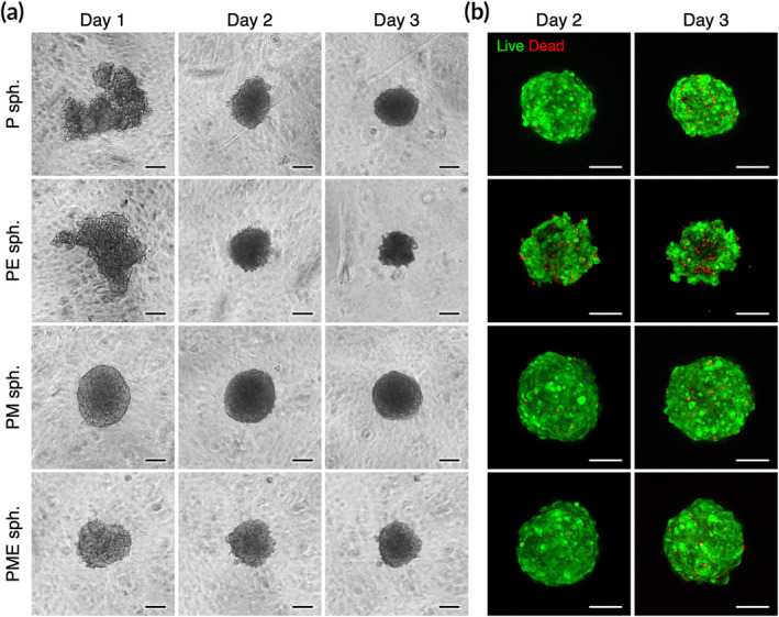
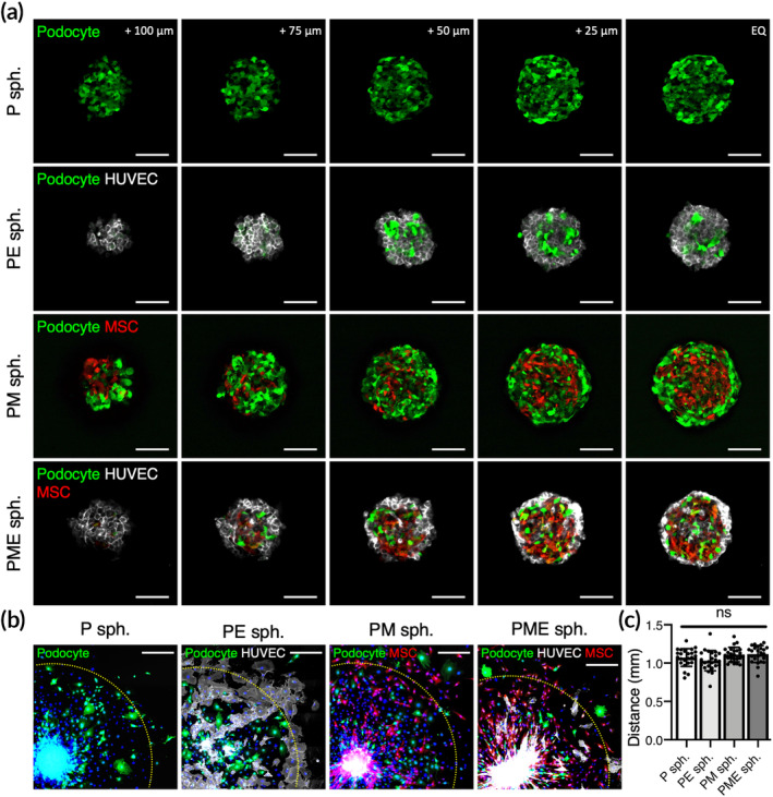
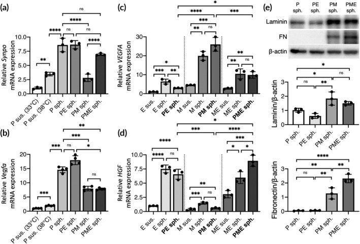
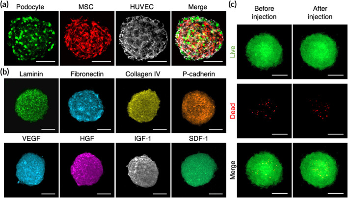
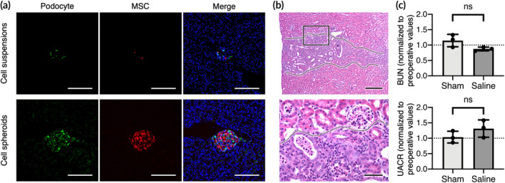
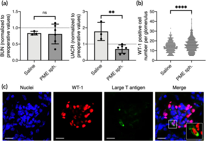
Podocytes are highly differentiated epithelial cells that are crucial for maintaining the glomerular filtration barrier in the kidney. Podocyte injury followed by depletion is the major cause of pathological progression of kidney diseases. Although cell therapy has been considered a promising alternative approach to kidney transplantation for the treatment of kidney injury, the resultant therapeutic efficacy in terms of improved renal function is limited, possibly owing to significant loss of engrafted cells. Herein, hybrid three-dimensional (3D) cell spheroids composed of podocytes, mesenchymal stem cells, and vascular endothelial cells were designed to mimic the glomerular microenvironment and as a cell delivery vehicle to replenish the podocyte population by cell transplantation. After creating a native glomerulus-like condition, the expression of multiple genes encoding growth factors and basement membrane factors that are strongly associated with podocyte maturation and functionality was significantly enhanced. Our in vivo results demonstrated that intrarenal transplantation of podocytes in the form of hybrid 3D cell spheroids improved engraftment efficiency and replenished glomerular podocytes. Moreover, the proteinuria of the experimental mice with hypertensive nephropathy was effectively reduced. These data clearly demonstrated the potential of hybrid 3D cell spheroids for repairing injured kidneys.
Keywords: cell spheroids; cell therapy; glomerulus; kidney injury; podocyte.
© 2021 The Authors. Bioengineering & Translational Medicine published by Wiley Periodicals LLC on behalf of The American Institute of Chemical Engineers.
Conflict of interest statement
The authors have declared no competing interest.
Figures
Similar articles
-
Mesenchymal stem cells ameliorate podocyte injury and proteinuria in a type 1 diabetic nephropathy rat model.Biol Blood Marrow Transplant. 2013 Apr;19(4):538-46. doi: 10.1016/j.bbmt.2013.01.001. Epub 2013 Jan 4. Biol Blood Marrow Transplant. 2013. PMID: 23295166
-
Mesenchymal stem cell therapy promotes renal repair by limiting glomerular podocyte and progenitor cell dysfunction in adriamycin-induced nephropathy.Am J Physiol Renal Physiol. 2012 Nov 1;303(9):F1370-81. doi: 10.1152/ajprenal.00057.2012. Epub 2012 Sep 5. Am J Physiol Renal Physiol. 2012. PMID: 22952284
-
Molecular and cellular events mediating glomerular podocyte dysfunction and depletion in diabetes mellitus.Front Endocrinol (Lausanne). 2014 Sep 25;5:151. doi: 10.3389/fendo.2014.00151. eCollection 2014. Front Endocrinol (Lausanne). 2014. PMID: 25309512 Free PMC article. Review.
-
Three-dimensional podocyte-endothelial cell co-cultures: Assembly, validation, and application to drug testing and intercellular signaling studies.Eur J Pharm Sci. 2016 Apr 30;86:1-12. doi: 10.1016/j.ejps.2016.02.013. Epub 2016 Feb 23. Eur J Pharm Sci. 2016. PMID: 26924225
-
Podocyte injury of diabetic nephropathy: Novel mechanism discovery and therapeutic prospects.Biomed Pharmacother. 2023 Dec;168:115670. doi: 10.1016/j.biopha.2023.115670. Epub 2023 Oct 13. Biomed Pharmacother. 2023. PMID: 37837883 Review.
Cited by
-
Light-controlled scaffold- and serum-free hard palatal-derived mesenchymal stem cell aggregates for bone regeneration.Bioeng Transl Med. 2022 May 13;8(1):e10334. doi: 10.1002/btm2.10334. eCollection 2023 Jan. Bioeng Transl Med. 2022. PMID: 36684075 Free PMC article.
-
Optimization strategies of mesenchymal stem cell-based therapy for acute kidney injury.Stem Cell Res Ther. 2023 Apr 30;14(1):116. doi: 10.1186/s13287-023-03351-2. Stem Cell Res Ther. 2023. PMID: 37122024 Free PMC article. Review.
-
A platform for automated and label-free monitoring of morphological features and kinetics of spheroid fusion.Front Bioeng Biotechnol. 2022 Aug 26;10:946992. doi: 10.3389/fbioe.2022.946992. eCollection 2022. Front Bioeng Biotechnol. 2022. PMID: 36091464 Free PMC article.
-
Efficacy of stem cells versus microvesicles in ameliorating chronic renal injury in rats (histological and biochemical study).Sci Rep. 2024 Jul 18;14(1):16589. doi: 10.1038/s41598-024-66299-0. Sci Rep. 2024. PMID: 39025899 Free PMC article.
-
3D Culture of MSCs for Clinical Application.Bioengineering (Basel). 2024 Nov 27;11(12):1199. doi: 10.3390/bioengineering11121199. Bioengineering (Basel). 2024. PMID: 39768017 Free PMC article. Review.
References
-
- Rinschen MM, Schroeter CB, Koehler S, et al. Quantitative deep mapping of the cultured podocyte proteome uncovers shifts in proteostatic mechanisms during differentiation. Am J Physiol Cell Physiol. 2016;311(3):C404‐C417. - PubMed
LinkOut - more resources
Full Text Sources
Other Literature Sources
