Non-invasive in vivo monitoring of transplanted stem cells in 3D-bioprinted constructs using near-infrared fluorescent imaging
- PMID: 34027098
- PMCID: PMC8126817
- DOI: 10.1002/btm2.10216
Non-invasive in vivo monitoring of transplanted stem cells in 3D-bioprinted constructs using near-infrared fluorescent imaging
Abstract
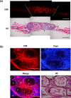
Cell-based tissue engineering strategies have been widely established. However, the contributions of the transplanted cells within the tissue-engineered scaffolds to the process of tissue regeneration remain poorly understood. Near-infrared (NIR) fluorescence imaging systems have great potential to non-invasively monitor the transplanted cell-based tissue constructs. In this study, labeling mesenchymal stem cells (MSCs) using a lipophilic pentamethine indocyanine (CTNF127, emission at 700 nm) as a NIR fluorophore was optimized, and the CTNF127-labeled MSCs (NIR-MSCs) were printed embedding in gelatin methacryloyl bioink. The NIR-MSCs-loaded bioink showed excellent printability. In addition, NIR-MSCs in the 3D constructs showed high cell viability and signal stability for an extended period in vitro. Finally, we were able to non-invasively monitor the NIR-MSCs in constructs after implantation in a rat calvarial bone defect model, and the transplanted cells contributed to tissue formation without specific staining. This NIR-based imaging system for non-invasive cell monitoring in vivo could play an active role in validating the cell fate in cell-based tissue engineering applications.
Keywords: near‐infrared fluorescence; non‐invasive monitoring; scaffold monitoring; stem cell tracking.
© 2021 The Authors. Bioengineering & Translational Medicine published by Wiley Periodicals LLC on behalf of American Institute of Chemical Engineers.
Conflict of interest statement
Authors declare no conflicts of interest.
Figures


Similar articles
-
NIR fluorescence for monitoring in vivo scaffold degradation along with stem cell tracking in bone tissue engineering.Biomaterials. 2020 Nov;258:120267. doi: 10.1016/j.biomaterials.2020.120267. Epub 2020 Aug 6. Biomaterials. 2020. PMID: 32781325 Free PMC article.
-
Near-infrared optical imaging for monitoring the regeneration of osteogenic tissue-engineered constructs.Biores Open Access. 2013 Jun;2(3):186-91. doi: 10.1089/biores.2013.0005. Biores Open Access. 2013. PMID: 23741629 Free PMC article.
-
Hybrid biofabrication of 3D osteoconductive constructs comprising Mg-based nanocomposites and cell-laden bioinks for bone repair.Bone. 2022 Jan;154:116198. doi: 10.1016/j.bone.2021.116198. Epub 2021 Sep 15. Bone. 2022. PMID: 34534709
-
Recent Advances in Tracking the Transplanted Stem Cells Using Near-Infrared Fluorescent Nanoprobes: Turning from the First to the Second Near-Infrared Window.Adv Healthc Mater. 2018 Oct;7(20):e1800497. doi: 10.1002/adhm.201800497. Epub 2018 Jul 17. Adv Healthc Mater. 2018. PMID: 30019509 Review.
-
Recent Advances in Endocrine, Metabolic and Immune Disorders: Mesenchymal Stem Cells (MSCs) and Engineered Scaffolds.Endocr Metab Immune Disord Drug Targets. 2018;18(5):466-469. doi: 10.2174/1871530318666180423102905. Endocr Metab Immune Disord Drug Targets. 2018. PMID: 29692270 Review.
Cited by
-
De novo design of a nanoregulator for the dynamic restoration of ovarian tissue in cryopreservation and transplantation.J Nanobiotechnology. 2024 Jun 11;22(1):330. doi: 10.1186/s12951-024-02602-5. J Nanobiotechnology. 2024. PMID: 38862987 Free PMC article. Review.
-
Bioprinting of gelatin-based materials for orthopedic application.Front Bioeng Biotechnol. 2024 Mar 13;12:1357460. doi: 10.3389/fbioe.2024.1357460. eCollection 2024. Front Bioeng Biotechnol. 2024. PMID: 38544981 Free PMC article. Review.
-
Doubly Strapped Zwitterionic NIR-I and NIR-II Heptamethine Cyanine Dyes for Bioconjugation and Fluorescence Imaging.Angew Chem Int Ed Engl. 2023 Jul 10;62(28):e202305062. doi: 10.1002/anie.202305062. Epub 2023 May 31. Angew Chem Int Ed Engl. 2023. PMID: 37163228 Free PMC article.
-
3D digital light process bioprinting: Cutting-edge platforms for resolution of organ fabrication.Mater Today Bio. 2024 Oct 2;29:101284. doi: 10.1016/j.mtbio.2024.101284. eCollection 2024 Dec. Mater Today Bio. 2024. PMID: 39430572 Free PMC article. Review.
-
Biohybrid neural interfaces: improving the biological integration of neural implants.Chem Commun (Camb). 2023 Dec 14;59(100):14745-14758. doi: 10.1039/d3cc05006h. Chem Commun (Camb). 2023. PMID: 37991846 Free PMC article. Review.
References
-
- da Rocha DN, Marçal RLSB, Barbosa RM, Ferreira JRM, da Silva MHP. Mesenchymal stem cells associated with bioceramics for bone tissue regeneration. Biomater Med Appl. 2017;1:2.
-
- Arbab AS, Yocum GT, Kalish H, et al. Efficient magnetic cell labeling with protamine sulfate complexed to ferumoxides for cellular MRI. Blood. 2004;104(4):1217‐1223. - PubMed
-
- Lalande C, Miraux S, Derkaoui S, et al. Magnetic resonance imaging tracking of human adipose derived stromal cells within three‐dimensional scaffolds for bone tissue engineering. Eur Cell Mater. 2011;21(1):341‐354. - PubMed
Grants and funding
LinkOut - more resources
Full Text Sources
Other Literature Sources
Miscellaneous

