Review
. 2021 May 8;6(12):4470-4490.
doi: 10.1016/j.bioactmat.2021.04.033. eCollection 2021 Dec.

Antibacterial approaches in tissue engineering using metal ions and nanoparticles: From mechanisms to applications

Affiliations
Review

Antibacterial approaches in tissue engineering using metal ions and nanoparticles: From mechanisms to applications

Maria Godoy-Gallardo et al. Bioact Mater. .

Abstract

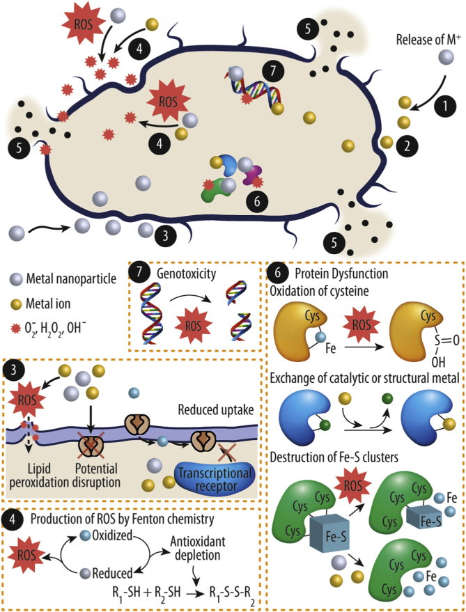
Bacterial infection of implanted scaffolds may have fatal consequences and, in combination with the emergence of multidrug bacterial resistance, the development of advanced antibacterial biomaterials and constructs is of great interest. Since decades ago, metals and their ions had been used to minimize bacterial infection risk and, more recently, metal-based nanomaterials, with improved antimicrobial properties, have been advocated as a novel and tunable alternative. A comprehensive review is provided on how metal ions and ion nanoparticles have the potential to decrease or eliminate unwanted bacteria. Antibacterial mechanisms such as oxidative stress induction, ion release and disruption of biomolecules are currently well accepted. However, the exact antimicrobial mechanisms of the discussed metal compounds remain poorly understood. The combination of different metal ions and surface decorations of nanoparticles will lead to synergistic effects and improved microbial killing, and allow to mitigate potential side effects to the host. Starting with a general overview of antibacterial mechanisms, we subsequently focus on specific metal ions such as silver, zinc, copper, iron and gold, and outline their distinct modes of action. Finally, we discuss the use of these metal ions and nanoparticles in tissue engineering to prevent implant failure.

Keywords: Antibacterial activity; Biomaterials applications; Mechanism of action; Metal ions; Metal nanoparticles; Tissue engineering.


Conflict of interest statement

All authors declare no competing financial interests.

Figures

Image 1
Graphical abstract
Fig. 1
Fig. 1
Cell wall schematic of (A) Gram-positive and (B) Gram-negative bacteria. IMP: Integral membrane protein; LP: lipoprotein; LPS: lipopolysaccharide; LTA: lipoteichoic acid; OMP: Outer membrane protein; PGN: Peptidoglycan; TA: wall teichoic acid. Note: the schematic is not to scale.
Fig. 2
Fig. 2
Antibacterial mechanisms of metal ions and nanoparticles. The central modes of action are: (1) release of metal ions from the metal nanoparticles and (2) direct interaction of the metal ions and/or (3) metal nanoparticles with the cell wall through electrostatic interactions, leading to impaired membrane function and impaired nutrient assimilation; (4) formation of extracellular and intracellular reactive oxygen species (ROS), and damage of lipids, proteins and DNA by oxidative stress; (5) high-levels of metal-binding to the cell envelope and high ROS levels can cause damage to the plasma membrane and thus lead to the leakage of the cell content; (6, 7) upon metal uptake, metal nanoparticles and metal ions can directly interfere with both proteins and DNA, impairing their function and disturbing the cellular metabolism in addition to metal-mediated ROS production.
Fig. 3
Fig. 3
(A) Transmission electron microscopy (TEM) (i-ii) and scanning electron microscopy (SEM) (iii-iv) analysis of Escherichia coli (E. coli) upon treatment with silver nanoparticles (Ag-NPs) (i and iii: controls; ii and iv: treated samples). (B) Dual immunofluorescence and reactive oxygen species (ROS) staining images of Staphylococcus aureus (S. aureus) (i-vi) and Klebsiella pneumoniae (K. pneumoniae) (vii-xii) treated with zinc nanoparticles and zinc chloride (0.35 mM) under dark conditions. Reprinted with the permission from Elseiver [78,81].
Fig. 4
Fig. 4
(A) Field emission scanning electron microscope (FESEM) images of Escherichia coli (E. coli) (i-iii) and Staphylococcus aureus (S. aureus) (iv-vi) treated with copper at 0h (i and iv), 2h (ii and v) and 24h (iii and vi). (B) Fenton and Haber-Weiss reaction for generation of hydroxyl radical. Intracellularly, hydroxyl radicals are primarily produced by iron-catalyzed Haber-Weiss/Fenton reaction. (C) (i) Cell viability of Bacillus subtilis (B. subtilis) and E. coli after treatment with negative iron oxide nanoparticles (nFeO-NPs) (left) and positive iron oxide nanoparticles (pFeO-NPs) (right) at different concentrations; (ii) Fluorescence microscopy images of B. subtilis and E. coli in absence and presence of nFeO-NPs and pFeO-NPs using the LIVE/DEAD BacLight fluorescence kit (green fluorescence: viable cells; red fluorescence: dead cells). Reprinted with the permission from Royal Society of Chemistry [8], CellPress [134], and Nature [135].
Fig. 5
Fig. 5
Schematic overview of different biomaterials modified with metal ions and/or metallic nanoparticles (red dots represent metal ions or metallic nanoparticles). Metal ions and nanoparticles can be entrapped in or coated on materials such as polymers, ceramics, metals, and composites, where they are hold in place e.g. by electrostatic interactions or covalent bonding, and thereby augment the material with antibacterial activity.
Fig. 6
Fig. 6
(A) Confocal laser scanning microcopy (CLSM) of bacterial biofilms treated with manufactured titanium scaffolds (TS) and after functionalization with nanosilver encapsulated silk fibrin (m-SFAg). The overlap of green and red signal yields green yellow. On the right, a schematic of the experimental design and results is depicted. Topical reactive oxygen species (ROS) and silver ions (Ag+) species are released from m-SFAg scaffolds and diffuse into vicinities of the biofilms and further degrade the biofilm EPS, thus exposing the embedded biofilm bacteria and inactivating them. (B) Schematic illustration of the antibacterial and osteogenic processes of Copper (Cu)-modified carboxymethyl chitosan (CMC) and alginate (Alg) (Cu-CMC/Alg) and (CMC/Alg) scaffolds in vivo. When the Cu2+ ions released from the Cu nanoparticles gradually cross-linked the polymer mixtures, which was further turned into a Cu-CMC/Alg scaffold with an interconnected porous structure by freeze-drying. The in vivo study demonstrated that the Cu-CMC/Alg scaffolds induced the formation of vascularized new bone tissue and avoided the clinical bacterial infection. (C) Schematic of the role of zinc (Zn) and calcium (Ca) in 80S (80SiO2–15CaO–5P2O5 in mol%) glass. Because of the Zn2+ substituted in the glass, it release was limited and no anti-methicillin-resistant staphylococcus aureus was detected. (D) Schematic illustrator of synergistic antibacterial mechanism of Lysozyme/Chitosan/Silver/Hydroxyapatite hybrid coating on Ti. In the early stage, lysosome hydrolyze the β-1,4-glycosidic bond of peptidoglycan on the cell wall of bacteria, resulting in the rupture of cell wall and the cytoplasm as well as the spilling of other intracellular substances. At the same time, chitosan (CS) can adsorb the negatively charged protein of cell wall, blocking the cell wall pore channels and leading to bacteria apoptosis with no nutrition exchange. The Ag+ released from Ag-NPs can penetrate into the bacterial cell and destroy or damage DNA by generation of intracellular ROS or direct contact. Reprinted with the permission from ACS Publications [173,174] and Elseiver [180,181].

References

    1. Costa F., Carvalho I.F., Montelaro R.C., Gomes P., Cristina M., Martins L. Covalent immobilization of antimicrobial peptides (AMPs) onto biomaterial surfaces. Acta Biomater. 2011;7:1431–1440. doi: 10.1016/j.actbio.2010.11.005. - DOI - PubMed
    1. Masters E.A., Trombetta R.P., de Mesy Bentley K.L., Boyce B.F., Gill A.L., Gill S.R., Nishitani K., Ishikawa M., Morita Y., Ito H., Bello-Irizarry S.N., Ninomiya M., Brodell J.D., Lee C.C., Hao S.P., Oh I., Xie C., Awad H.A., Daiss J.L., Owen J.R., Kates S.L., Schwarz E.M., Muthukrishnan G. Evolving concepts in bone infection: redefining “biofilm”, “acute vs. chronic osteomyelitis”, “the immune proteome” and “local antibiotic therapy. Bone Res. 2019;7:1–18. doi: 10.1038/s41413-019-0061-z. - DOI - PMC - PubMed
    1. Wang L., Hu C., Shao L. The antimicrobial activity of nanoparticles: present situation and prospects for the future. Int. J. Nanomed. 2017;12:1227–1249. doi: 10.2147/IJN.S121956. - DOI - PMC - PubMed
    1. Salas Orozco M.F., Niño-Martínez N., Martínez-Castañón G.A., Méndez F.T., Ruiz F. Molecular mechanisms of bacterial resistance to metal and metal oxide nanoparticles. Int. J. Mol. Sci. 2019;20:2808. doi: 10.3390/ijms20112808. - DOI - PMC - PubMed
    1. Lemire J.A., Harrison J.J., Turner R.J. Antimicrobial activity of metals: mechanisms, molecular targets and applications. Nat. Rev. Microbiol. 2013;11:371–384. doi: 10.1038/nrmicro3028. - DOI - PubMed