. 2021 May 11;6(12):4506-4516.
doi: 10.1016/j.bioactmat.2021.04.039. eCollection 2021 Dec.

Capturing dynamic biological signals via bio-mimicking hydrogel for precise remodeling of soft tissue

Affiliations

Capturing dynamic biological signals via bio-mimicking hydrogel for precise remodeling of soft tissue

Zhengwei Cai et al. Bioact Mater. .

Abstract

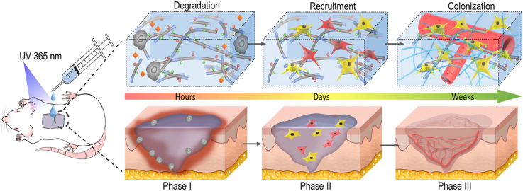
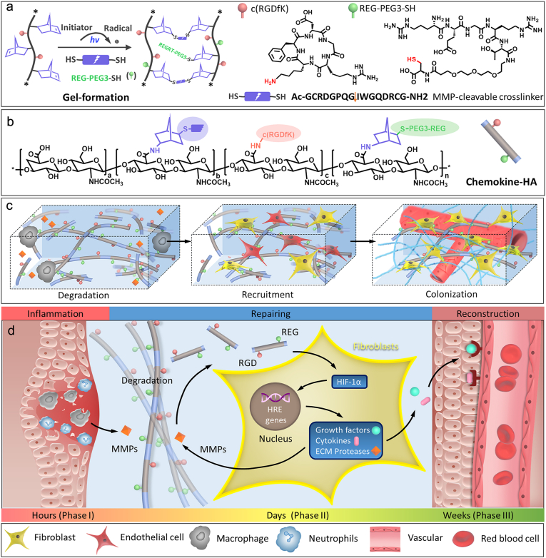
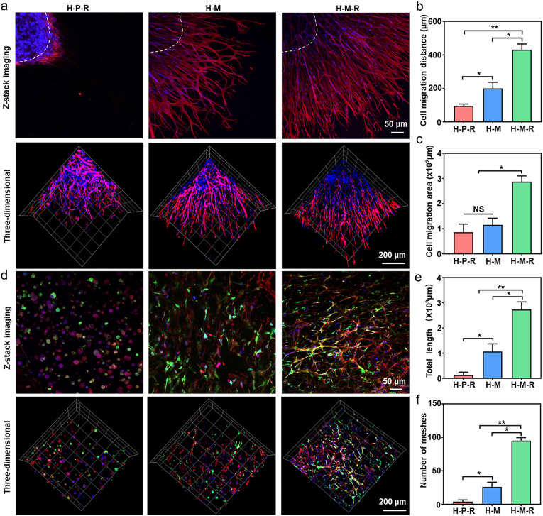
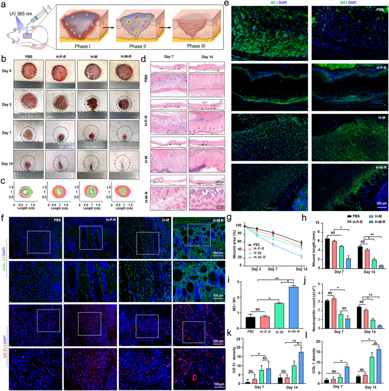
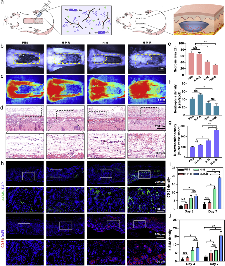
Soft tissue remodeling is a sophisticated process that sequentially provides dynamic biological signals to guide cell behavior. However, capturing these signals within hydrogel and directing over time has still been unrealized owing to the poor comprehension of physiological processes. Here, a bio-mimicking hydrogel is designed via thiol-ene click reaction to capture the early physical signal triggered by inflammation, and the chemical signals provided with chemokine and natural adhesion sites, which guaranteed the precise soft tissue remodeling. This bio-mimicking hydrogel efficiently facilitated cell anchoring, migration, and invasion in the 3D matrix due to the permissive space and the interaction with integrin receptors. Besides, the covalently grafted chemokine-like peptide is optimal for colonization and functional differentiation of endothelial cells through a HIF-1α dependent signal pathway. Furthermore, the early polarization of macrophages, collagen deposition and angiogenesis in rat acute wound model, and the increased blood perfusion in mouse skin flap model have confirmed that the bio-mimicking hydrogel realized precise soft tissue remodeling and opens new avenues for the phased repair of different tissues such as nerve, myocardium, and even bone.

Keywords: Biological signals; Chemotactic; Hydrogel; Physiological phase; Soft tissue remodeling.


Conflict of interest statement

The authors declare no conflict of interest.

Figures

Image 1
Graphical abstract
Fig. 1
Fig. 1
Schematic illustrations of the hydrogel formation and matching the phases (I-III) of soft tissue regeneration. a) The degradation-recruitment-colonization integrated hydrogel were constructed by HA-NB-RGD, MMP-cleavable crosslinker, and chemokine-like peptide REG-PEG3-SH with thiol-ene click reaction. b) Enzymatic degradation and release of chemotactic hyaluronic acid fragments (chemokine-HA). c) Early inflammatory cells infiltrated hydrogel and released inflammatory mediators MMPs and the degraded hydrogel provided space for the recruitment, colonization, and functional differentiation of regenerative cells. d) Degradation, recruitment, and colonization of hydrogel were precisely matched with the inflammation of phase I, repairing of phase II, and reconstruction of phase III in soft tissue injury.
Fig. 2
Fig. 2
Synthesis, characterization, and biocompatibility of MMPs sensitive hydrogels for Phase I. a) 1H NMR spectrum analysis (600 MHz, D2O) of HA, HA-NB, and HA-NB-RGD. b) Rheology analysis and a photograph of hydrogel formation for HA-NB-RGD with MMP-cleavable crosslinker SH-MMP-SH (H–M) or non-degradable crosslinker SH-PEG-SH (H–P) (SH: NB = 2:3). c) Swelling weight ratio. SEM analysis d), porosity analysis e), photograph visualization f) and rheological analysis of H-M g) and H–P h) hydrogel degradation. Cytoskeleton staining i), cell aspect ratio j) and cell count k) of the HFVs on the H–P and H-M hydrogel.
Fig. 3
Fig. 3
In vitro cell angiogenesis and cell migration activity analysis of chemokine-like hydrogels for Phase II. a) The confocal images of vascular tube-formation with HUVEC at 24 h and 96 h after culturing on H-M hydrogel grafted with different concentrations of chemokine-like peptide. Quantification of total length (b) and the number of junctions (c). d) Schematic diagram showing the co-culture model of macrophages with H-M/H–P hydrogel grafted with 0.1 mM REG-PEG3-SH (H-M-R or H–P-R hydrogel) to form a conditioned medium (CM) for cell migration and the mechanism of the hydrogel degradation with macrophages. e) HVF (red) and HUVEC (green) wound scratch assay with H-M, H–P-R, and H-M-R hydrogel conditioned medium. Quantification of the wound healing rate f) and cell number g) of migrated cells. *p < 0.05, **p < 0.01.
Fig. 4
Fig. 4
Cell invasion and tubule formation in 3D hydrogels for Phase III. a) Z-stack and 3D confocal images of actin/nuclei (red/blue) stained HVF clusters encapsulated in H–P-R hydrogel (non-degradable, adhesive, and chemotactic), H-M hydrogel (degradable and adhesive), and H-M-R hydrogel (degradable, adhesive, and chemotactic) cultured for 14 d. And the quantification of the cell migration distance b) and cell migration area c). d) Z-stack and 3D confocal images of HVFs (red) and HUVECs (green) co-cultured in H–P-R, H-M, and H-M-R hydrogels for 14 days. e, f) The quantification of total length and number of meshes. *p < 0.05, **p < 0.01.
Fig. 5
Fig. 5
Bio-mimicking hydrogel promoted wound healing in vivo by matching the different phases of soft tissue regeneration. a) The schematic diagram illustrating the mechanism of accelerating wound closure based on H-M-R hydrogel (n = 6). b) The representative photographs of wound healing of rats treated with PBS, H–P-R, H-M, and H-M-R hydrogels treatments on days 0, 3, 7, 14. c) Wound closure boundaries during 14 days in vivo for each treatment. d) HE stained sections of skin tissue on days 7 and 14 (above: cross-section; below: longitudinal section), the black arrow means the length of the wound in the HE staining. e) Immunofluorescence images of explanted samples stained for, iNOS (M1), CD206 (M2). f) The immunofluorescence staining for Col I and CD31(CD31-positive endothelial cells) at 14 days. g, h) Wound area and length of each group at different time points. i) The ratio of M2/M1 macrophages in all groups on day 3. j) The quantification of neutrophilic cells of different groups at 7 and 14 days. k, l) The quantification of immunofluorescence intensity of Col I and α-SMA at 7 and 14 days. *p < 0.05, **p < 0.01.
Fig. 6
Fig. 6
Bio-mimicking hydrogel promoted skin flap angiogenesis and regeneration. a) Schematic diagram of hydrogel injection below skin flap (n = 6). b) Photographic images of a necrotic area in the flaps. c) The real-time blood flow images of each group at days 7 were detected with laser speckle contrast. The Red signal represents the perfusion of blood perfusion. d) H&E staining sections of the junction of skin flap necrosis and survival in each group at days 7. e) The necrosis area proportion of flaps in different groups. f, g) The quantification of the neutrophilic density and microvascular density in different groups. h) Images of immunofluorescence staining for α-SMA and CD31 at 7 days. And the quantification of immunofluorescence intensity of α-SMA i) and CD31 j) at 3 and 7 days. *p < 0.05, **p < 0.01.

Similar articles

Cited by

References

    1. Li X., Cho B., Martin R., Seu M., Zhang C., Zhou Z., Choi J.S., Jiang X., Chen L., Walia G., Yan J., Callanan M., Liu H., Colbert K., Morrissette-McAlmon J., Grayson W., Reddy S., Sacks J.M., Mao H.Q. Nanofiber-hydrogel composite–mediated angiogenesis for soft tissue reconstruction. Sci. Transl. Med. 2019;11(490) - PubMed
    1. Raghow R. The role of extracellular matrix in postinflammatory wound healing and fibrosis. Faseb. J. 1994;8(11):823–831. - PubMed
    1. Olczyk P., Mencner K., Komosinska-Vassev K. The role of the extracellular matrix components in cutaneous wound healing. BioMed Res. Int. 2014;2014 - PMC - PubMed
    1. Slaughter B.V., Khurshid S.S., Fisher O.Z., Khademhosseini A., Peppas N.A. Hydrogels in regenerative medicine. Adv. Mater. 2009;21:3307–3329. - PMC - PubMed
    1. Zhang Y.S., Khademhosseini A. Advances in engineering hydrogels. Science. 2017;356 - PMC - PubMed

LinkOut - more resources