. 2021 Feb 5;5(5):846-864.
doi: 10.1002/hep4.1679. eCollection 2021 May.

Exosome-Like Nanoparticles From Lactobacillus rhamnosus GG Protect Against Alcohol-Associated Liver Disease Through Intestinal Aryl Hydrocarbon Receptor in Mice

Affiliations

Exosome-Like Nanoparticles From Lactobacillus rhamnosus GG Protect Against Alcohol-Associated Liver Disease Through Intestinal Aryl Hydrocarbon Receptor in Mice

Zelin Gu et al. Hepatol Commun. .

Abstract

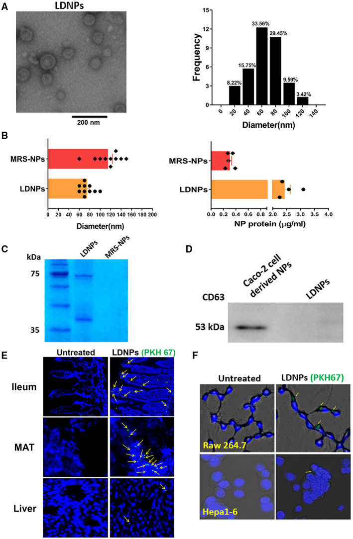
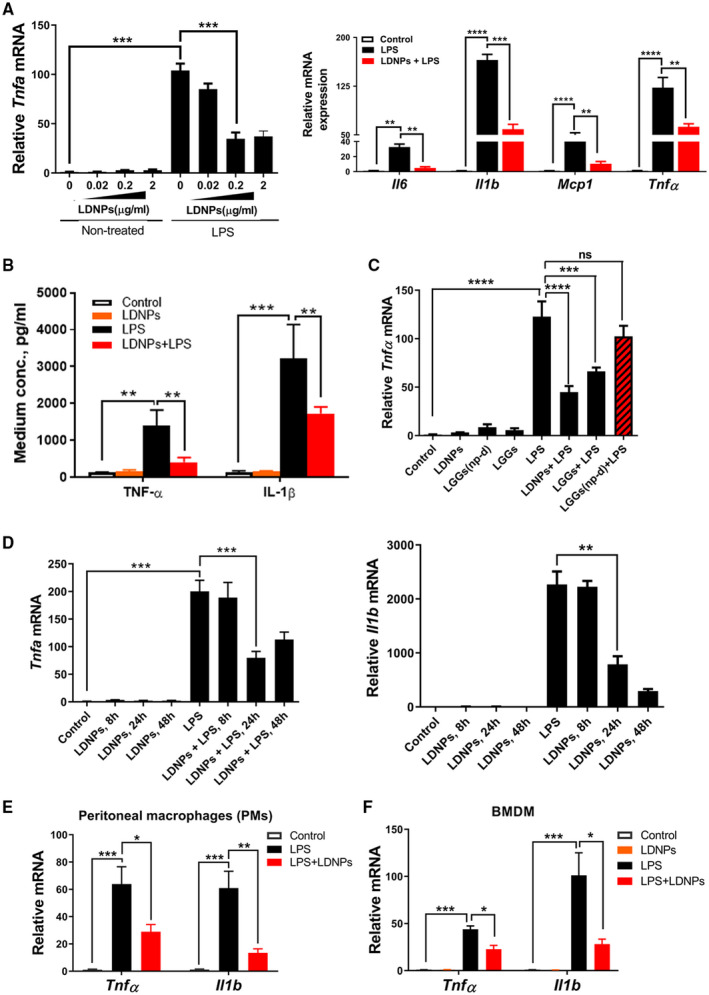
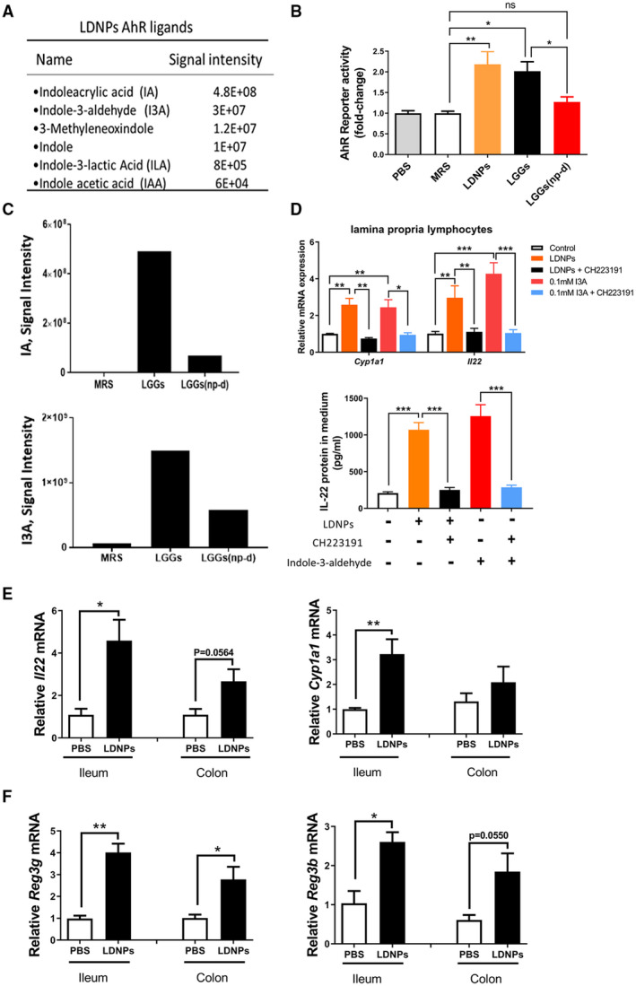
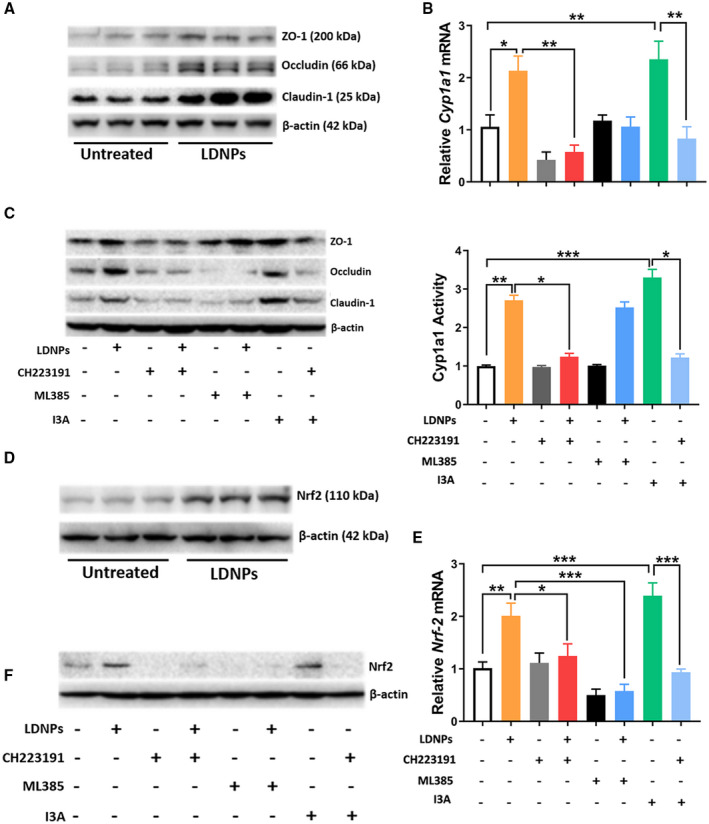
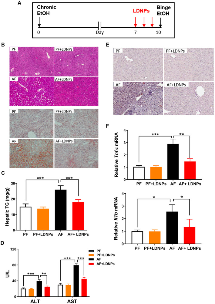
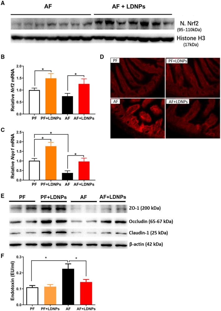
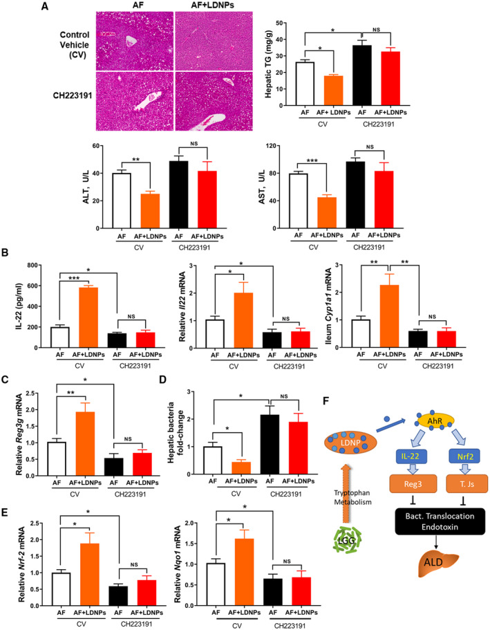
Alcohol-associated liver disease (ALD) is a major cause of mortality. Gut barrier dysfunction-induced bacterial translocation and endotoxin release contribute to the pathogenesis of ALD. Probiotic Lactobacillus rhamnosus GG (LGG) is known to be beneficial on experimental ALD by reinforcing the intestinal barrier function. In this study, we aim to investigate whether the protective effects of LGG on intestinal barrier function is mediated by exosome-like nanoparticles (ELNPs) released by LGG. Intestinal epithelial cells and macrophages were treated with LGG-derived ELNPs (LDNPs) isolated from LGG culture. LDNPs increased tight junction protein expression in epithelial cells and protected from the lipopolysaccharide-induced inflammatory response in macrophages. Three-day oral application of LDNPs protected the intestine from alcohol-induced barrier dysfunction and the liver from steatosis and injury in an animal model of ALD. Co-administration of an aryl hydrocarbon receptor (AhR) inhibitor abolished the protective effects of LDNPs, indicating that the effects are mediated, at least in part, by intestinal AhR signaling. We further demonstrated that LDNP administration increased intestinal interleukin-22-Reg3 and nuclear factor erythroid 2-related factor 2 (Nrf2)-tight junction signaling pathways, leading to the inhibition of bacterial translocation and endotoxin release in ALD mice. This protective effect was associated with LDNP enrichment of bacterial tryptophan metabolites that are AhR agonists. Conclusions: Our results suggest that the beneficial effects of LGG and their supernatant in ALD are likely mediated by bacterial AhR ligand-enriched LDNPs that increase Reg3 and Nrf2 expression, leading to the improved barrier function. These findings provide a strategy for the treatment of ALD and other gut barrier dysfunction-associated diseases.


Figures

FIG. 1
FIG. 1
Characterization of LDNPs. (A) Representative transmission electron microscopy image of LDNPs and the frequency of observed NPs by diameter. (B) Size (left panel) and protein concentration (right panel) comparison between LDNPs and MRS‐derived NPs. (C) Coomassie blue staining of protein bands on sodium dodecyl sulfate–polyacrylamide gel electrophoresis. (D) Western blot for CD63 protein in LDNPs and Caco‐2‐derived NPs. (E) Uptake of (PKH67‐labeled) LDNPs in the ileum, mesenteric adipose tissue, and liver tissue. (F) Uptake of LDNPs in macrophages RAW264.7 and hepatocytes Hepa1‐6. Yellow arrows indicate PKH67‐positive staining of LDNPs. 4´,6‐diamidino‐2‐phenylindole (blue) was used for nucleic counter staining. Abbreviations: MAT, mesenteric adipose tissue; and MRS‐NP, MRS‐derived NP.
FIG. 2
FIG. 2
LDNPs inhibited LPS‐induced inflammation in macrophages. (A‐D) RAW264.7 cells. Dose‐dependent effects of LDNPs on relative Tnfα mRNA expression with or without LPS stimulation (A, left panel); relative mRNA expression of inflammatory mediators (A, right panel) and protein levels of TNF‐α and IL‐1β (B) after LDNPs and LPS treatment. (C) Effects of LDNPs depletion in LGGs on LPS‐induced Tnf α mRNA expression. (D) Effects of LDNPs on Tnf α and Il‐1 β mRNA expression are time‐dependent. LDNPs inhibited LPS‐induced Tnf α and Il‐1 β mRNA expression in PMs (E) and BMDMs (F). Data shown represent the mean ± SEM of at least three independent experiments performed in triplicate. *P < 0.05, **P < 0.01, ***P < 0.001, ****P < 0.0001. Abbreviations: Mcp1, monocyte chemoattractant protein 1; ns, not significant.
FIG. 3
FIG. 3
LDNPs increased AhR reporter activity and intestinal downstream signaling. (A) Signal intensity of representative enriched bacterial metabolites of tryptophan in LDNPs analyzed by LC‐MS. (B) AhR reporter activity of MRS, LDNPs, LGGs, and LGGs(np‐d). (C) Signal intensity of IA and I3A. The standard curve study by LC‐MS showed a linear representation in the signal range shown in the y‐axes. (D) Upper panel: The effects of the AhR inhibitor, CH229131, on LDNPs‐induced up‐regulation of Cyp1a1 and Il‐22 mRNA expression in lamina propria lymphocytes; lower panel: IL‐22 protein level in the culture medium of LPLs. AhR ligand I3A: positive control. (E) Relative mRNA expression of Il‐22 and Cyp2E1 in mouse ileum and colon. (F) Relative mRNA expression of Reg3γ and Reg3β in mouse ileum and colon. Data are presented as the mean ± SEM (n = 5). *P < 0.05, **P < 0.01, ***P < 0.001. Abbreviations: IAA, indole acetic acid; LPL, lamina propria lymphocyte.
FIG. 4
FIG. 4
LDNPs increased intestinal tight junction expression in Caco‐2 cells. (A) Western blot for ZO‐1, occludin, and claudin‐1 protein in Caco‐2 cell lysates. (B) Relative Cyp1a1 mRNA expression (upper panel) and Cyp1a1 activity (lower panel) in Caco‐2 cells treated with LDNPs, CH223191 (AhR inhibitor), ML385 (Nrf2 inhibitor), and I3A. (C) Western blot for tight junction proteins in Caco‐2 cell lysates. (D) Western blot for Nrf2 protein in cell lysates of LDNPs‐treated Caco‐2 cells. (E) Relative mRNA expression of Nrf‐2 in Caco‐2 cells treated with LDNPs, CH223191, ML385, and I3A. I3A was used as an AhR ligand control. (F) Effects of CH223191 or ML385 on Nrf2 protein level in Caco‐2 cells treated with LDNPs. Data shown represent the mean ± SEM of at least three independent experiments performed in triplicate for cell culture studies. *P < 0.05, **P < 0.01, ***P < 0.001.
FIG. 5
FIG. 5
LDNPs reversed/prevented ALD. (A) Experimental design of animal treatment. (B) Representative microphotographs of H&E (upper panel) and Oil Red O (lower panel) stained mouse liver sections. (C) Hepatic triglyceride levels. (D) Serum ALT and AST levels. (E) Representative microphotographs of terminal deoxynucleotidyl transferase–mediated deoxyuridine triphosphate nick‐end labeling stained mouse liver sections. (F) Hepatic Tnf α and Il‐1 β mRNA expression. Data are expressed as mean ± SEM (n = 5‐7 mice/group). *P < 0.05, **P < 0.01, ***P < 0.001.
FIG. 6
FIG. 6
LDNPs increased intestinal AhR activity and decreased hepatic bacterial translocation. Relative ileum Cyp1a1 mRNA expression (A) and activity (B) in LDNPs‐treated PF‐fed or AF‐fed mice. (C) Relative ileum Il‐22 mRNA expression (upper panel) and serum IL‐22 protein level (lower panel). (D) Effects of LDNPs on ileum mRNA expression of Reg3b and Reg3g. (E) Upper panel: Representative microphotographs of immunofluorescence staining for lysozyme on mouse ileum tissue. Lower panel: Quantification of lysozyme‐positive stained Paneth cells (red). 4´,6‐diamidino‐2‐phenylindole (blue): nucleic counter stain. (F) Fold change of bacteria load in the livers of LDNPs‐treated PF‐fed or AF‐fed mice. Data are expressed as the mean ± SEM (n = 5‐7 mice/group). *P < 0.05, **P < 0.01, ***P < 0.001.
FIG. 7
FIG. 7
LDNP treatment decreased circulating endotoxin level through Nrf2 activation. (A) Western blot for nuclear Nrf2 protein in ileum tissues of LDNPs‐treated mice fed with alcohol. Histone H3 serves as a loading control. (B,C) Relative mRNA level of Nrf2 and Nqo1 in mouse ileum. (D) Dihydroethidium staining for the measurement of ROS in the ileum tissues. (E) Western blots for tight junction proteins in the ileum tissue. (F) Serum endotoxin levels. Data are expressed as the mean ± SEM (n = 5‐7 mice/group). *P < 0.05.
FIG. 8
FIG. 8
Effects of LDNPs in ALD are regulated by the AhR signaling pathway. (A) Upper panel: H&E staining of liver tissues (left) and hepatic triglyceride levels (right) of alcohol‐fed mice that co‐administered with LDNPs and control vehicle or AhR inhibitor CH223191. Lower panel: serum ALT and AST. (B) Serum IL‐22 protein levels (left panel); relative ileum mRNA expression of Il‐22 and Cyp1a1 (middle and right panel). (C) Ileum Reg3g mRNA expression. (D) Fold change of hepatic bacterial load. (E) Ileum mRNA expression of Nrf2 and Nqo1. Data are expressed as the mean ± SEM (n = 5‐7 mice/group). (F) Proposed model of LDNP action on intestinal AhR signaling in ALD. *P < 0.05, **P < 0.01, ***P < 0.001. Abbreviation: CV, control vehicle.

References

    1. Sanders ME, Merenstein DJ, Reid G, Gibson GR, Rastall RA. Probiotics and prebiotics in intestinal health and disease: from biology to the clinic. Nat Rev Gastroenterol Hepatol 2019;16:605‐616. - PubMed
    1. Brussow H. Probiotics and prebiotics in clinical tests: an update. F1000Res 2019;8:1157. - PMC - PubMed
    1. Knudsen C, Neyrinck AM, Lanthier N, Delzenne NM. Microbiota and nonalcoholic fatty liver disease: promising prospects for clinical interventions? Curr Opin Clin Nutr Metab Care 2019;22:393‐400. - PubMed
    1. Han SH, Suk KT, Kim DJ, Kim MY, Baik SK, Kim YD, et al. Effects of probiotics (cultured Lactobacillus subtilis/Streptococcus faecium) in the treatment of alcoholic hepatitis: randomized‐controlled multicenter study. Eur J Gastroenterol Hepatol 2015;27:1300‐1306. - PubMed
    1. Sorbara MT, Pamer EG. Interbacterial mechanisms of colonization resistance and the strategies pathogens use to overcome them. Mucosal Immunol 2019;12:1‐9. - PMC - PubMed