. 2021 Mar 21;3(3):100278.
doi: 10.1016/j.jhepr.2021.100278. eCollection 2021 Jun.

Single-cell RNA sequencing of human liver reveals hepatic stellate cell heterogeneity

Affiliations

Single-cell RNA sequencing of human liver reveals hepatic stellate cell heterogeneity

Valéry L Payen et al. JHEP Rep. .

Abstract

Background & aims: The multiple vital functions of the human liver are performed by highly specialised parenchymal and non-parenchymal cells organised in complex collaborative sinusoidal units. Although crucial for homeostasis, the cellular make-up of the human liver remains to be fully elucidated. Here, single-cell RNA-sequencing was used to unravel the heterogeneity of human liver cells, in particular of hepatocytes (HEPs) and hepatic stellate cells (HSCs).

Method: The transcriptome of ~25,000 freshly isolated human liver cells was profiled using droplet-based RNA-sequencing. Recently published data sets and RNA in situ hybridisation were integrated to validate and locate newly identified cell populations.

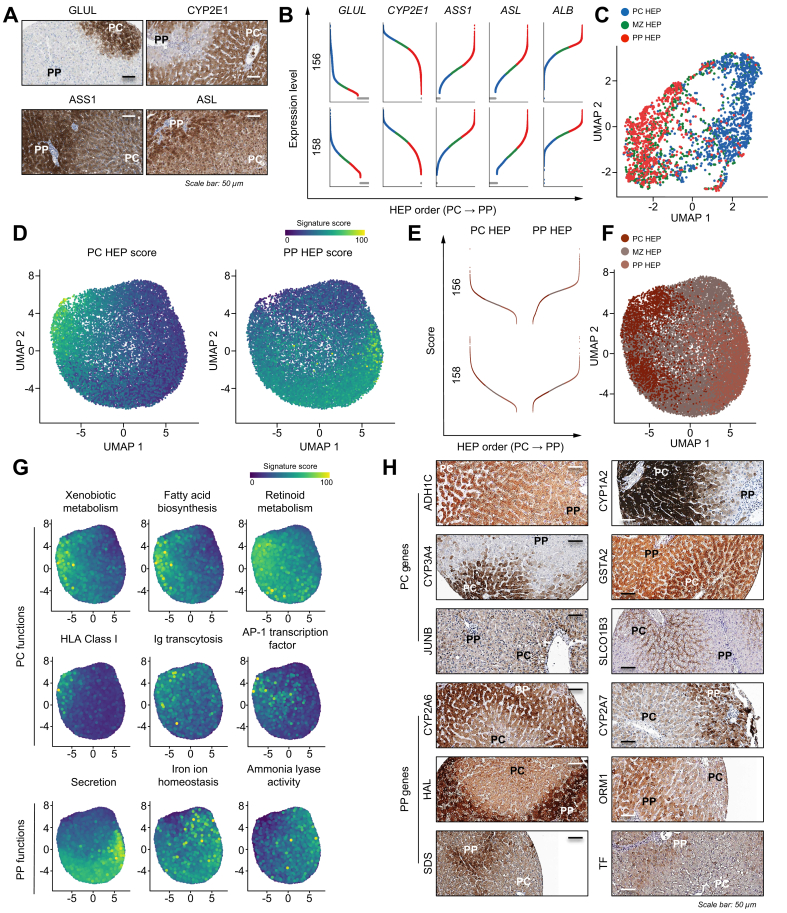
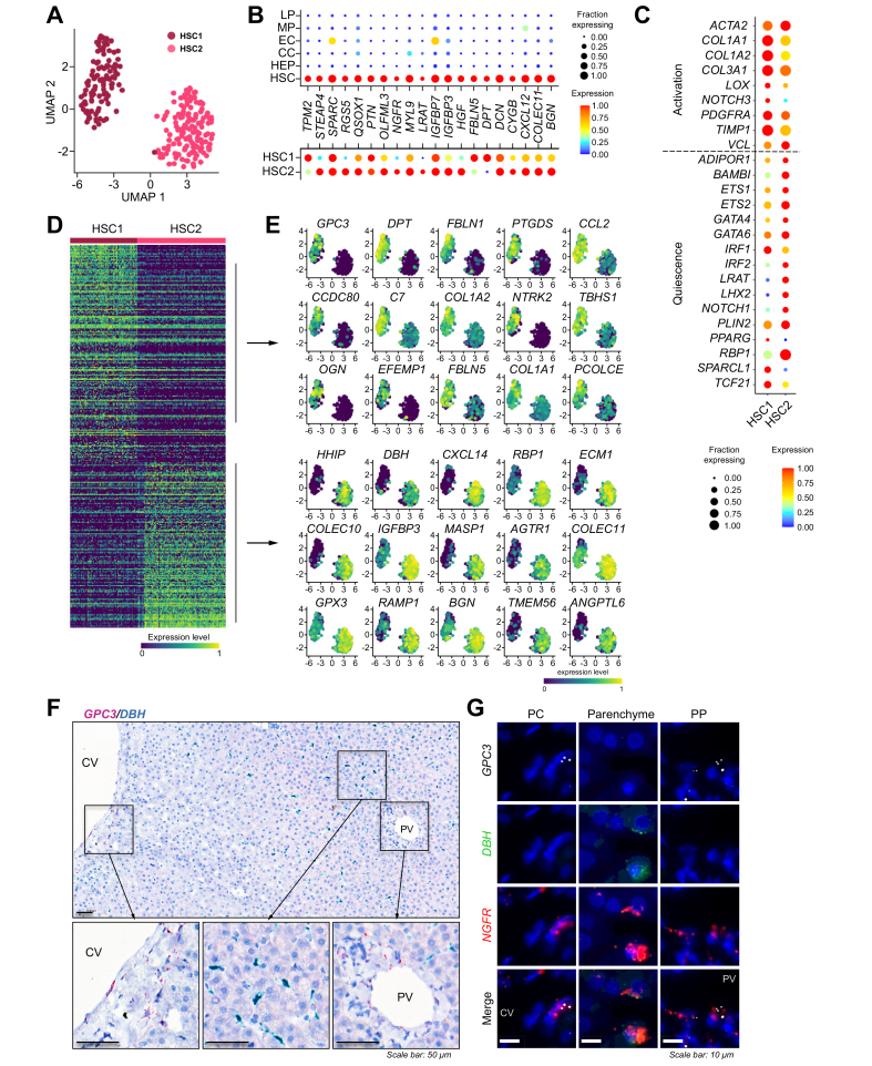
Results: In total, 22 cell populations were annotated that reflected the heterogeneity of human parenchymal and non-parenchymal liver cells. More than 20,000 HEPs were ordered along the portocentral axis to confirm known, and reveal previously undescribed, zonated liver functions. The existence of 2 subpopulations of human HSCs with unique gene expression signatures and distinct intralobular localisation was revealed (i.e. portal and central vein-concentrated GPC3 + HSCs and perisinusoidally located DBH + HSCs). In particular, these data suggest that, although both subpopulations collaborate in the production and organisation of extracellular matrix, GPC3 + HSCs specifically express genes involved in the metabolism of glycosaminoglycans, whereas DBH + HSCs display a gene signature that is reminiscent of antigen-presenting cells.

Conclusions: This study highlights metabolic zonation as a key determinant of HEP transcriptomic heterogeneity and, for the first time, outlines the existence of heterogeneous HSC subpopulations in the human liver. These findings call for further research on the functional implications of liver cell heterogeneity in health and disease.

Lay summary: This study resolves the cellular landscape of the human liver in an unbiased manner and at high resolution to provide new insights into human liver cell biology. The results highlight the physiological heterogeneity of human hepatic stellate cells.

Keywords: BSA, bovine serum albumin; CC, cholangiocyte; CV, central vein; DEG, differentially expressed gene; EC, endothelial cell; ECM, extracellular matrix; Extracellular matrix; FFPE, formaldehyde-fixed paraffin embedded; GAG, glycosaminoglycan; GEO, Gene Expression Omnibus; GO, gene ontology; HEP, hepatocyte; HLA, human leukocyte antigen; HRP, horseradish peroxidase; HSC, hepatic stellate cell; Hepatocyte; ISH, in situ hybridisation; KLR, killer lectin-like receptor; LP, lymphoid cell; Liver cell atlas; MP, macrophage; MZ, midzonal; PC, pericentral; PP, periportal; PV, portal vein; TBS, Tris buffered saline; TSA, tyramide signal amplification; UMAP, uniform manifold approximation and projection; UMI, unique molecular identifier; VIM, vimentin; Zonation; scRNA-seq, single-cell RNA-sequencing.


Conflict of interest statement

The authors declare no conflicts of interest that pertain to this work. Please refer to the accompanying ICMJE disclosure forms for further details.

Figures

None
Graphical abstract
Fig. 1
Fig. 1
Clustering and annotation of healthy human liver cell transcriptomes. (A) Whole-cell suspensions extracted from 2 freshly explanted human livers were centrifuged and prepared for droplet-based scRNA-seq using the 10X Chromium technology. (B) UMAP visualisation of 25,325 human liver cells clustered into 22 cell subpopulations. (C) Cell-type annotation of each subpopulation based on the differential expression of liver cell type-specific genes. (D) Heatmap displaying the expression level of established liver cell type-specific markers in each cell subpopulation. (E) Top 5 genes most highly expressed in each cell type. scRNA-seq, single cell RNA sequencing; UMAP, uniform manifold approximation and projection.
Fig. 2
Fig. 2
Liver zonation drives the clustering of human hepatocytes. (A) Immunostaining for GLUL, CYP2E1, ASS1 and ASL (images from the Human Protein Atlas). (B) HEPs ordered along the portocentral axis according to the expression of GLUL, CYP2E1, ASS1, ASL, and ALB, per donor. (C) Annotation of HEPs as PC, MZ, or PP HEPs based on the expression of ≥4 of the zonation markers GLUL, CYP2E1, ASS1, ASL, and ALB. (D) Distribution of the expression of genes significantly upregulated in (i) PC or (ii) PP HEPs. (E) HEPs ordered along the portocentral axis according to the expression of genes significantly upregulated in PC or PP HEPs. (F) Annotation of all HEPs as PC, MZ, or PP based on the expression of genes significantly upregulated in PC or PP HEPs. (G) Distribution of the expression of genes associated with zonated functions in HEPs. (H) Protein-level validation of a selection of differentially expressed genes from the PP and PC HEP gene signatures (images from the Human Protein Atlas). HEP, hepatocytes; MZ, midzonal; PC, pericentral; PP: periportal.
Fig. 3
Fig. 3
Transcriptomic and spatial heterogeneity of human HSCs. (A) UMAP clustering of 246 HSCs. (B) Expression levels of HSC-specific genes in LPs, MPs, ECs, CCs and HEPs, as well as in the HSC subpopulations HSC1 and HSC2. (C) Expression levels of key HSC activation and quiescence marker genes in HSC1 and HSC2. (D) Heatmap of the significantly differentially expressed genes between HSC1 and HSC2. (E) UMAP plots for a selection of 30 HSC1- and HSC2-specific genes. (F) Representative pictures of brightfield in situ hybridisation using GPC3- and DBH-specific probes in human liver tissue. (G) Representative pictures of fluorescent in situ hybridisation using GPC3-, DBH- and NGFR-specific probes in human liver tissue. CC, cholangiocyte; CV, central vein; EC, endothelial cell; HEP, hepatocyte; HSCs, hepatic stellate cells; LP, lymphoid cell; MP, macrophage; PC, pericentral; PP, periportal; PV, portal vein; UMAP, uniform manifold approximation and projection.
Fig. 4
Fig. 4
Functional heterogeneity of human hepatic stellate cells. (A) Distribution of the expression of genes associated with selected functions in HSCs. (B) Representative pictures of VIM and HLA-DR immunostaining in liver tissue. (C) Representative pictures of CD45 and HLA-DR immunostaining in liver tissue. (D) Distribution of the expression of CD80 and CD86 in HSCs. (E,F) Representative pictures of GAG and Elastin staining in liver tissue. (G) Expression levels of liver extracellular matrix glycoproteins, collagens and proteoglycans in LPs, MPs, ECs, CCs, and HEPs, as well as in the HSC subpopulations HSC1 and HSC2. (H) Expression levels of relevant growth factors and cytokines in HSC1 and HSC2. CC, cholangiocyte; CV, central vein; EC, endothelial cell; GAGs, glycosaminoglycans; HEP, hepatocyte; HLA, human leukocyte antigen; HSCs, hepatic stellate cells; LP, lymphoid cell; MP, macrophage; PC, pericentral; PP, periportal; PV, portal vein; VIM, vimentin.

References

    1. El Taghdouini A., Najimi M., Sancho-Bru P., Sokal E., van Grunsven L.A. In vitro reversion of activated primary human hepatic stellate cells. Fibrogenesis Tissue Repair. 2015;8:14. - PMC - PubMed
    1. El Taghdouini A., Sorensen A.L., Reiner A.H., Coll M., Verhulst S., Mannaerts I. Genome-wide analysis of DNA methylation and gene expression patterns in purified, uncultured human liver cells and activated hepatic stellate cells. Oncotarget. 2015;6:26729–26745. - PMC - PubMed
    1. Mederacke I., Dapito D.H., Affo S., Uchinami H., Schwabe R.F. High-yield and high-purity isolation of hepatic stellate cells from normal and fibrotic mouse livers. Nat Protoc. 2015;10:305–315. - PMC - PubMed
    1. D'Ambrosio D.N., Walewski J.L., Clugston R.D., Berk P.D., Rippe R.A., Blaner W.S. Distinct populations of hepatic stellate cells in the mouse liver have different capacities for retinoid and lipid storage. PLoS One. 2011;6 - PMC - PubMed
    1. Macosko E.Z., Basu A., Satija R., Nemesh J., Shekhar K., Goldman M. Highly parallel genome-wide expression profiling of individual cells using nanoliter droplets. Cell. 2015;161:1202–1214. - PMC - PubMed