Review
. 2021 Jul;95(7):2235-2253.
doi: 10.1007/s00204-021-03069-1. Epub 2021 May 24.

Infections at the nexus of metabolic-associated fatty liver disease

Affiliations
Review

Infections at the nexus of metabolic-associated fatty liver disease

Joost Boeckmans et al. Arch Toxicol. 2021 Jul.

Abstract

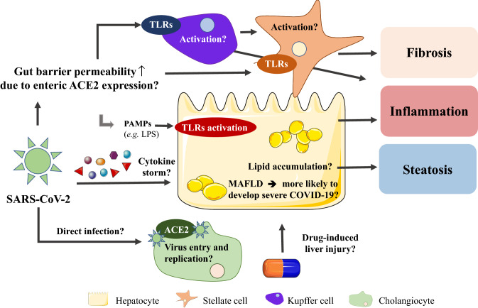
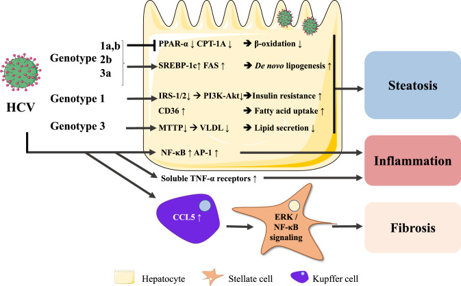
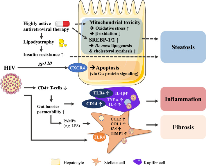
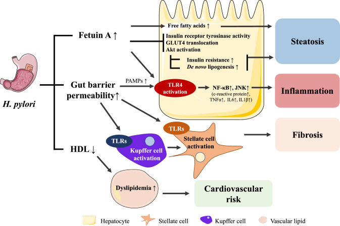
Metabolic-associated fatty liver disease (MAFLD) is a chronic liver disease that affects about a quarter of the world population. MAFLD encompasses different disease stadia ranging from isolated liver steatosis to non-alcoholic steatohepatitis (NASH), fibrosis, cirrhosis and hepatocellular carcinoma. Although MAFLD is considered as the hepatic manifestation of the metabolic syndrome, multiple concomitant disease-potentiating factors can accelerate disease progression. Among these risk factors are diet, lifestyle, genetic traits, intake of steatogenic drugs, male gender and particular infections. Although infections often outweigh the development of fatty liver disease, pre-existing MAFLD could be triggered to progress towards more severe disease stadia. These combined disease cases might be underreported because of the high prevalence of both MAFLD and infectious diseases that can promote or exacerbate fatty liver disease development. In this review, we portray the molecular and cellular mechanisms by which the most relevant viral, bacterial and parasitic infections influence the progression of fatty liver disease and steatohepatitis. We focus in particular on how infectious diseases, including coronavirus disease-19, hepatitis C, acquired immunodeficiency syndrome, peptic ulcer and periodontitis, exacerbate MAFLD. We specifically underscore the synergistic effects of these infections with other MAFLD-promoting factors.

Keywords: Helicobacter pylori; Hepatitis C; Human immunodeficiency virus; Infectious diseases; Klebsiella pneumoniae; Lipid metabolism; Liver; Metabolic-associated fatty liver disease (MAFLD); Non-alcoholic steatohepatitis (NASH); SARS-CoV-2.


Conflict of interest statement

The authors declare that they have no conflict of interest.

Figures

Fig. 1
Fig. 1
Possible interconnections between COVID-19 and MAFLD. Disturbed liver laboratory parameters are frequently observed in severe COVID-19 cases. Patients infected by SARS-CoV-2 often exhibit also liver steatosis, while MAFLD seems to aggravate COVID-19. SARS-CoV-2 could induce PAMP translocation from the gut to the liver to initiate Kupffer cell and stellate cell activation and hence promote an acute inflammatory response. Further, the liver could get damaged or primed for secondary hits by cytokines during cytokine storms. Until today, cholangiocytes are the only liver cells known to express the virus entry receptor ACE2, although specific consequences remain to be investigated
Fig. 2
Fig. 2
HCV-mediated mechanisms leading to hepatic steatosis and inflammation. HCV infection leads to SREBP-1c-mediated lipid accumulation and PPAR-α/PPARA and CPTA downregulation, suggesting impaired fatty acid β-oxidation and reduced repression of inflammation. HCV genotypes 1 and 4 infections are associated with insulin resistance, while HCV genotype 3 infection impairs VLDL-mediated triglyceride secretion. HCV-exposed Kupffer cells secrete CCL5 resulting in hepatic stellate cell activation
Fig. 3
Fig. 3
HIV- and HAART-mediated mechanisms leading to hepatic steatosis, inflammation and fibrosis. HIV itself can induce liver inflammation via TLRs, hepatocyte apoptosis through CXCR4 and fibrosis by directly (via envelope glycoprotein gp120) and indirectly (via LPS) activating stellate cells. HAART-associated lipodystrophy induces insulin resistance and hepatic steatosis while also evoking mitochondrial toxicity
Fig. 4
Fig. 4
H. Pylori-infection-mediated mechanisms leading to hepatic steatosis inflammation and fibrosis. H. Pylori infection relates to increased serum fetuin A levels. Fetuin A can impair insulin signaling by interrogating with, among others, insulin receptor tyrosine kinase activity and promote inflammation through TLR4 activation. H. Pylori infection probably also relates to increased gut barrier permeability which can result in translocation of PAMPs to the liver through the gut-liver axis and initiate inflammation and fibrosis

References

    1. Abu-Shanab A, Quigley EMM. The role of the gut microbiota in nonalcoholic fatty liver disease. Nat Rev Gastroenterol Hepatol. 2010;7:691–701. doi: 10.1038/nrgastro.2010.172. - DOI - PubMed
    1. Adinolfi LE, Gambardella M, Andreana A, et al. Steatosis accelerates the progression of liver damage of chronic hepatitis C patients and correlates with specific HCV genotype and visceral obesity. Hepatology. 2001;33:1358–1364. doi: 10.1053/jhep.2001.24432. - DOI - PubMed
    1. Adinolfi LE, Rinaldi L, Guerrera B, et al. NAFLD and NASH in HCV infection: prevalence and significance in hepatic and extrahepatic manifestations. Int J Mol Sci. 2016;17:1–12. doi: 10.3390/ijms17060803. - DOI - PMC - PubMed
    1. Alakhali MS, Al-Maweri SA, Al-Shamiri HM, et al. The potential association between periodontitis and non-alcoholic fatty liver disease: a systematic review. Clin Oral Investig. 2018;22:2965–2974. doi: 10.1007/s00784-018-2726-1. - DOI - PubMed
    1. Alazawi W, Bernabe E, Tai D, et al. Periodontitis is associated with significant hepatic fibrosis in patients with non-alcoholic fatty liver disease. PLoS ONE. 2017;12:1–13. doi: 10.1371/journal.pone.0185902. - DOI - PMC - PubMed

MeSH terms