

Line-field confocal optical coherence tomography of xanthogranuloma: Correlation with vertical and horizontal histopathology
- PMID: 34028070
- PMCID: PMC8453847
- DOI: 10.1111/cup.14067
Line-field confocal optical coherence tomography of xanthogranuloma: Correlation with vertical and horizontal histopathology
Abstract
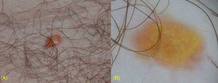
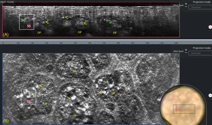
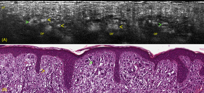
Line-field confocal optical coherence tomography (LC-OCT) is a new noninvasive technique for a real-time, vertical, and horizontal imaging of the skin at cellular resolution. A 47-year-old female presented with a 6-month history of an asymptomatic yellowish papule. LC-OCT evaluation was able to show the diagnostic microscopic features of xanthogranuloma and showed an excellent correlation with vertical and horizontal histopathological sections by revealing enlarged dermal papillae containing multiple, bright roundish giant cells, corresponding to foamy histiocytes, and giant cells characterized by a dark center surrounded by a highly hyper-refractile peripheral ring, corresponding to Touton cells. LC-OCT may represent a valid, noninvasive alternative to histopathological examination in clinically atypical cases of xanthogranuloma.
Keywords: Touton cells; foamy histiocytes; histopathology; line-field confocal optical coherence tomography; xanthogranuloma.
© 2021 The Authors. Journal of Cutaneous Pathology published by John Wiley & Sons Ltd.
Conflict of interest statement
The authors declare no potential conflict of interest.
Figures

Similar articles
-
Line-field confocal optical coherence tomography of lentigo maligna with horizontal and vertical histopathologic correlations.J Cutan Pathol. 2023 Feb;50(2):118-122. doi: 10.1111/cup.14321. Epub 2022 Sep 19. J Cutan Pathol. 2023. PMID: 36056910 Free PMC article.
-
Analysis of the characteristics of reflectance confocal microscopy images of xanthogranuloma and xanthoma.Int J Dermatol. 2025 Jan;64(1):155-163. doi: 10.1111/ijd.17265. Epub 2024 Jun 16. Int J Dermatol. 2025. PMID: 38880994
-
Morphological evaluation of melanocytic lesions with three-dimensional line-field confocal optical coherence tomography: correlation with histopathology and reflectance confocal microscopy. A pilot study.Clin Exp Dermatol. 2022 Dec;47(12):2222-2233. doi: 10.1111/ced.15383. Epub 2022 Oct 20. Clin Exp Dermatol. 2022. PMID: 35988042
-
Role of In Vivo Reflectance Confocal Microscopy in the Analysis of Melanocytic Lesions.Acta Dermatovenerol Croat. 2018 Apr;26(1):64-67. Acta Dermatovenerol Croat. 2018. PMID: 29782304 Review.
-
[Xanthogranulomas].Ann Dermatol Venereol. 2011 Feb;138(2):156-8. doi: 10.1016/j.annder.2010.10.016. Epub 2010 Nov 24. Ann Dermatol Venereol. 2011. PMID: 21333830 Review. French. No abstract available.
Cited by
-
Diagnostic Accuracy of Line-Field Confocal Optical Coherence Tomography for the Diagnosis of Skin Carcinomas.Diagnostics (Basel). 2023 Jan 18;13(3):361. doi: 10.3390/diagnostics13030361. Diagnostics (Basel). 2023. PMID: 36766466 Free PMC article.
-
Line-Field Confocal Optical Coherence Tomography May Enhance Monitoring of Superficial Basal Cell Carcinoma Treated with Imiquimod 5% Cream: A Pilot Study.Cancers (Basel). 2021 Sep 30;13(19):4913. doi: 10.3390/cancers13194913. Cancers (Basel). 2021. PMID: 34638396 Free PMC article.
-
Line-field confocal optical coherence tomography of lentigo maligna with horizontal and vertical histopathologic correlations.J Cutan Pathol. 2023 Feb;50(2):118-122. doi: 10.1111/cup.14321. Epub 2022 Sep 19. J Cutan Pathol. 2023. PMID: 36056910 Free PMC article.
-
Line-Field Confocal Optical Coherence Tomography Evaluation of Eyelid Skin Lesions.Diagnostics (Basel). 2023 Dec 3;13(23):3590. doi: 10.3390/diagnostics13233590. Diagnostics (Basel). 2023. PMID: 38066831 Free PMC article.
-
Line-Field Confocal Optical Coherence Tomography for the Diagnosis of Skin Carcinomas: Real-Life Data over Three Years.Curr Oncol. 2023 Sep 28;30(10):8853-8864. doi: 10.3390/curroncol30100639. Curr Oncol. 2023. PMID: 37887539 Free PMC article.
References
-
- Janssen D, Harms D. Juvenile xanthogranuloma in childhood and adolescence: a clinicopathologic study of 129 patients from the Kiel pediatric tumor registry. Am J Surg Pathol. 2005;29(1):21‐28. - PubMed
-
- Micali G, Verzì AE, Quattrocchi E, Ng CY, Lacarrubba F. Dermatoscopy of common lesions in pediatric dermatology. Dermatol Clin. 2018;36(4):463‐472. - PubMed
-
- Dubois A, Levecq O, Azimani H, et al. Line‐field confocal optical coherence tomography for high‐resolution noninvasive imaging of skin tumors. J Biomed Opt. 2018;23(10):1‐9. - PubMed
-
- Ruini C, Schuh S, Pellacani G, French L, Welzel J, Sattler E. In vivo imaging of Sarcoptes scabiei infestation using line‐field confocal optical coherence tomography. J Eur Acad Dermatol Venereol. 2020;34(12):e808‐e809. - PubMed
Publication types
MeSH terms
LinkOut - more resources
Full Text Sources
Other Literature Sources
