Review
. 2021 May;5(5):e2000180.
doi: 10.1002/adbi.202000180. Epub 2021 Jan 14.

Steering Molecular Activity with Optogenetics: Recent Advances and Perspectives

Affiliations
Review

Steering Molecular Activity with Optogenetics: Recent Advances and Perspectives

Teak-Jung Oh et al. Adv Biol (Weinh). 2021 May.

Abstract

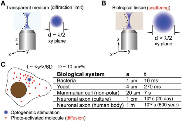
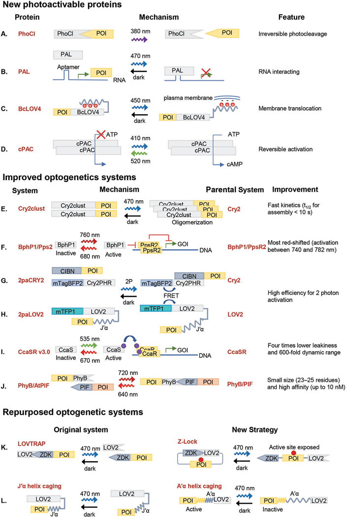
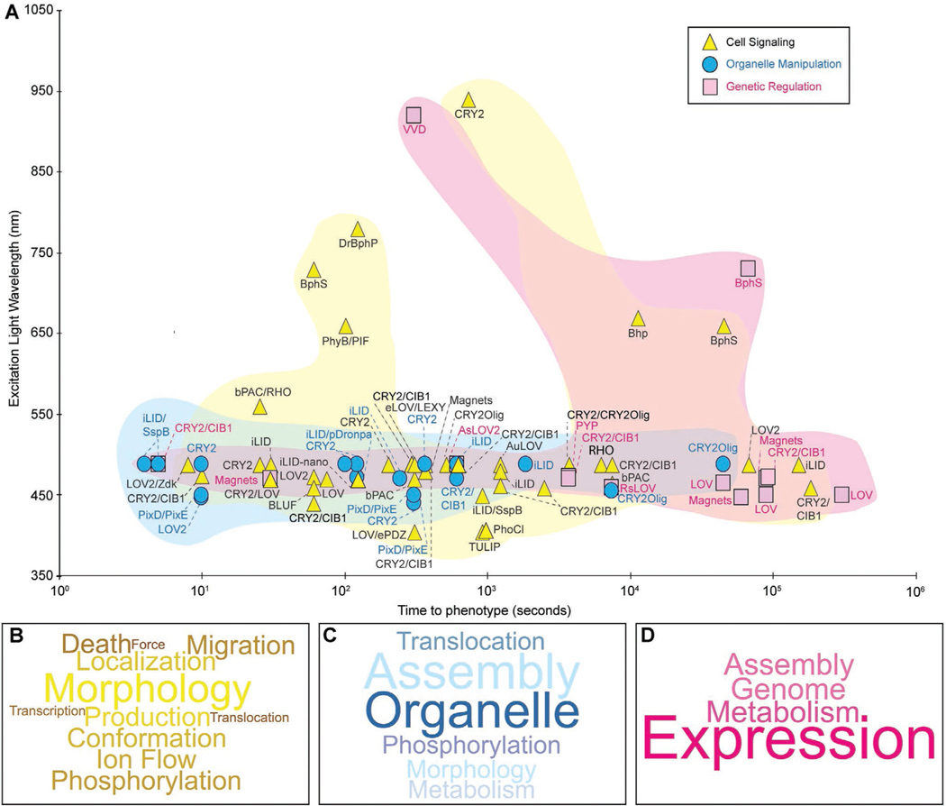
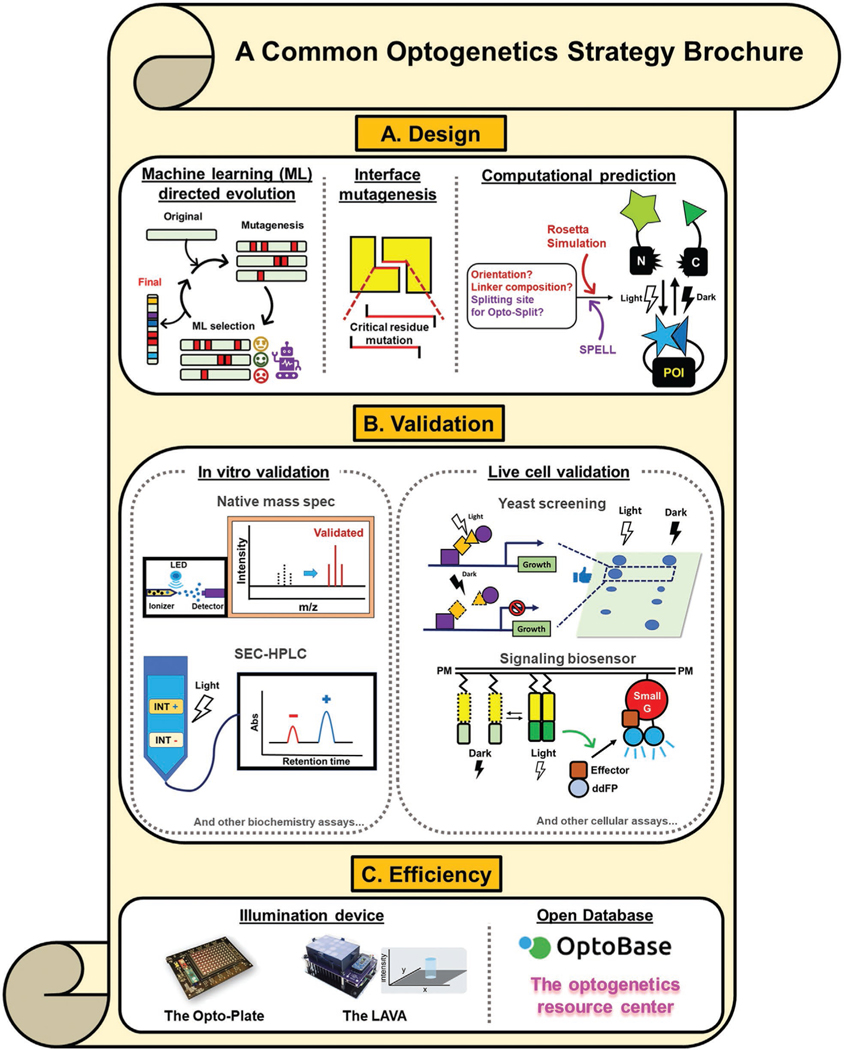
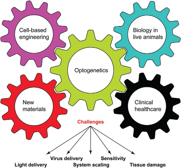
Optogenetics utilizes photosensitive proteins to manipulate the localization and interaction of molecules in living cells. Because light can be rapidly switched and conveniently confined to the sub-micrometer scale, optogenetics allows for controlling cellular events with an unprecedented resolution in time and space. The past decade has witnessed an enormous progress in the field of optogenetics within the biological sciences. The ever-increasing amount of optogenetic tools, however, can overwhelm the selection of appropriate optogenetic strategies. Considering that each optogenetic tool may have a distinct mode of action, a comparative analysis of the current optogenetic toolbox can promote the further use of optogenetics, especially by researchers new to this field. This review provides such a compilation that highlights the spatiotemporal accuracy of current optogenetic systems. Recent advances of optogenetics in live cells and animal models are summarized, the emerging work that interlinks optogenetics with other research fields is presented, and exciting clinical and industrial efforts to employ optogenetic strategy toward disease intervention are reported.

Keywords: cross-disciplinary interface; gene regulation; optogenetics; organelle manipulation; signal transduction.


Conflict of interest statement

Conflict of Interest

The authors declare no conflict of interest.

Figures

Figure 1.
Figure 1.
A) Light diffraction limits the size of a coherent beam to approximately half of the excitation wavelength in lens-based optical microscopy. B) In opaque biological tissues, light scattering varies the coherent beam’s wavevector randomly and causes an expansion of the effective focal volume. C) molecular diffusion in cells further compromises the spatial resolution of the optogenetic stimulation. A back-of-envelop estimation of the traversing time of a small protein across a cell is presented. The typical value of a small protein’s diffusion coefficient in the cytoplasm is selected based on experimental measurement.[3]
Figure 2.
Figure 2.
Recent advancements of optogenetic systems. A–D) Recently developed new photoactivatable protein systems. E–J) Improvement of existing optogenetic systems. K–L) Existing optogenetic systems with repurposed functions. References of each photoactivatable protein are listed as follows: A) PhoCl,[10] B) PAL,[11] C) BcLOV4,[12] D) cPAC,[14] E) CRY2clust,[16] F) BphP1/Pps2,[19] G,H) FRAPA,[23] I) CcaSR v3.0,[26] J) PhyB/AtPIF,[29] K) Z-Lock,[31] and L) usage of the A’α helix of LOV.[33]
Figure 3.
Figure 3.
Applications of optogenetics in steering molecular activity in cells and organisms. A) A scattered plot depicting selected work of optogenetic control of molecular activities in the past three years. Strategies are grouped into three categories: cell signaling (yellow),[,,–,,–,,,–,–83,66] organelle manipulation (cyan),[–,–100] and genetic regulation (magenta).[,,–,–118] Excitation wavelength refers to the light used to activate the optogenetic proteins, which are individually labeled. The time to phenotype was chosen based on reported timescales outlined in Table 2. Word clouds indicating the frequency of each outcome per category: signaling B), organelle manipulation C), and genetic regulation D). The outcomes are also summarized in Table 2. The word clouds were generated in Wordcloud.com.
Figure 4.
Figure 4.
Collection of commonly used optogenetic strategies. a) Optically active protein design and engineering approaches. POI: Protein of interest. b) Live cell or in vitro validation modules. INT+: Positive interaction; INT−, negative interaction; ddFP, dimerization-dependent fluorescence protein c) Efficient resources for conducting an experiment or collecting data. References of each strategy are listed as follows: machine learning directed evolution,[119,120] interface directed mutagenesis,[122] computational prediction,[31,123,124] native mass spectrometry,[128] SEC-HPLC,[129] yeast screening,[122,130] signaling biosensor,[131] optoplate,[132] LAVA plate,[133] optobase,[1a] and the optogenetic resource center.[1] The image of the optoplate is reproduced with permission.[51b] Copyright 2018, The American Association for the Advancement of Science. The image of the LAVA plate is reproduced with permission.[133] Copyright 2020, Elsevier.
Figure 5.
Figure 5.
The cross-disciplinary interplay between optogenetics and other fields. Optogenetics could be applied to facilitate cell-based engineering (e.g., tissue engineering) or generate new biomaterials. By overcoming technical challenges, such as light and virus delivery, sensitivity, toxicity, and scaling up to large animals, optogenetic tools would be increasingly applied in the live animals. Advanced technologies, such as the two-photon optogenetic stimulation and light upconversion, will continue pushing optogenetic technology toward clinical settings in the healthcare sector.

References

    1. Kolar K, Knobloch C, Stork H, Znidaric M, Weber W, ACS Synth. Biol 2018, 7, 1825; - PubMed
    2. Deisseroth K, Optogenetics Resource Center https://web.stanford.edu/group/dlab/optogenetics/ (accessed: December 2020).
    1. Tichy AM, Gerrard EJ, Legrand JMD, Hobbs RM, Janovjak H, J. Mol. Biol 2019, 431, 3046; - PubMed
    2. Muhlhauser WWD, Weber W, Radziwill G, ACS Synth. Biol 2019, 8, 1679. - PubMed
    1. Kumar M, Mommer MS, Sourjik V, Biophys. J 2010, 98, 552. - PMC - PubMed
    1. Harris JM, Wang AY, Boulanger-Weill J, Santoriello C, Foianini S, Lichtman JW, Zon LI, Arlotta P, Dev. Cell 2020, 53, 577. - PMC - PubMed
    1. Wang Q, Fan H, Li F, Skeeters SS, Krishnamurthy VV, Song Y, Zhang K, Elife 2020, 9, e57395. - PMC - PubMed

Publication types

LinkOut - more resources