. 2021 Jul;297(1):100821.
doi: 10.1016/j.jbc.2021.100821. Epub 2021 May 23.

Arginine methylation of SARS-Cov-2 nucleocapsid protein regulates RNA binding, its ability to suppress stress granule formation, and viral replication

Affiliations

Arginine methylation of SARS-Cov-2 nucleocapsid protein regulates RNA binding, its ability to suppress stress granule formation, and viral replication

Ting Cai et al. J Biol Chem. 2021 Jul.

Abstract

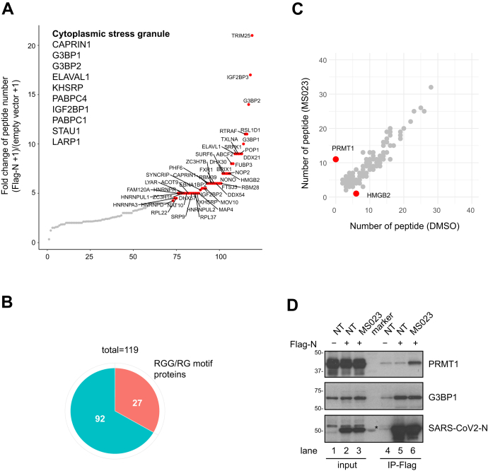
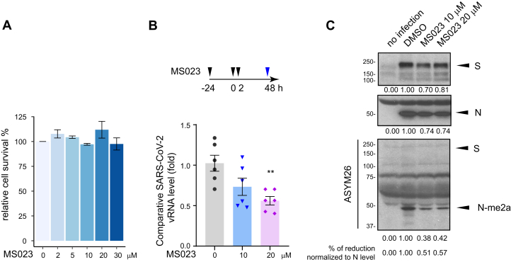
Viral proteins are known to be methylated by host protein arginine methyltransferases (PRMTs) necessary for the viral life cycle, but it remains unknown whether severe acute respiratory syndrome coronavirus 2 (SARS-CoV-2) proteins are methylated. Herein, we show that PRMT1 methylates SARS-CoV-2 nucleocapsid (N) protein at residues R95 and R177 within RGG/RG motifs, preferred PRMT target sequences. We confirmed arginine methylation of N protein by immunoblotting viral proteins extracted from SARS-CoV-2 virions isolated from cell culture. Type I PRMT inhibitor (MS023) or substitution of R95 or R177 with lysine inhibited interaction of N protein with the 5'-UTR of SARS-CoV-2 genomic RNA, a property required for viral packaging. We also defined the N protein interactome in HEK293 cells, which identified PRMT1 and many of its RGG/RG substrates, including the known interacting protein G3BP1 as well as other components of stress granules (SGs), which are part of the host antiviral response. Methylation of R95 regulated the ability of N protein to suppress the formation of SGs, as R95K substitution or MS023 treatment blocked N-mediated suppression of SGs. Also, the coexpression of methylarginine reader Tudor domain-containing protein 3 quenched N protein-mediated suppression of SGs in a dose-dependent manner. Finally, pretreatment of VeroE6 cells with MS023 significantly reduced SARS-CoV-2 replication. Because type I PRMT inhibitors are already undergoing clinical trials for cancer treatment, inhibiting arginine methylation to target the later stages of the viral life cycle such as viral genome packaging and assembly of virions may represent an additional therapeutic application of these drugs.

Keywords: PRMT1; RGG/RG motif; RNA binding; SARS-CoV-2; arginine methylation; condensate; nucleocapsid (N) protein; stress granules; type I PRMT inhibitor.


Conflict of interest statement

Conflict of interest The authors declare that they have no conflicts of interest with the contents of this article.

Figures

Figure 1
Figure 1
R95 and R177 within SARS-CoV-2 N RGG/RG motifs are methylated by PRMT1.A, schematic diagram of N protein with its N-terminal domain (NTD) and C-terminal domain (CTD) and its NIDR and CIDR for N- and C-terminal intrinsic disordered regions and finally the linker region between NTD and CTD known for its SR-rich sequences. Note R95GG and R177GG are conserved in SARS-CoV and SARS-CoV-2, but not MERS-CoV. BD, recombinant GST–N protein fragments were subjected to in vitro methylation assays with recombinant (B) GST–PRMT1, (C) PRMT5/MEP50, and (D) GST–PRMT6. Coomassie Blue staining and fluorography images are presented. GST alone and GST–RGG were used as negative and positive controls, respectively. Blue arrowheads indicate the migration of the GST–N protein fragments. The migration of GST–PRMT1, PRMT5, and GST–PRMT6 is shown on the right with a black arrow. The molecular mass markers are shown in kDa on the left. E and F, GST–N protein fragments with arginine to lysine substitution were subjected to in vitro methylation assays. Coomassie Blue staining and fluorography images are presented. G, HEK293 cells were transfected with control (-) or Flag-N (+) expression vectors for 24 h and incubated with or without (NT) 1 μM MS023 for another 24 h. The cell lysates were subjected to immunoprecipitation with anti-Flag-M2 beads and immunoblotting with anti-asymmetrical dimethylarginine antibody ASYM26 (upper panels) and anti-SARS-CoV-2 N protein antibody (lower panels). The band of the asymmetrically dimethylated N protein (N-me2) is marked by a black arrowhead on the right. The molecular mass markers are shown in kDa on the left. H, HEK293 cells were transfected with siRNA targeting firefly luciferase (siCTL) or siPRMT1 for 24 h and subsequently transfected with Flag-N vector for another 24 h. The cell lysates were subjected to immunoprecipitation with anti-Flag-M2 beads and then immunoblotting with anti-SARS-CoV-2 N protein and anti-ASYM26 antibodies. The migration of the methylated N protein is indicated. Flag-N, Flag-epitope N protein; MERS-CoV, Middle East respiratory syndrome coronavirus; N, nucleocapsid; PRMTs, protein arginine methyltransferases; SARS-CoV-2, severe acute respiratory syndrome coronavirus 2.
Figure 2
Figure 2
N protein interactome with and without MS023: association with many RGG/RG proteins and PRMT1. HEK293 cells were transfected with control or Flag-N, and the next day, Flag-N-transfected cells were subsequently treated with or without 1 μM MS023 for 24 h. Cell lysates were subjected to immunoprecipitation using anti-Flag-M2 beads. The bound proteins were identified by MS (AC). A, interactors were ranked by fold change of unique peptides detected from Flag-N-transfected cells and control plasmid-transfected cells (Flag-N + 1)/(empty vector + 1). Proteins with FC >4 are highlighted in red. Immunoprecipitated proteins known to be localized in stress granule are listed. B, correlation analysis between MS023 and DMSO-treated N protein interactome is shown. Proteins with a significant fold change (>3 or <3−1) after MS023 treatment are highlighted in red. C, the pie chart represents the number of RGG/RG motif containing proteins among N protein interactors. D, HEK293 cells were transfected with control (-) or Flag-N (+) and subsequentially treated with or without (NT) 1 μM MS023 for 24 h. Cell lysates were immunoprecipitated with anti-Flag antibodies, and the associated proteins separated by SDS-PAGE and immunoblotted with anti-PRMT1, anti-G3BP1, and anti-SARS-CoV-2 N antibodies. The asterisk denotes nonspecific recognition of a molecular mass marker protein. DMSO, dimethylsulfoxide; PRMTs, protein arginine methyltransferases; SARS-CoV-2, severe acute respiratory syndrome coronavirus 2.
Figure 3
Figure 3
SARS-CoV-2 N protein regulates G3BP1 stress granule formation in an arginine methylation-dependent manner.A, Huh-7 cells were transfected with control vector or Flag-N for 24 h and subsequently incubated with 1 mM sodium arsenite for 2 h. Cells were fixed with 4% PFA and coimmunostained with anti-Flag and anti-G3BP1 antibodies. A typical image is shown. The scale bar represents 20 μm. Arrowheads indicate FLAG-N-transfected cells, and the empty arrowheads indicate cells with FLAG-N and G3BP1 colocalization. B, Huh-7 cells were transfected with control vector or Flag-N for 24 h and subsequently incubated with 0.5 mM sodium arsenite for 1 h. Cells were fixed with 4% PFA and coimmunostained with anti-Flag and anti-G3BP1 antibodies. A typical image is shown. The scale bar represents 20 μm. Arrowheads indicate FLAG-N-transfected cells, and transfected cells with SGs (>5 G3BP1 foci) are highlighted in magenta. The percentage of cells harboring SGs (>5 G3BP1 foci) are quantified and shown in the bar plot on the right. n = 15 fields from three independent experiments are shown. Welch's t test. ∗∗∗∗p < 0.0001. C, Huh-7 cells were transfected with Flag-N overnight and treated with or without 5 μM MS023 for another 24 h. Then, the cells were incubated with 0.5 mM sodium arsenite for another hour and fixed with 4% PFA. Cells were coimmunostained with anti-Flag and anti-G3BP1 antibodies. A typical image is shown in the left panel. The scale bar represents 20 μm. Arrowheads indicate Flag-N-transfected cells, and transfected cells with SGs (>5 G3BP1 foci) are highlighted in magenta. The percentage of cells harboring SGs (>5 G3BP1 foci) was quantified in the transfected cells (Flag-N positive) and nontransfected cells (Flag-N negative), respectively, and shown in the bar plot on the right. Twenty fields from two independent experiments are shown. Welch's t test. ∗p < 0.05. Flag-N, Flag-epitope N protein; N, nucleocapsid; PFA, paraformaldehyde; SARS-CoV-2, severe acute respiratory syndrome coronavirus 2; SGs, stress granules.
Figure 4
Figure 4
R95 is required for N protein–regulating G3BP1 stress granule, and methylarginine reader protein TDRD3 is involved in this process.A, Huh-7 cells were transfected with Flag-N (WT) and its mutants for 24 h and subsequently incubated with 0.5 mM sodium arsenite for another hour. Cells were fixed with 4% PFA and coimmunostained with anti-Flag and anti-G3BP1 antibodies. A typical image is shown in the left panel. The scale bar represents 20 μm. Arrowheads indicate transfected cells, and transfected cells with SGs (>5 G3BP1 foci) are highlighted in magenta. The percentage of cells harboring SGs (>5 G3BP1 foci) in the Flag-N-positive cell group was quantified and shown in the bar plot on the right. n = 15 fields from three independent experiments are shown. Welch's t test. ∗p < 0.05, ∗∗p < 0.01. B, HEK293 cells were cotransfected with Flag-N and myc-SMN or myc-TDRD3. Twenty-four hours later, cell lysates were immunoprecipitated with anti-Flag antibodies and the associated proteins separated by SDS-PAGE and immunoblotted with anti-SMN, anti-TDRD3, and anti-Flag antibodies. The asterisk denotes nonspecific recognition of a molecular mass marker protein. C, Huh-7 cells were cotransfected with Flag-N and myc-TDRD3 with indicated plasmid ratios. Twenty-four hours later, cells were incubated with 0.5 mM sodium arsenite for another hour. Cells were fixed with 4% PFA and coimmunostained with anti-Flag and G3BP1 antibodies. A typical image is shown on the left. The scale bar represents 20 μm. Arrowheads indicate transfected cells, and transfected cells with SGs (>5 G3BP1 foci) are highlighted in magenta. The percentage of the transfected cells (Flag-N positive) harboring SGs (>5 G3BP1 foci) are quantified and shown in the bar plot on the right. n > 15 fields from two independent experiments are shown. Welch's t test. ∗∗p < 0.01. Flag-N, Flag-epitope N protein; PFA, paraformaldehyde; SGs, stress granules; SMN, survival of motor neuron; TDRD3, Tudor domain-containing protein 3.
Figure 5
Figure 5
Arginine methylation of N R95 and R177 is a requirement for SARS-CoV-2 N binding to the 5’-UTR of its genomic RNA.A, HEK293 cells were cotransfected with the plasmid expressing Flag-N and the plasmid expressing a 400 bp RNA fragment of the SARS-CoV-2 5’-UTR region (p5’-UTR:CoV-2, NC_045512: 1–400 bp). The cells were incubated with 5 μM MS023 or DMSO for 24 h. Cells were cross-linked with 1% formaldehyde and subjected to RIP using IgG or anti-Flag antibodies. The immunoprecipitated RNA was extracted, and RT-qPCR was used to assess the bound RNA. Data are shown as the percentage of input from two independent experiments. Welch's t test. ∗∗p < 0.01. B and C, HEK293 cells were cotransfected with plasmids expressing WT and RK N proteins along with the plasmid expressing p5’-UTR:CoV-2 RNA. Then, the cells were treated with 4-thiouridine for 16 h and subjected to PAR-CLIP analysis (B). RT-qPCR with primers targeting consecutive regions, shown in the diagram, were used to assess the bound RNA. Data are shown as the percentage of input from two independent experiments. Welch's t test. ∗p < 0.05, ∗∗p < 0.01. Immunoblotting for expression of indicated proteins from two independent experiments is shown, respectively, in the top and bottom panels (C). DMSO, dimethylsulfoxide; IgG, immunoglobulin G; PAR-CLIP, photoactivatable ribonucleoside–enhanced crosslinking and immunoprecipitation; RIP, RNA immunoprecipitation; SARS-CoV-2, severe acute respiratory syndrome coronavirus 2.
Figure 6
Figure 6
SARS-CoV-2 replication is impaired by type I PRMT inhibitor MS023.A, VeroE6 cells were treated with the indicated dose of MS023 for 3 days as described in the “Experimental procedures” section and cell viability was determined by MTT assay. Data represent the percentage of survival compared with the control (DMSO alone) from three independent experiments. B, VeroE6 cells were pretreated with MS023 or vehicle DMSO as control for 24 h. Then, the cells were infected with SARS-CoV-2 at an MOI of 0.1. Two hours later, the medium was refreshed, and cells were incubated with the same concentration of MS023 or DMSO, respectively. Forty-eight hours later, the cell medium containing the released SARS-CoV-2 virions was collected and lysed in TRIzol immediately. Viral RNA was isolated, and TaqMan probe base RT-qPCR was used to assess the viral load. Data represent the fold change against control samples from two independent experiments. Welch's t test. ∗∗p < 0.01. C, viral proteins were extracted from the organic phase of samples in panel B and immunoblotted with anti-S, anti-N, and anti-ASYM26 antibodies. The density of the protein bands was calculated using ImageJ. DMSO, dimethylsulfoxide; MOI, multiplicity of infection; PRMT, protein arginine methyltransferase; SARS-CoV-2, severe acute respiratory syndrome coronavirus 2.

Similar articles

Cited by

References

    1. Kim D., Lee J.Y., Yang J.S., Kim J.W., Kim V.N., Chang H. The architecture of SARS-CoV-2 transcriptome. Cell. 2020;181:914–921. - PMC - PubMed
    1. McBride R., van Zyl M., Fielding B.C. The coronavirus nucleocapsid is a multifunctional protein. Viruses. 2014;6:2991–3018. - PMC - PubMed
    1. Ray M., Sarkar S., Rath S.N. Druggability for COVID-19: In silico discovery of potential drug compounds against nucleocapsid (N) protein of SARS-CoV-2. Genomics Inform. 2020;18 - PMC - PubMed
    1. Kang S., Yang M., Hong Z., Zhang L., Huang Z., Chen X., He S., Zhou Z., Zhou Z., Chen Q., Yan Y., Zhang C., Shan H., Chen S. Crystal structure of SARS-CoV-2 nucleocapsid protein RNA binding domain reveals potential unique drug targeting sites. Acta Pharm. Sin. B. 2020;10:1228–1238. - PMC - PubMed
    1. Gordon D.E., Jang G.M., Bouhaddou M., Xu J., Obernier K., White K.M., O’Meara M.J., Rezelj V.V., Guo J.Z., Swaney D.L., Tummino T.A., Huttenhain R., Kaake R.M., Richards A.L., Tutuncuoglu B. A SARS-CoV-2 protein interaction map reveals targets for drug repurposing. Nature. 2020;583:459–468. - PMC - PubMed

Publication types

MeSH terms