. 2021 Apr:98:49-63.
doi: 10.1016/j.matbio.2021.05.004. Epub 2021 May 21.

Organization of the laminin polymer node

Affiliations

Organization of the laminin polymer node

Karen K McKee et al. Matrix Biol. 2021 Apr.

Abstract

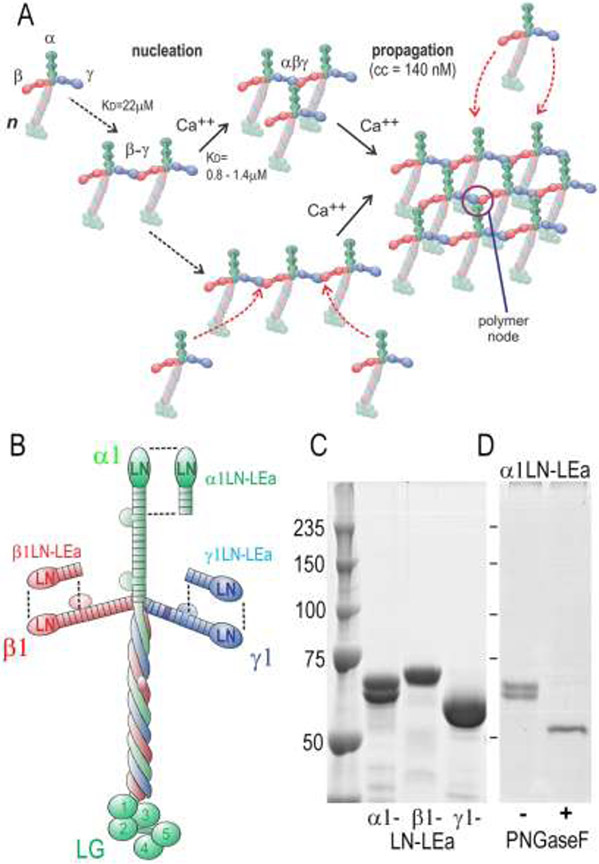
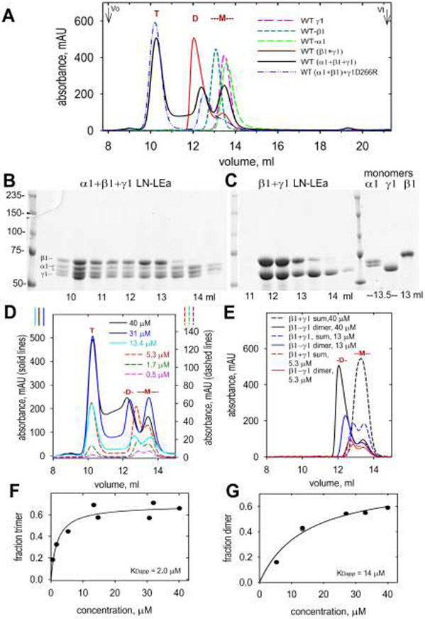
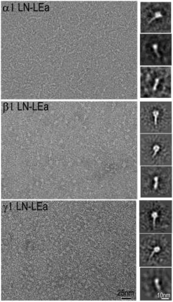
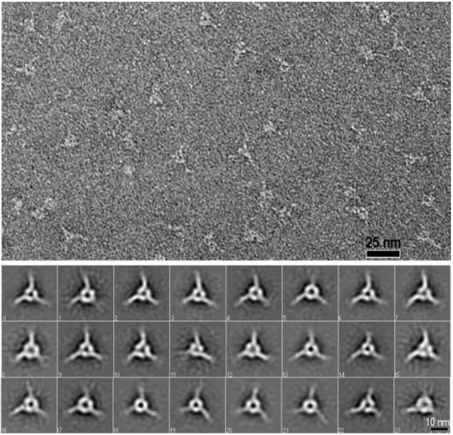
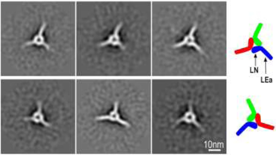
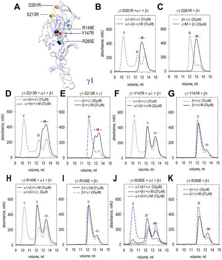
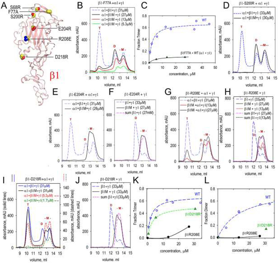
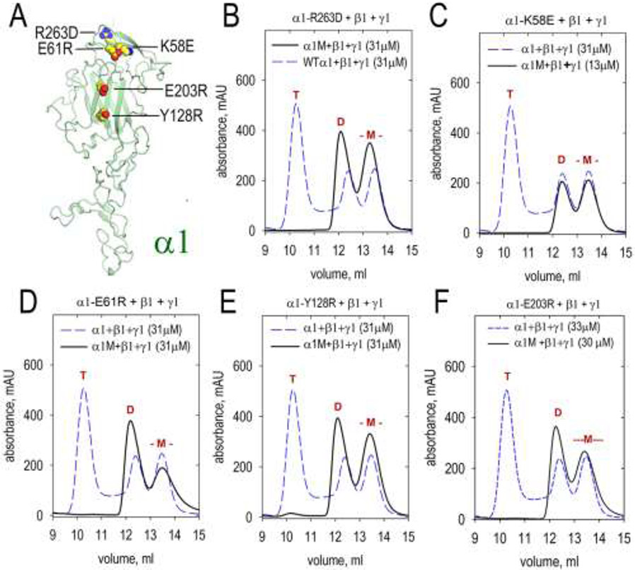
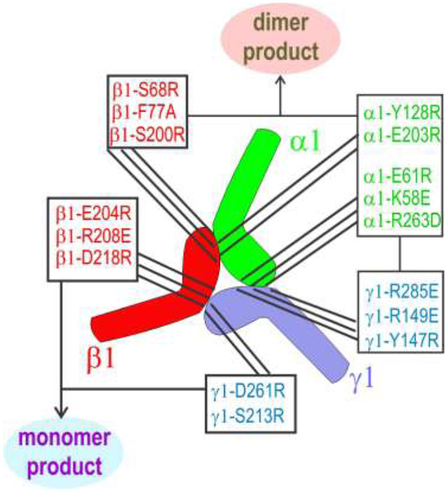
Laminin polymerization is a key step of basement membrane assembly that depends on the binding of α, β and γ N-terminal LN domains to form a polymer node. Nodal assembly can be divided into two steps consisting of β- and γ-LN dimerization followed by calcium-dependent addition of the α-LN domain. The assembly and structural organization of laminin-111 LN-LEa segments was examined by size-exclusion chromatography (SEC) and electron microscopy. Triskelion-like structures were observed in negatively-stained images of purified α1/β1/γ1 LN-LEa trimers. Image averaging of these revealed a heel-to-toe organization of the LN domains with angled outward projections of the LEa stem-like domains. A series of single-amino acid substitutions was introduced into the polymerization faces of the α1, β1 and γ1 LN domains followed by SEC analysis to distinguish between loss of β-γ mediated dimerization and loss of α-dependent trimerization (with intact β-γ dimers). Dimer-blocking mutations were confined to the γ1-toe and the β1-heel, whereas the trimer-only-blocking mutations mapped to the γ1-heel, β1-toe and the α1-toe and heel. Thus, in the polymer node the γ1-toe pairs with the β1-heel, the β1-toe pairs with the α1-heel, and the α1-toe pairs with the γ1-heel.

Keywords: Basement membrane; image averaging; ln mutations; self-assembly; triskelion.


Figures

Figure 1.
Figure 1.. Laminin-111 assembly, protein domains and LN-LEa segments.
A. Model of laminin polymerization based on previous studies. Laminin first assembles into a β-γ dimer through LN domain interactions. The α-LN domain of a third laminin molecule binds to the β-γ dimer in a calcium-dependent interaction, forming the polymer node. Additional laminin molecules bind to the initial complex, forming a sheet-like polymer (cc, critical concentration of polymerization). B. Domain organization of laminin-111 with corresponding LN-LEa segments. C. Coomassie Blue-stained gel (SDS-PAGE, 7.5%, reduced) of recombinant. α1-, β1- and γ1-LN-LEa glycoproteins. D. PNGaseF de-glycosylation of α1-LN-LEa. After enzyme treatment, a single band, migrating at 55 kDa, is seen.
Figure 2.
Figure 2.. Size-exclusion chromatography of WT LN-LEa monomers, dimers and trimers.
A. α1-LN-LEa, β1-LN-LEa and γ1-LN-LEa were analyzed by SEC (1 ml/min flow rate) each alone, following incubation of the β1 and γ1 components, or following incubation of the α1, β1 and γ1 components together, at 27°C for 1 hr. Monomer (-M-), dimer (D) and trimer (T) elution positions are marked. B, C. Aliquots of 0.5 ml fractions were analyzed by SDS-PAGE (7.5%, reducing). Gels were stained with Coomassie Blue. These confirmed the monomer, dimer and trimer assignments. D, E. SEC profiles for trimer (D) and dimer (E) fractions shown for different initial concentrations (three lower concentration plots use right-sided y-axis values). The “sum” is the arithmetic sum of β1 and γ1 monomers. F, G. Concentration-dependency for trimer and dimer assembly (data points were fitted for simple binding).
Figure 3.
Figure 3.. Negative-stain images of α1-, β1- and γ1-LN-LEa monomers.
Representative micrograph field shown in left panels. Class-averaged images shown in right side panels. Note the monomer consist of an oblong globule attached to a narrow stem-like structure. The oblong globules were noted to be positioned as wider and narrower presentations, thought to correspond to the known flat shape as determined by crystallography [26, 47].
Fig. 4.
Fig. 4.. Negative-stain images of LN-LEa trimer complexes.
Top panel: Negative stain electron micrograph of a SEC-purified trimer fraction resulting from the incubation of γ1-D266R-LN-LEa with WT α1- and β1-LN-LEa (60,000 x magnification). Fields contained trimeric particles with narrow projections from the corners and central clearing. Bottom panels. Full set of class averages derived from analysis of 11,721 particles (74x74 pixel box size) that reveal the triskelion consists of three segments that contact each other. Each segment consists of a foot-like inner portion in a heel-to-toe arrangement attached to rod-like ankle projecting out.
Figure 5.
Figure 5.. LN orientations.
Triskelions can be arranged in either a clockwise heel-to-toe (upper row) or a counter-clockwise (lower row) orientation. This suggests that these structures are flat, lying either on one side or the other on the carbon support film.
Fig. 6.
Fig. 6.. SEC analysis of dimer and trimer assembly involving γ1-LN-LEa mutants.
A. The front face of the γ1 LN domain was modified with the indicated single amino acid substitutions. The mutated γ1-LN-LEa proteins (labeled “γ1M” in graphs) were analyzed by SEC in combination with WT α1-LN-LEa and β1-LN-LEa or with β1-LN-LEa alone to determine whether trimer or dimer assembly was altered. Elution volumes of trimers (T), dimers (D) and monomers (-M-) marked in red above peaks. The mutated γ1 monomers eluted at the same volume as WT γ1 monomer. B, C. γ1-D261R produced neither dimer nor trimer. D, E. γ1-S213R produced neither dimer nor trimer. F, G. γ1-Y147R produced dimer but no trimer. H, I. γ1-R149E produced dimer but no trimer. J, K. γ1-R285E produced dimer but no trimer.
Fig. 7.
Fig. 7.. SEC analysis of dimer and trimer assembly involving β1-LN-LEa mutants.
A. The front face of the β1 LN domain was modified with the indicated single amino acid substitutions. The mutated β1 proteins (labeled “β1M” in graphs) were analyzed by SEC in combination with WT α1-LN-LEa and γ1-LN-LEa or with γ1-LN-LEa alone to determine whether trimer or dimer assembly was altered. Elution volumes of trimers (T), dimers (D) and monomers (-M-) marked in red above peaks. The mutated β1 monomers eluted at the same volume as WT β1 monomer. B. β1-F77A produced dimer and small amounts of trimer. C. Concentration-dependency of trimer containing β1-F77A (the WT data are the same as in Fig. 2F). D. β1-S200R produced dimer but no trimer. E, F. β1-E204R produced neither dimer nor trimer. G, H. β1-R208E produced little or no dimer and small amounts of trimer. I, J. β1-D218R produced very little dimer and reduced amounts of trimer. In (F), (H) and (J), the arithmetic sums of β1 and γ1 monomers are shown for comparison. K,L. Concentration-dependency of trimer and dimer containing β1-R208E and β1-D218R (the WT data are the same as in Fig. 2F).
Fig. 8.
Fig. 8.. SEC analysis of dimer and trimer assembly involving β1-LN-LEa mutants.
A. The front face of the β1 LN domain was modified with the indicated single amino acid substitutions. The mutated β1 proteins (labeled “β1M” in graphs) were analyzed by SEC in combination with WT β1-LN-LEa and γ1-LN-LEa to determine whether trimer assembly was altered. Elution volumes of trimers (T), dimers (D) and monomers (-M-) marked in red above peaks. The mutated α1 monomers eluted at the same volume as WT α1 monomer. All mutants produced dimer but no trimer: B. α1-R263D C. α1-R263L D. α1-E61R. E. α1-Y128R F. α1-E203R.
Fig. 9.
Fig. 9.. Summary of mutations that prevented or greatly reduced trimer or dimer assembly.
The LN-LEa segments of the laminin α1, β1 and γ1 chains have the shape of a foot, with the toes corresponding to the tip of the LN domain and the heel corresponding to the LN region near the juncture with the first LEa domain. Modifications in the γ1-toe and the β1-heel produce only monomers. All other modifications produce β1-γ1 dimers but no trimers. From this it is deduced that the γ1-toe bind to the β1-heel, the γ1-heel binds to the α1-toe, and the β1-toe binds to the α1-heel. This pseudo-symmetric arrangement places the front faces of the three LN domains in the center of the triskelion, with polymerization-critical residues facing inward towards each other.

References

    1. Yurchenco PD, Basement membranes: cell scaffoldings and signaling platforms, Cold Spring Harb Perspect Biol 3(2) (2011) a004911. - PMC - PubMed
    1. Pozzi A, Yurchenco PD, Iozzo RV, The nature and biology of basement membranes, Matrix Biol 57-58 (2017) 1–11. - PMC - PubMed
    1. Morrissey MA, Sherwood DR, An active role for basement membrane assembly and modification in tissue sculpting, J Cell Sci 128(9) (2015) 1661–8. - PMC - PubMed
    1. Ramos-Lewis W, Page-McCaw A, Basement membrane mechanics shape development: Lessons from the fly, Matrix Biol 75-76 (2019) 72–81. - PMC - PubMed
    1. Hohenester E, Yurchenco PD, Laminins in basement membrane assembly, Cell Adh Migr 7(1) (2013) 56–63. - PMC - PubMed

Publication types

LinkOut - more resources