Review
. 2021 Jul:98:102220.
doi: 10.1016/j.ctrv.2021.102220. Epub 2021 May 11.

Coordination and optimization of FDG PET/CT and COVID-19 vaccination; Lessons learned in the early stages of mass vaccination

Affiliations
Review

Coordination and optimization of FDG PET/CT and COVID-19 vaccination; Lessons learned in the early stages of mass vaccination

Lacey J McIntosh et al. Cancer Treat Rev. 2021 Jul.

Abstract

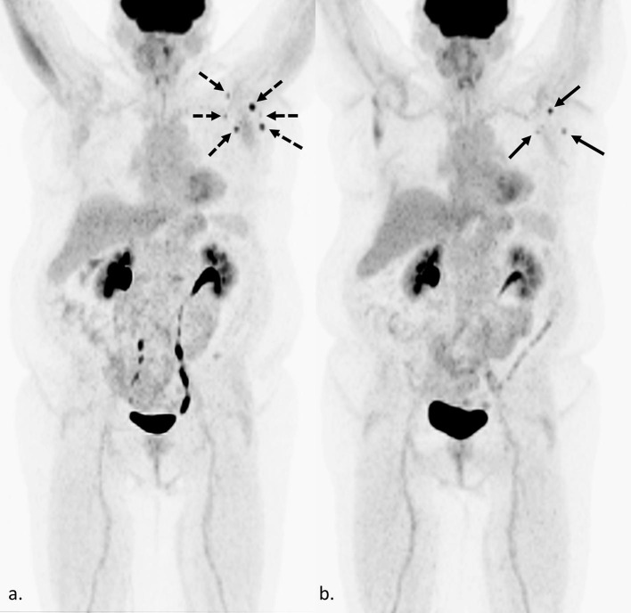
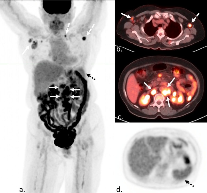
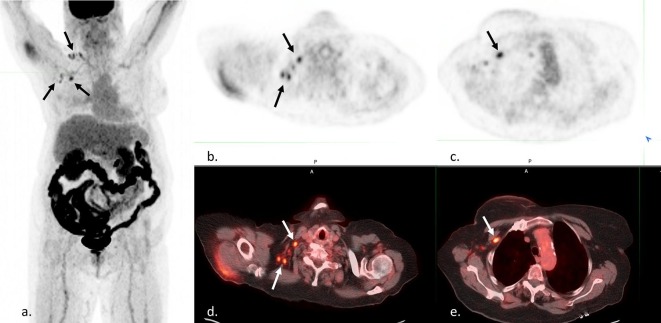
As the world embarks on mass vaccination for COVID-19, we are beginning to encounter unintended dilemmas in imaging oncology patients; particularly with regards to FDG PET/CT. In some cases, vaccine-related lymphadenopathy and FDG uptake on PET/CT can mimic cancer and lead to confounding imaging results. These cases where findings overlap with cancer pose a significant dilemma for diagnostic purposes, follow-up, and management leading to possible treatment delays, unnecessary repeat imaging and sampling, and patient anxiety. These cases can largely be avoided by optimal coordination between vaccination and planned imaging as well as preemptive selection of vaccine administration site. This coordination hinges on patient, oncologist, and radiologists' awareness of this issue and collaboration. Through close communication and patient education, we believe this will eliminate significant challenges for our oncology patients as we strive to end this pandemic.

Keywords: COVID-19 vaccine; FDG PET/CT; Oncologic imaging.


Figures

Fig. 1
Fig. 1
Confounding findings on FDG PET/CT in a 57-year-old woman with metastatic breast cancer 4 weeks after Moderna COVID-19 vaccination. Maximum intensity projection (MIP) (a.) shows an increase in size, number, and degree of FDG uptake in left axillary lymph nodes (dashed black arrows), compared to the previous FDG PET/CT performed 6 months prior (b. black arrows). As this is her only site of metastatic disease, and she received the Moderna vaccine 4 weeks prior, it is unclear if the increase is due to recent vaccination or an increase in her tumor burden. In this case, an increase in tumor burden was favored based on trends of prior imaging and tumor markers.
Fig. 2
Fig. 2
Confounding findings on FDG PET/CT in a 51 year-old man with new Hodgkin’s lymphoma after recent vaccination. MIP (a), axial fusion (b and c), and axial attenuation correction (d) images demonstrate FDG avid lymphadenopathy above and below the diaphragm (white arrows). It is unclear if the left axillary and supraclavicular uptake (white dashed arrows) are from a recent COVID-19 vaccination to the left deltoid three weeks prior, but would not result in a change in stage. However, increased uptake in the spleen (black dashed arrows), more avid than liver, could be due to either vaccination or disease involvement and would result in a change in stage.
Fig. 3
Fig. 3
Confounding findings on FDG PET/CT in a 73 year-old woman with history of breast cancer to evaluate for recurrence. MIP (a), axial PET (b and c), and axial fusion (d and e) images demonstrate FDG-avid right axillary and supraclavicular lymph nodes (black and white arrows). This patient reported a recent Moderna COVID-19 vaccination in the left deltoid 5 days prior at a mass vaccination site. No records about vaccine administration were available to the reading radiologist or primary care physician. Initially it was unclear if the findings represented a recurrence of a right breast cancer or incorrect reporting/recording of vaccination side. Ultimately, the patient was able to be reached, and a right-sided vaccination was confirmed.
Fig. 4
Fig. 4
Confounding findings on FDG PET/CT in a 43 year-old man with lymphadenopathy and suspected diagnosis of lymphoma. MIP (a), and axial fusion (b, c, and d) FDG PET/CT shows an FDG avid retroperitoneal mass (white arrows), internal mammary lymphadenopathy (white dashed arrows), and left greater than right axillary lymphadenopathy (black arrows). This patient reported receiving a Moderna COVID-19 vaccine 1 week prior to the left deltoid. It is unclear if the findings in the left axilla are related to recent vaccination or lymphoma. While these findings would not change disease stage, this site should be avoided for biopsy due to potential confounding findings, and another more technically challenging and invasive site must be considered for highest yield sampling of representative disease.

References

    1. Shirone N., Shinkai T., Yamane T., et al. Axillary lymph node accumulation on FDG-PET/CT after influenza vaccination. Ann Nucl Med. 2012;26:248–252. doi: 10.1007/s12149-011-0568-x. - DOI - PubMed
    1. Thomassen A., Lerberg Nielsen A., Gerke O., et al. Duration of 18F-FDG avidity in lymph nodes after pandemic H1N1v and seasonal influenza vaccination. Eur J Nucl Med Mol Imaging. 2011;38:894–898. doi: 10.1007/s00259-011-1729-9. - DOI - PubMed
    1. Panagiotidis E., Exarhos D., Housianakou, et al. FDG uptake in axillary lymph nodes after vaccination against pandemic (H1N1) Eur Radiol. 2010;20:1251–1253. doi: 10.1007/s00330-010-1719-5. - DOI - PubMed
    1. Coates E.E., Costner P.J., Nason M.C., et al. Lymph node activation by PET/CT following vaccination with licensed vaccines for human papilloma viruses. Clin Nucl Med. 2017;42(5):329–334. doi: 10.1097/RLU.0000000000001603. - DOI - PubMed
    1. Burger I.A., Husmann L., Hany T.F., Schmid D.T., Schaefer N.G. Incidence and intensity of F-18 FDG uptake after vaccination with H1N1 vaccine. Clin Nucl Med. 2011;36(10):848–853. - PubMed

MeSH terms