

Regulation of TRPML1 channel activity and inflammatory exosome release by endogenously produced reactive oxygen species in mouse podocytes
- PMID: 34030116
- PMCID: PMC8163985
- DOI: 10.1016/j.redox.2021.102013
Regulation of TRPML1 channel activity and inflammatory exosome release by endogenously produced reactive oxygen species in mouse podocytes
Abstract
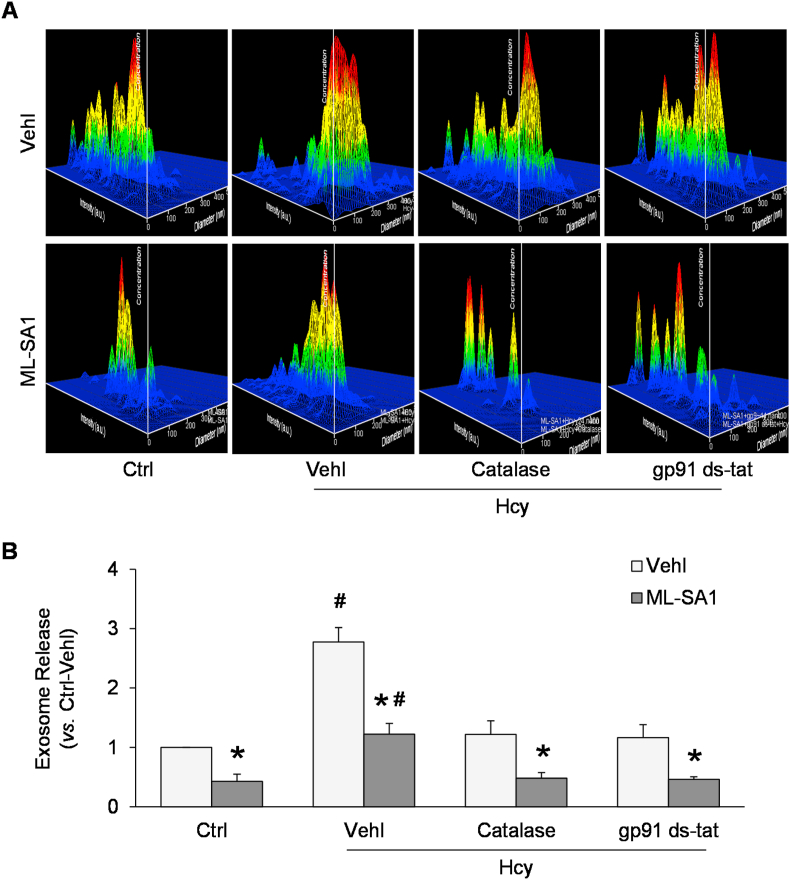
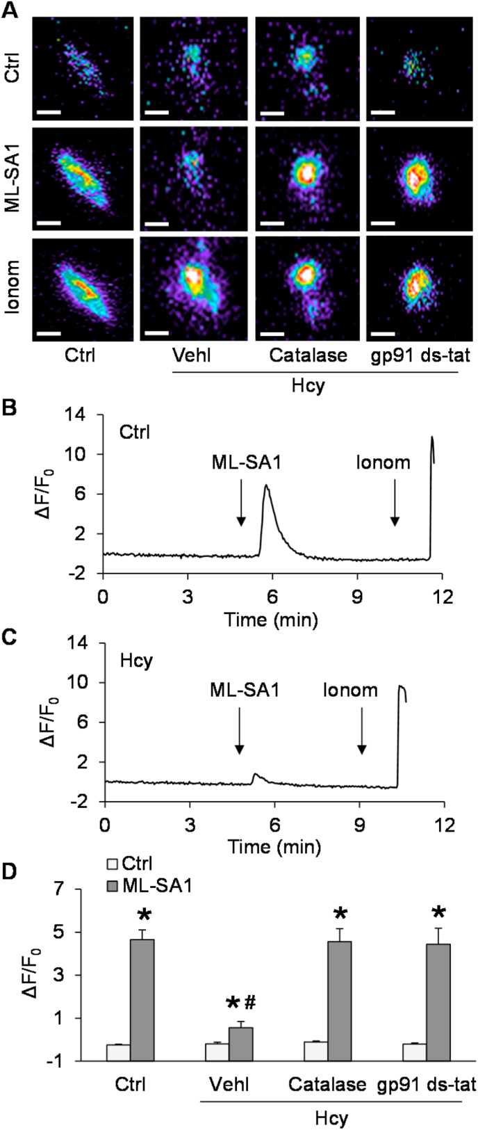
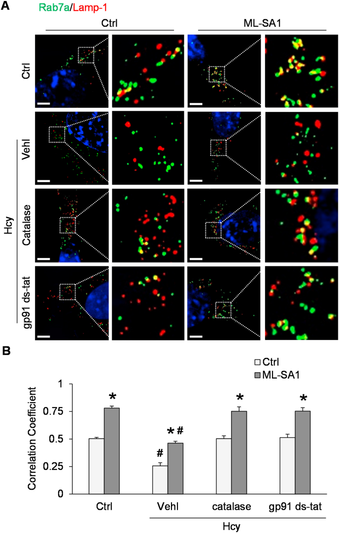
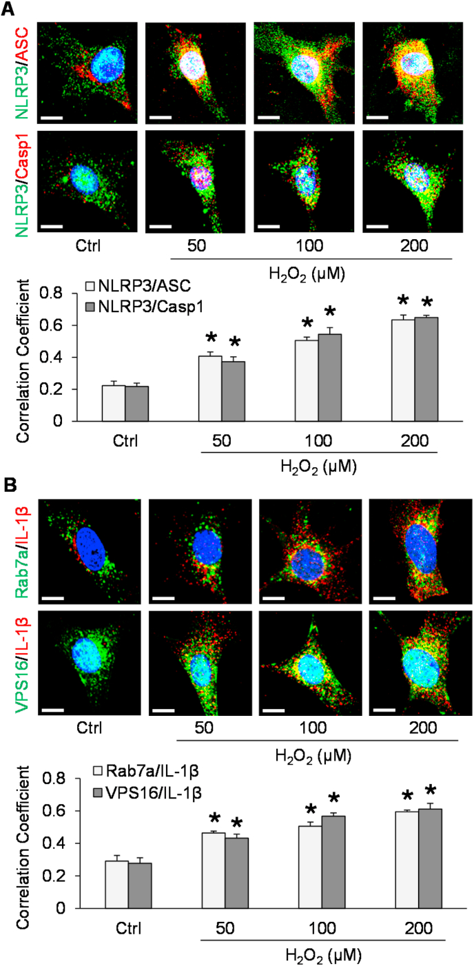
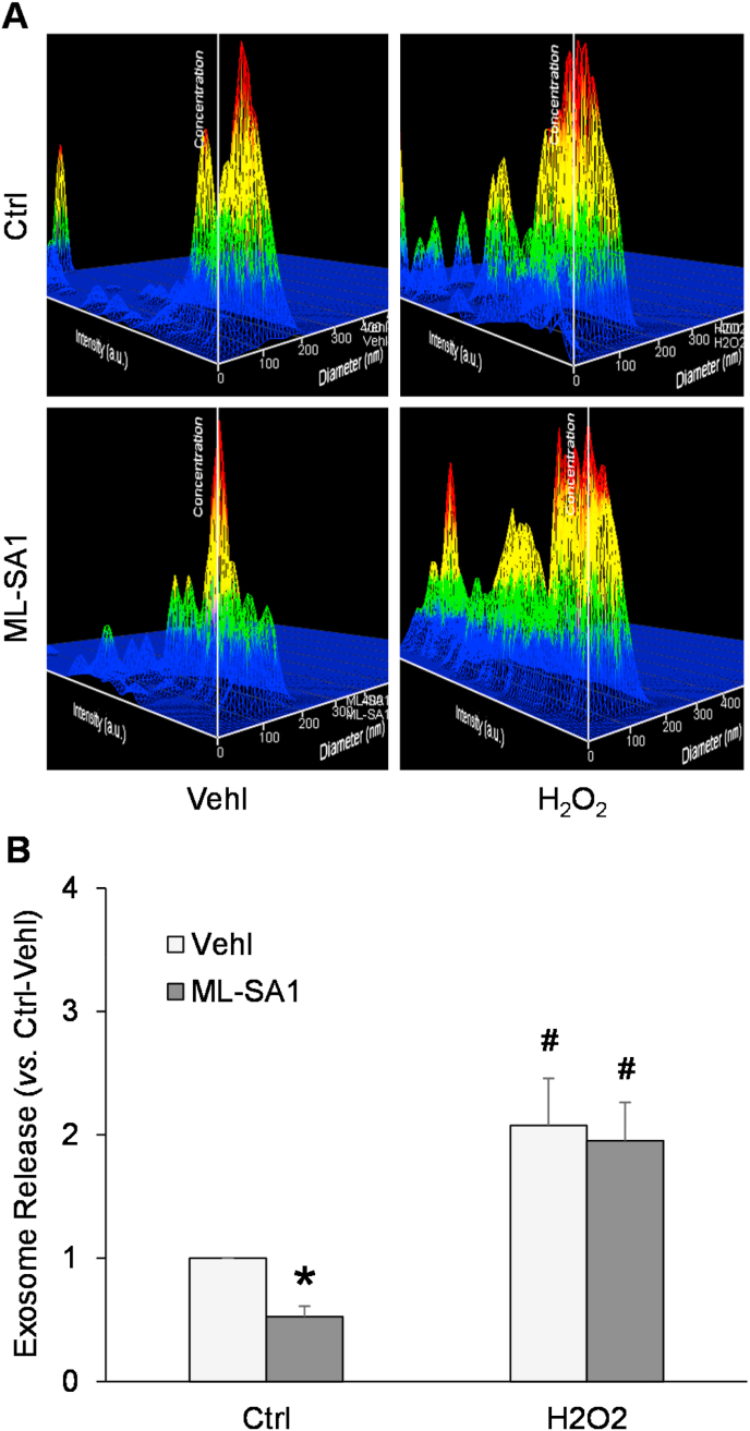
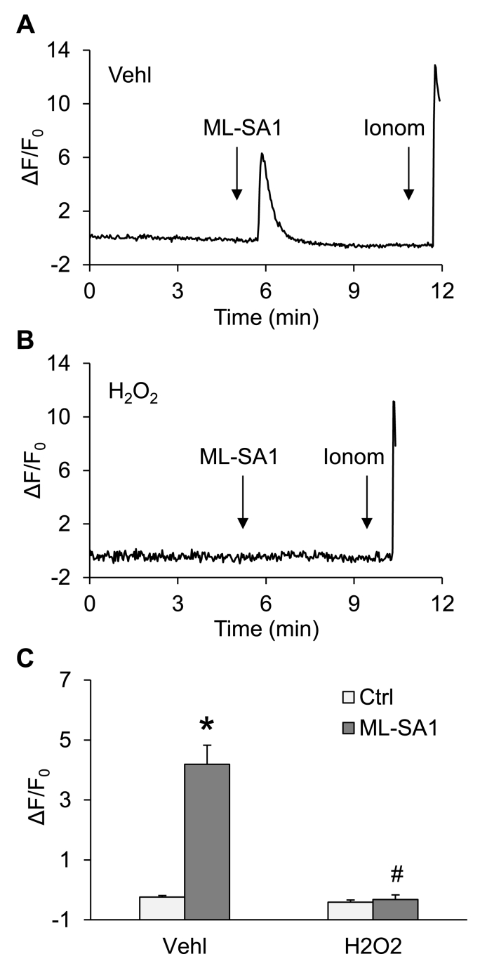
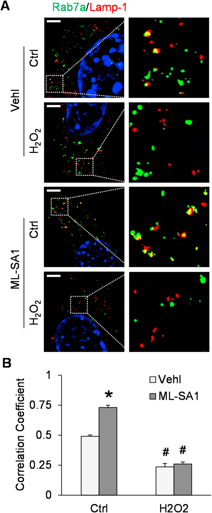
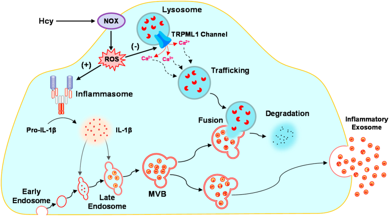
The nucleotide-binding oligomerization domain-like receptor containing pyrin domain 3 (NLRP3) inflammasome in podocytes has been implicated in the initiation of glomerular inflammation during hyperhomocysteinemia (hHcy). However, the mechanism by which NLRP3 inflammasome products are released from podocytes remains unknown. The present study tested whether exosome secretion from podocytes is enhanced by NADPH oxidase-produced reactive oxygen species (ROS), which may serve as a pathogenic mechanism mediating the release of inflammatory cytokines produced by the NLRP3 inflammasome in podocytes after Hcy stimulation. We first demonstrated the remarkable elevation of endogenously produced ROS in podocytes treated with Hcy compared with control podocytes, which was abolished by pre-treatment with the NADPH oxidase inhibitors, gp91 ds-tat peptide and diphenyleneiodonium (DPI). In addition, Hcy induced activation in podocytes of NLRP3 inflammasomes and the formation of multivesicular bodies (MVBs) containing inflammatory cytokines, which were prevented by treatment with gp91 ds-tat or the ROS scavenger, catalase. Given the importance of the transient receptor potential mucolipin 1 (TRPML1) channel in Ca2+-dependent lysosome trafficking and consequent lysosome-MVB interaction, we tested whether lysosomal Ca2+ release through TRPML1 channels is inhibited by endogenously produced ROS in podocytes after Hcy stimulation. By GCaMP3 Ca2+ imaging, we confirmed the inhibition of TRPML1 channel activity by Hcy which was remarkably ameliorated by catalase and gp91 ds-tat peptide. By structured illumination microscopy (SIM) and nanoparticle tracking analysis (NTA), we found that ML-SA1, a TRPML1 channel agonist, significantly enhanced lysosome-MVB interaction and reduced exosome release in podocytes, which were attenuated by Hcy. Pre-treatment of podocytes with catalase or gp91 ds-tat peptide restored ML-SA1-induced changes in lysosome-MVB interaction and exosome secretion. Moreover, we found that hydrogen peroxide (H2O2) mimicked the effect of Hcy on TRPML1 channel activity, lysosome-MVB interaction, and exosome secretion in podocytes. Based on these results, we conclude that endogenously produced ROS importantly contributes to inflammatory exosome secretion from podocytes through inhibition of TRPML1 channel activity, which may contribute to the initiation of glomerular inflammation during hHcy.
Keywords: Exosome; Homocysteine; Lysosome; Podocyte; Reactive oxygen species; TRPML1 channel.
Copyright © 2021 The Authors. Published by Elsevier B.V. All rights reserved.
Conflict of interest statement
None of the authors have conflict of interest.
Figures


Similar articles
-
Exosome Biogenesis and Lysosome Function Determine Podocyte Exosome Release and Glomerular Inflammatory Response during Hyperhomocysteinemia.Am J Pathol. 2022 Jan;192(1):43-55. doi: 10.1016/j.ajpath.2021.10.005. Epub 2021 Oct 27. Am J Pathol. 2022. PMID: 34717894 Free PMC article.
-
Abnormal podocyte TRPML1 channel activity and exosome release in mice with podocyte-specific Asah1 gene deletion.Biochim Biophys Acta Mol Cell Biol Lipids. 2021 Feb;1866(2):158856. doi: 10.1016/j.bbalip.2020.158856. Epub 2020 Nov 19. Biochim Biophys Acta Mol Cell Biol Lipids. 2021. PMID: 33221496 Free PMC article.
-
Podocyte-specific silencing of acid sphingomyelinase gene to abrogate hyperhomocysteinemia-induced NLRP3 inflammasome activation and glomerular inflammation.Am J Physiol Renal Physiol. 2024 Jun 1;326(6):F988-F1003. doi: 10.1152/ajprenal.00195.2023. Epub 2024 Apr 18. Am J Physiol Renal Physiol. 2024. PMID: 38634138 Free PMC article.
-
Inflammasome Activation in Chronic Glomerular Diseases.Curr Drug Targets. 2017;18(9):1019-1029. doi: 10.2174/1389450117666160817103435. Curr Drug Targets. 2017. PMID: 27538510 Free PMC article. Review.
-
Redox regulation of NLRP3 inflammasomes: ROS as trigger or effector?Antioxid Redox Signal. 2015 May 1;22(13):1111-29. doi: 10.1089/ars.2014.5994. Epub 2015 Jan 19. Antioxid Redox Signal. 2015. PMID: 25330206 Free PMC article. Review.
Cited by
-
Uncovering the role of transient receptor potential channels in pterygium: a machine learning approach.Inflamm Res. 2023 Mar;72(3):589-602. doi: 10.1007/s00011-023-01693-4. Epub 2023 Jan 24. Inflamm Res. 2023. PMID: 36692516
-
Impaired autophagic flux and dedifferentiation in podocytes lacking Asah1 gene: Role of lysosomal TRPML1 channel.Biochim Biophys Acta Mol Cell Res. 2023 Jan;1870(1):119386. doi: 10.1016/j.bbamcr.2022.119386. Epub 2022 Oct 24. Biochim Biophys Acta Mol Cell Res. 2023. PMID: 36302466 Free PMC article.
-
The pathological role of damaged organelles in renal tubular epithelial cells in the progression of acute kidney injury.Cell Death Discov. 2022 May 2;8(1):239. doi: 10.1038/s41420-022-01034-0. Cell Death Discov. 2022. PMID: 35501332 Free PMC article. Review.
-
Exosome Biogenesis and Lysosome Function Determine Podocyte Exosome Release and Glomerular Inflammatory Response during Hyperhomocysteinemia.Am J Pathol. 2022 Jan;192(1):43-55. doi: 10.1016/j.ajpath.2021.10.005. Epub 2021 Oct 27. Am J Pathol. 2022. PMID: 34717894 Free PMC article.
-
Predictors of delayed encephalopathy after acute carbon monoxide poisoning: a literature review.Front Med (Lausanne). 2025 Mar 26;12:1559264. doi: 10.3389/fmed.2025.1559264. eCollection 2025. Front Med (Lausanne). 2025. PMID: 40206479 Free PMC article. Review.
References
-
- Colombo M., Raposo G., Thery C. Biogenesis, secretion, and intercellular interactions of exosomes and other extracellular vesicles. Annu. Rev. Cell Dev. Biol. 2014;30:255–289. - PubMed
Publication types
MeSH terms
Substances
Grants and funding
LinkOut - more resources
Full Text Sources
Other Literature Sources
Miscellaneous
