. 2021 Jul:43:102008.
doi: 10.1016/j.redox.2021.102008. Epub 2021 May 13.

Analysis of genes regulated by DUX4 via oxidative stress reveals potential therapeutic targets for treatment of facioscapulohumeral dystrophy

Affiliations

Analysis of genes regulated by DUX4 via oxidative stress reveals potential therapeutic targets for treatment of facioscapulohumeral dystrophy

Anna Karpukhina et al. Redox Biol. 2021 Jul.

Abstract

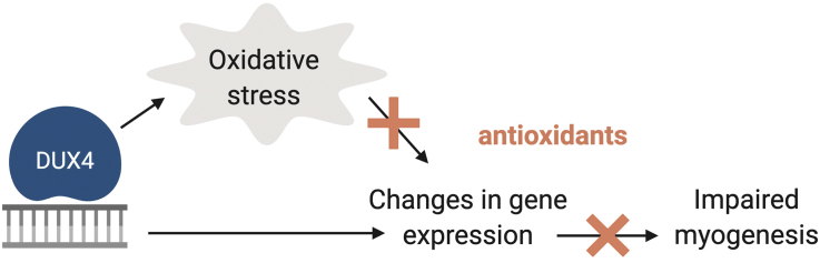
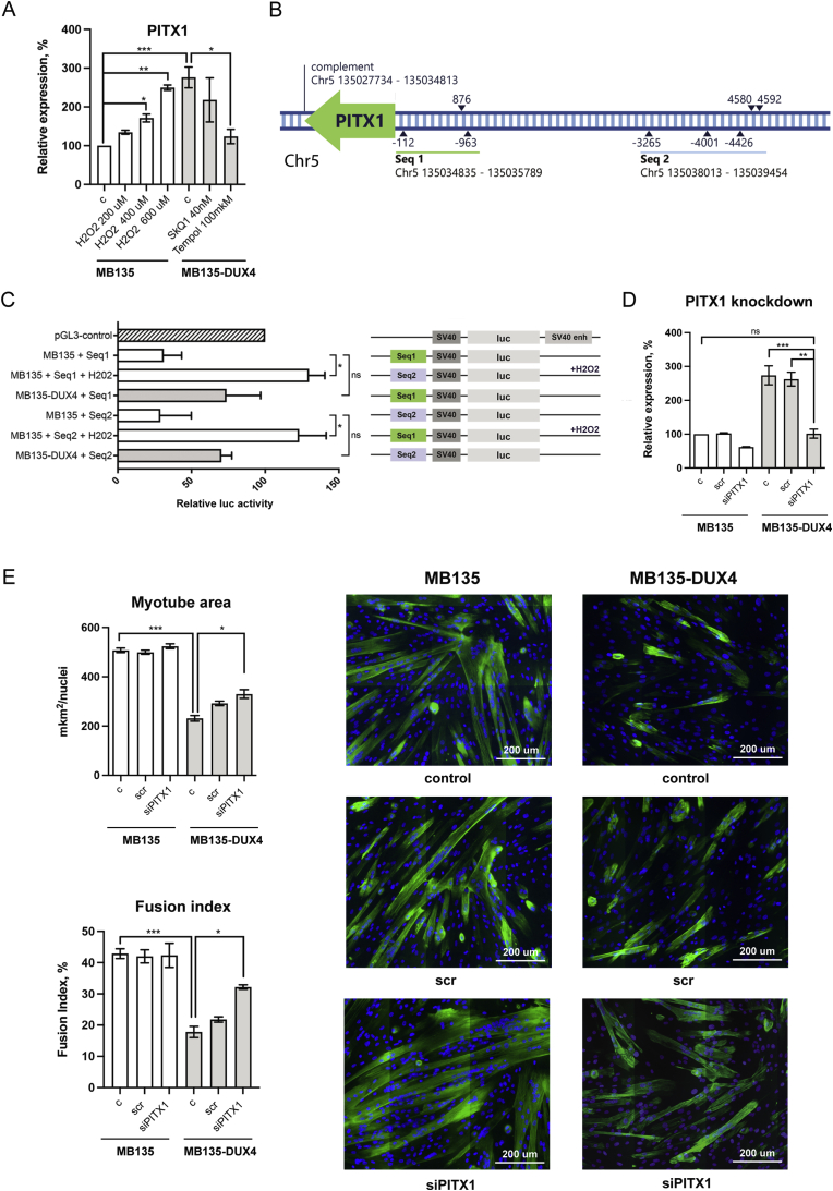
Muscles of patients with facioscapulohumeral dystrophy (FSHD) are characterized by sporadic DUX4 expression and oxidative stress which is at least partially induced by DUX4 protein. Nevertheless, targeting oxidative stress with antioxidants has a limited impact on FSHD patients, and the exact role of oxidative stress in the pathology of FSHD, as well as its interplay with the DUX4 expression, remain unclear. Here we set up a screen for genes that are upregulated by DUX4 via oxidative stress with the aim to target these genes rather than the oxidative stress itself. Immortalized human myoblasts expressing DUX4 (MB135-DUX4) have an increased level of reactive oxygen species (ROS) and exhibit differentiation defects which can be reduced by treating the cells with classic (Tempol) or mitochondria-targeted antioxidants (SkQ1). The transcriptome analysis of antioxidant-treated MB135 and MB135-DUX4 myoblasts allowed us to identify 200 genes with expression deregulated by DUX4 but normalized upon antioxidant treatment. Several of these genes, including PITX1, have been already associated with FSHD and/or muscle differentiation. We confirmed that PITX1 was indeed deregulated in MB135-DUX4 cells and primary FSHD myoblasts and revealed a redox component in PITX1 regulation. PITX1 silencing partially reversed the differentiation defects of MB135-DUX4 myoblasts. Our approach can be used to identify and target redox-dependent genes involved in human diseases.

Keywords: DUX4; FSHD; Mitochondrial ROS; Muscle differentiation; Oxidative stress; PITX1.


Conflict of interest statement

Declare that they have no conflict of interests.

Figures

Image 1
Graphical abstract
Fig. 1
Fig. 1
Oxidative stress and differentiation defects in MB135-DUX myoblasts. (A) ROS production in MB135 and MB135-DUX4 cells, treated or non-treated with antioxidants. Cells were incubated in the presence of 40 nM SkQ1 or 100 uM Tempol for four days and then stained with a ROS indicator CM-H2DCFD for 30 min. CM-H2DCFD fluorescence was detected by flow cytometry. The data are presented as means ± SEM, N = 5–16. Mean value for the untreated MB135-DUX4 cells is set to 100%. (B) Cardiolipin peroxidation in MB135 and MB135-DUX4 cells, treated or non-treated with antioxidants. Cells were incubated in the presence of 40 nM SkQ1 or 100 uM Tempol for four days and stained with MitoCLox for 5 h. MitoCLox fluorescence in the FITC and PE channels was then detected by flow cytometry. The data are presented as mean ± SEM, N = 4–6. The representative histograms show the fluorescence of MitoCLox-stained cells in the FITC and PE channels. The rightmost peak in the FITC channel corresponds to the cells with the oxidized MitoCLox. (C) Analysis of myotubes formed by MB135 and MB135-DUX4 myoblasts after four days of differentiation. Cells were treated with H202 for four days and then seeded on glass coverslips in 100% confluence. Myogenic differentiation was induced the next day by serum starvation and the cells were left to differentiate for four days. The myotubes were stained with monoclonal antibodies against skeletal troponin T (green) and DAPI (blue). To create a large image of the specimen, the images from adjacent fields were captured and stitched together using the Cartograph software (Microvision). Differentiation efficiency was assessed by measuring the troponin T - positive area normalized to the number of nuclei and the fusion index in 5–6 large images per sample. The fusion index is defined as the percentage of nuclei residing inside the troponin T-positive area. The data are presented as mean ± SEM, N = 3–5. (D) Apoptosis assessment in MB135 and MB135-DUX4 myoblasts treated with H202. Cells were treated with 200 or 400 uM H202 for four days, stained with Annexin V - FITC and PI and analyzed by flow cytometry. The percentage of viable (annexin V -, PI -), early apoptotic (annexin V +, PI -), late apoptotic (annexin V +, PI +) and necrotic (annexin V -, PI +) calls was calculated, N = 3. (For interpretation of the references to color in this figure legend, the reader is referred to the Web version of this article.)
Fig. 2
Fig. 2
Antioxidant pre-treatment improves differentiation of MB135-DUX4 myoblasts. (A) Myotubes formed by MB135 and MB135-DUX4 myoblasts pre-treated with antioxidants before differentiation induction. Representative myotubes are marked with white errors. Cells were treated with 40 nM SkQ1 or 100 uM Tempol for four days and then seeded in six-well plates in 100% confluence. The next day differentiation was induced by serum starvation and the cells were left to differentiate for two days. The myotubes were stained with May-Grunwald Giemsa histological dye and 5 microscopic fields per specimen were captured. The differentiation efficiency was assessed by measuring the areas of myotubes in each of the 5 fields captured for each sample. The data is presented as mean myotube area ±SEM, N = 3. (B) Myotubes formed by MB135-DUX4 myoblasts treated with antioxidants during differentiation. The non-treated cells were seeded in 6-well plates in 100% confluence and induced to differentiate the next day. Antioxidants (40 nM SkQ1 or 100 uM Tempol) were added directly to the differentiation medium. The cells were left to differentiate for two days and then stained and analyzed as in (A). The data is presented as mean myotube area ±SEM, N = 3.
Fig. 3
Fig. 3
Identification of genes deregulated by DUX4 via oxidative stress. (A) Volcano plot and heatmap for the genes differentially expressed between MB135-DUX4 and MB135 myoblasts. Experiments were performed in biological duplicates. The genes with padj <0.01 and |log2Fold| > 1 were considered significant. The heatmap shows 50 significant genes with the lowest padj. The color scale represents relative expression levels as row mean-centered rlog normalized counts. Hierarchical clustering was performed using Euclidean distance. The volcano plot shows all the genes differentially expressed between MB135-DUX4 and MB135, with the genes passing the significance threshold of padj < 0.01 colored blue (log2Fold < −1, downregulated) or orange (log2Fold > 1, upregulated). Darker colors correspond to lower padj values. (B) Schematic representation of the DUX4-driven gene expression deregulation and the venn diagram showing the numbers of differentially expressed genes in different comparisons (with padj < 0.01 and |log2Fold| > 1). 200 genes that were differentially expressed between MB135 and MB135-DUX4, but not between MB135 and MB135-DUX4 treated with SkQ1 or Tempol, were considered antioxidant-sensitive. (C) Gene Ontology Slim annotation of the 200 genes falling into the antioxidant-sensitive group. (D) Gene Ontology overrepresentation analysis of the 200 genes falling into the antioxidant-sensitive group. (E) Heatmap for 10 selected differentially expressed genes from the antioxidant-sensitive group. The genes are significantly differentially expressed between MB135 and MB135-DUX4 cells (with padj < 0.01 and |log2Fold| > 1), but not between the MB135 and MB135-DUX4 cells treated with antioxidants. The color scale represents relative expression levels as row mean-centered rlog normalized counts. Hierarchical clustering was performed using Euclidean distance. (For interpretation of the references to color in this figure legend, the reader is referred to the Web version of this article.)
Fig. 4
Fig. 4
PITX1 expression is redox-sensitive and can be silenced to improve the differentiation of MB135-DUX4 myoblasts. (A)PITX1 expression assessed by RT-qPCR. Myoblasts were treated with 40 nM SkQ1 or 100 uM Tempol for four days or 200–600 uM H2O2 for 24 h. The data are represented as mean relative expression ± SEM, N = 4–10. Mean value for the MB135 cells is set to 100%. (B) Regions upstream of PITX1 contain potential ARE sequences. Blue triangles mark potential AREs, numbers indicate the distances in base pairs from the start of PITX1 gene. Seq1 and Seq2 are the sequences used for reporter assays. Genomic coordinates are for GRCh38. (C) Dual luciferase reporter assay for enhancer activity of potential ARE-containing sequences. Seq1 and Seq2 sequences were cloned into pGL3 luciferase reporter plasmid upstream of the SV40 promoter. MB135 and MB135-DUX4 cells were treated with 400 uM H202 for 24 h and then transfected with the pGL3-Seq1/Seq2 plasmids and pCMV-Red Firefly Luc plasmid, used as a as a normalization control. The data are presented as mean relative luciferase activity ± SEM, mean luciferase activity in the cells transfected with a pGL3 control vector, containing an SV40 enhancer, is set to 100%. N = 4–5. (D) PITX1 expression four days after siRNA transfection. Myoblasts were seeded on six-well plates (2 × 105 cells/well) and transfected with scr siRNA (negative control) or siRNA against PITX1 the next day. The cells were left to proliferate in siRNA-containing medium for 4 days and then PITX1 expression was assessed by RT-qPCR. The data are presented as mean relative expression ± SEM, mean value for non-transfected MB135 cells is set to 100%, N = 3. (E) Myotubes formed by MB135 and MB135-DUX4 myoblasts upon PITX1 siRNA knockdown. The cells were seeded and treated as described in (D). After 4 days of proliferation with siRNA, myoblasts were induced to differentiate by serum starvation. On the fourth day of differentiation, the cells were fixed and stained with monoclonal antibodies against skeletal troponin T (green) and DAPI (blue). To create a large image of the specimen, the images from adjacent fields were captured and stitched together using Cartograph software (Microvision). Differentiation efficiency was assessed by measuring the troponin T - positive area normalized to the number of nuclei and the fusion index in 5–6 large images per sample. The fusion index is defined as the percentage of nuclei residing inside the troponin T-positive area. The data are presented as mean ± SEM, N = 3. (For interpretation of the references to color in this figure legend, the reader is referred to the Web version of this article.)

Similar articles

Cited by

References

    1. Karpukhina A., Tiukacheva E., Dib C., Vassetzky Y.S. Control of DUX4 expression in facioscapulohumeral muscular dystrophy and cancer. Trends Mol. Med. 2021 doi: 10.1016/j.molmed.2021.03.008. cited 13 Apr 2021. - DOI - PubMed
    1. Jagannathan S., Shadle S.C., Resnick R., Snider L., Tawil R.N., van der Maarel S.M. Model systems of DUX4 expression recapitulate the transcriptional profile of FSHD cells. Hum. Mol. Genet. 2016;25:4419–4431. doi: 10.1093/hmg/ddw271. - DOI - PMC - PubMed
    1. Bosnakovski D., Xu Z., Gang E.J., Galindo C.L., Liu M., Simsek T. An isogenetic myoblast expression screen identifies DUX4-mediated FSHD-associated molecular pathologies. EMBO J. 2008;27:2766–2779. doi: 10.1038/emboj.2008.201. - DOI - PMC - PubMed
    1. Dmitriev P., Bou Saada Y., Dib C., Ansseau E., Barat A., Hamade A. DUX4-induced constitutive DNA damage and oxidative stress contribute to aberrant differentiation of myoblasts from FSHD patients. Free Radic. Biol. Med. 2016;99:244–258. doi: 10.1016/j.freeradbiomed.2016.08.007. - DOI - PubMed
    1. Turki A., Hayot M., Carnac G., Pillard F., Passerieux E., Bommart S. Functional muscle impairment in facioscapulohumeral muscular dystrophy is correlated with oxidative stress and mitochondrial dysfunction. Free Radic. Biol. Med. 2012;53:1068–1079. doi: 10.1016/j.freeradbiomed.2012.06.041. - DOI - PubMed

Publication types

Substances