

Cannabidiol has a unique effect on global brain activity: a pharmacological, functional MRI study in awake mice
- PMID: 34030718
- PMCID: PMC8142641
- DOI: 10.1186/s12967-021-02891-6
Cannabidiol has a unique effect on global brain activity: a pharmacological, functional MRI study in awake mice
Abstract
Background: The phytocannabinoid cannabidiol (CBD) exhibits anxiolytic activity and has been promoted as a potential treatment for post-traumatic stress disorders. How does CBD interact with the brain to alter behavior? We hypothesized that CBD would produce a dose-dependent reduction in brain activity and functional coupling in neural circuitry associated with fear and defense.
Methods: During the scanning session awake mice were given vehicle or CBD (3, 10, or 30 mg/kg I.P.) and imaged for 10 min post treatment. Mice were also treated with the 10 mg/kg dose of CBD and imaged 1 h later for resting state BOLD functional connectivity (rsFC). Imaging data were registered to a 3D MRI mouse atlas providing site-specific information on 138 different brain areas. Blood samples were collected for CBD measurements.
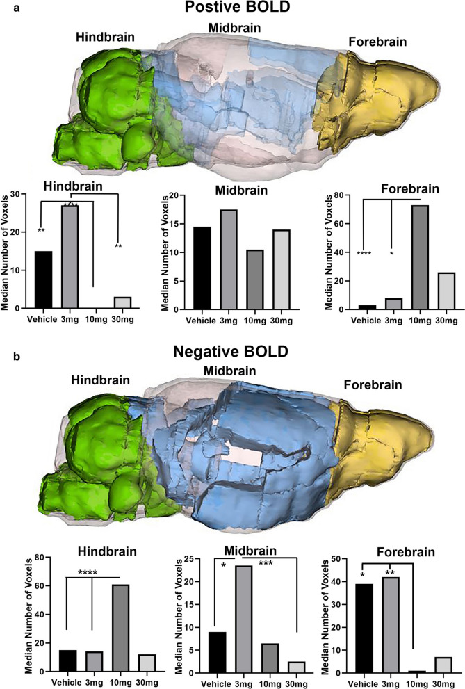
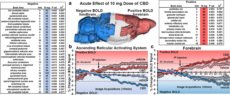
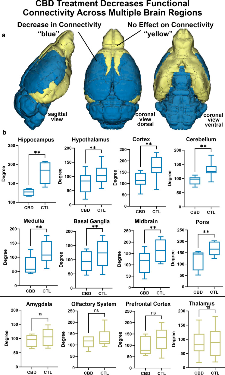
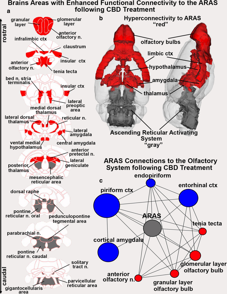
Results: CBD produced a dose-dependent polarization of activation along the rostral-caudal axis of the brain. The olfactory bulb and prefrontal cortex showed an increase in positive BOLD whereas the brainstem and cerebellum showed a decrease in BOLD signal. This negative BOLD affected many areas connected to the ascending reticular activating system (ARAS). The ARAS was decoupled to much of the brain but was hyperconnected to the olfactory system and prefrontal cortex.
Conclusion: The CBD-induced decrease in ARAS activity is consistent with an emerging literature suggesting that CBD reduces autonomic arousal under conditions of emotional and physical stress.
Keywords: Behavioral arrest; N-acyl-phosphatidylethanolamines-specific phospholipase D; Negative BOLD; Olfaction; PTSD; Reticular activating system; Tonic immobility.
Conflict of interest statement
Ferris has a financial interest in Animal Imaging Research, the company that makes the radiofrequency electronics and holders for animal imaging. Authors Kulkarni, Hohmann, Li, Sadaka, Ozuna, Ortiz, Cushing, Johnson, and Bradshaw have no biomedical financial interests or potential conflicts of interest.
Figures
Similar articles
-
Dose-dependent effects of esketamine on brain activity in awake mice: A BOLD phMRI study.Pharmacol Res Perspect. 2022 Dec;10(6):e01035. doi: 10.1002/prp2.1035. Pharmacol Res Perspect. 2022. PMID: 36504448 Free PMC article.
-
Effects of (-)-MBP, a novel 5-HT2C agonist and 5-HT2A/2B antagonist/inverse agonist on brain activity: A phMRI study on awake mice.Pharmacol Res Perspect. 2023 Oct;11(5):e01144. doi: 10.1002/prp2.1144. Pharmacol Res Perspect. 2023. PMID: 37837184 Free PMC article.
-
The impact of cannabidiol placebo on amygdala-based neural responses to an acute stressor.J Psychopharmacol. 2024 Nov;38(11):935-948. doi: 10.1177/02698811241287557. Epub 2024 Oct 14. J Psychopharmacol. 2024. PMID: 39400103 Free PMC article. Clinical Trial.
-
Cannabidiol and the corticoraphe circuit in post-traumatic stress disorder.IBRO Neurosci Rep. 2021 Aug 21;11:88-102. doi: 10.1016/j.ibneur.2021.08.001. eCollection 2021 Dec. IBRO Neurosci Rep. 2021. PMID: 34485973 Free PMC article. Review.
-
Regulatory Effects of Cannabidiol on Mitochondrial Functions: A Review.Cells. 2021 May 19;10(5):1251. doi: 10.3390/cells10051251. Cells. 2021. PMID: 34069407 Free PMC article. Review.
Cited by
-
Differences in Inhibitory Control and Resting Brain Metabolism between Older Chronic Users of Tetrahydrocannabinol (THC) or Cannabidiol (CBD)-A Pilot Study.Brain Sci. 2022 Jun 23;12(7):819. doi: 10.3390/brainsci12070819. Brain Sci. 2022. PMID: 35884627 Free PMC article.
-
Perinatal SSRI exposure impacts innate fear circuit activation and behavior in mice and humans.Nat Commun. 2025 May 6;16(1):4002. doi: 10.1038/s41467-025-58785-4. Nat Commun. 2025. PMID: 40328752 Free PMC article.
-
Examining Brain Activity Responses during Rat Ultrasonic Vocalization Playback: Insights from a Novel fMRI Translational Paradigm.eNeuro. 2024 Oct 3;11(10):ENEURO.0179-23.2024. doi: 10.1523/ENEURO.0179-23.2024. Print 2024 Oct. eNeuro. 2024. PMID: 39299806 Free PMC article.
-
Where do we stand on fMRI in awake mice?Cereb Cortex. 2024 Jan 14;34(1):bhad478. doi: 10.1093/cercor/bhad478. Cereb Cortex. 2024. PMID: 38100331 Free PMC article.
-
Applications in Awake Animal Magnetic Resonance Imaging.Front Neurosci. 2022 Apr 5;16:854377. doi: 10.3389/fnins.2022.854377. eCollection 2022. Front Neurosci. 2022. PMID: 35450017 Free PMC article. Review.
References
Publication types
MeSH terms
Substances
Grants and funding
LinkOut - more resources
Full Text Sources
Other Literature Sources
