. 2021 Jul 15;89(8):e0047120.
doi: 10.1128/IAI.00471-20. Epub 2021 Jul 15.

A Murine Model for Enhancement of Streptococcus pneumoniae Pathogenicity upon Viral Infection and Advanced Age

Affiliations

A Murine Model for Enhancement of Streptococcus pneumoniae Pathogenicity upon Viral Infection and Advanced Age

Basma H Joma et al. Infect Immun. .

Abstract

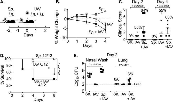
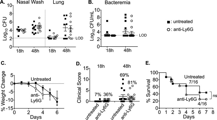
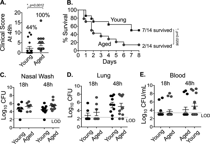
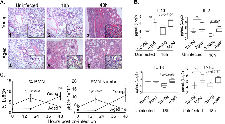
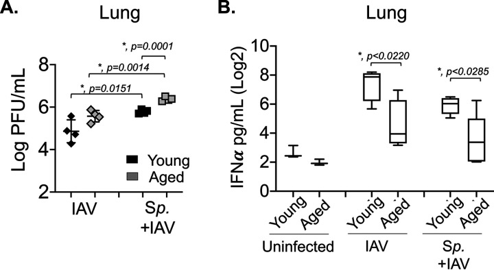
Streptococcus pneumoniae (pneumococcus) resides asymptomatically in the nasopharynx (NP) but can progress from benign colonizer to lethal pulmonary or systemic pathogen. Both viral infection and aging are risk factors for serious pneumococcal infections. Previous work established a murine model that featured the movement of pneumococcus from the nasopharynx to the lung upon nasopharyngeal inoculation with influenza A virus (IAV) but did not fully recapitulate the severe disease associated with human coinfection. We built upon this model by first establishing pneumococcal nasopharyngeal colonization, then inoculating both the nasopharynx and lungs with IAV. In young (2-month-old) mice, coinfection triggered bacterial dispersal from the nasopharynx into the lungs, pulmonary inflammation, disease, and mortality in a fraction of mice. In aged mice (18 to 24 months), coinfection resulted in earlier and more severe disease. Aging was not associated with greater bacterial burdens but rather with more rapid pulmonary inflammation and damage. Both aging and IAV infection led to inefficient bacterial killing by neutrophils ex vivo. Conversely, aging and pneumococcal colonization also blunted alpha interferon (IFN-α) production and increased pulmonary IAV burden. Thus, in this multistep model, IAV promotes pneumococcal pathogenicity by modifying bacterial behavior in the nasopharynx, diminishing neutrophil function, and enhancing bacterial growth in the lung, while pneumococci increase IAV burden, likely by compromising a key antiviral response. Thus, this model provides a means to elucidate factors, such as age and coinfection, that promote the evolution of S. pneumoniae from asymptomatic colonizer to invasive pathogen, as well as to investigate consequences of this transition on antiviral defense.

Keywords: Streptococcus pneumoniae; aging; coinfection; colonization; inflammation; influenza A; neutrophils; secondary bacterial pneumonia.


Figures

FIG 1
FIG 1
Combined intranasal/intratracheal influenza A virus (IAV) inoculation of Streptococcus pneumoniae (Sp)-colonized mice results in bacterial dissemination and disease. (A) Timeline of coinfection; 8- to 10-week-old male C57BL/6 (B6) mice were inoculated intranasally (i.n.) with 5 × 106 CFU of biofilm-grown S. pneumoniae TIGR4 to establish colonization in the nasopharynx. At 48 h later, the mice were either mock treated (Sp) or received 500 PFU of influenza A virus PR8 (IAV) i.n. and 20 PFU i.t. (B) Percent weight loss was monitored daily. (C) Blind clinical scoring was performed on day 2 and day 4 post IAV infection. The percentages denote the number of sick mice observed over the total number of mice. A score of 0 means no sign of sickness observed and a score greater than 1 indicates observable sickness. (D) Survival was monitored for 8 days post IAV infection, with fractions denoting the number of survivors over the total number of mice. (E) The bacterial burden in the nasopharynx and lung were determined at day 2 post IAV infection. Pooled data from four separate experiments are shown, in which one group of 12 mice in each experimental condition were monitored over time and another group of 6 mice per experimental condition were used for measuring bacterial burden. Statistically significant differences determined by Student’s t test for bacterial burden and clinical score and by the log rank (Mantel-Cox) test for survival are indicated by asterisks. #, Statistical significance (P < 0.05) by Fisher’s exact test between Sp and Sp plus IAV groups.
FIG 2
FIG 2
Polymorphonuclear leukocyte (PMN) depletion may have a small effect on the course of disease during IAV/S. pneumoniae coinfection. C57BL/6 mice (8 to 10 weeks old) were intraperitoneally (i.p.) injected with anti-Ly6G (clone 1A8) antibodies to deplete neutrophils or were mock treated. The antibodies were given daily from day −3 to day 1, and every other day from day 3 to the end of each experiment (with respect to IAV infection). (A) At 18 and 48 h post IAV infection, bacterial numbers in the nasal wash and lungs were determined. Pooled data from three separate experiments, with totals of n = 6 mice per experimental condition at 18 h and n = 9 mice per condition at 48 h, are shown. Bacteremia (B) and weight loss (C) were monitored over time. (D) Mice were scored in a blind manner for symptoms of diseases at 18 and 48 h post IAV-infection. The percentages indicate the number of mice with clinical sickness (clinical score above 1) over the total number of mice. (E) Survival was monitored over time; the fractions denote the number of survivors over the total number of mice at 8 days post IAV infection. (C to E) Data are pooled from three separate experiments, with n = 16 mice per group. Statistically significant differences were determined by Student’s t test for bacterial burden and clinical score and by the log-rank (Mantel-Cox). ns, not significant.
FIG 3
FIG 3
Aging increases susceptibility to IAV/S. pneumoniae coinfection. Young (8- to 10-week-old) and aged (18- to 24-month-old) C57BL/6 male mice were coinfected with S. pneumoniae TIGR4 i.n. and influenza A virus PR8 i.n. and i.t. (as shown in Fig. 1A). (A) Clinical score of coinfected young and aged mice at day 2 post IAV infection is shown; the percentage of mice with demonstrable illness is indicated. #, Statistical significance by Fisher’s exact test. (B) Survival of coinfected young and aged mice were monitored over time, with fractions denoting the number of survivors over the total number of mice. Data are pooled from four experiments, with n = 14 mice per group. Asterisks indicate statistical significance by the log rank (Mantel-Cox) test. (C to E) Bacterial burdens in the nasal wash, lungs, and blood were determined at 18 and 48 h post IAV inoculation. The mean ± standard error of the mean pooled from three separate experiments are shown with n = 7 mice per condition at 18 h and n = 10 mice per condition at 48 h. LOD, limit of detection. For clinical score (A), data shown are pooled from all mice from experiments shown in panels B to E that had survived up to that time point (i.e., n = 13 for old and n = 16 for young).
FIG 4
FIG 4
Aging is associated with more rapid lung inflammation. Young and aged C57BL/6 male mice were coinfected with S. pneumoniae and influenza A virus PR8. At 18 and 48 h following IAV infection (see experimental design in Fig. 1A), the lungs were harvested. (A) Lungs were stained with hematoxylin and eosin (H&E); shown are representative photographs at ×100 or ×400 (inset). (B) Cytokines in the supernatants of lung homogenates of young (n = 4) or aged (n = 5) mice at 18 h post coinfection were measured by multiplex enzyme-limited immunosorbent assay (ELISA). Asterisks represent statistical significance by Student’s t test. (C) The percentages and total numbers of PMNs (Ly6G+) in the lungs were measured by flow cytometry. Young mice are represented by open bars and aged mice by shaded bars. The mean ± SEM pooled from three separate experiments is shown where data are pooled from 12 mice per age group, except for 48 h post coinfection, at which point, due to the kinetics of disease, data from only 7 surviving old mice are shown. Statistically significant differences determined by Student’s t test are indicated by asterisks.
FIG 5
FIG 5
Aging and IAV infection diminish the ability of PMNs to kill S. pneumoniae ex vivo. PMNs were isolated from the bone marrow of young (8- to 10-week-old) and aged (18- to 24-months-old) C57BL/6 male mice that were mock infected (uninf.) or singly infected with IAV (i.n. + i.t.) for 2 days. PMNs were incubated with S. pneumoniae preopsonized with homologous serum from the same mouse for 45 or 90 min at 37°C. The percentages of S. pneumoniae killed upon incubation with PMNs were determined with respect to a no-PMN control. Data shown represent the means ± SEM pooled from two experiments (n = 3 mice per group per time point), where each condition was tested in quadruplicates per experiment. Asterisks represent statistical significance as determined by Student’s t test.
FIG 6
FIG 6
Aging and prior colonization with S. pneumoniae result in impaired IFN-α production and higher viral burden in the lungs. Young and aged C57BL/6 male mice were either coinfected with S. pneumoniae and influenza A virus PR8 (Sp + IAV), challenged with virus alone (IAV), or mock challenged with phosphate-buffered saline (PBS) (uninfected) (as in Fig. 1A). At 2 days post IAV-infection, (A) viral burdens in the lungs were determined and (B) levels of IFN-α in the supernatants of lung homogenates were measured by ELISA. Data from n = 4 mice per group are shown. Statistically significant differences were determined by Student’s t test.

Similar articles

Cited by

References

    1. Kadioglu A, Weiser JN, Paton JC, Andrew PW. 2008. The role of Streptococcus pneumoniae virulence factors in host respiratory colonization and disease. Nat Rev Microbiol 6:288–301. 10.1038/nrmicro1871. - DOI - PubMed
    1. Chao Y, Marks LR, Pettigrew MM, Hakansson AP. 2014. Streptococcus pneumoniae biofilm formation and dispersion during colonization and disease. Front Cell Infect Microbiol 4:194. 10.3389/fcimb.2014.00194. - DOI - PMC - PubMed
    1. Marks LR, Parameswaran GI, Hakansson AP. 2012. Pneumococcal interactions with epithelial cells are crucial for optimal biofilm formation and colonization in vitro and in vivo. Infect Immun 80:2744–2760. 10.1128/IAI.00488-12. - DOI - PMC - PubMed
    1. Marks LR, Davidson BA, Knight PR, Hakansson AP. 2013. Interkingdom signaling induces Streptococcus pneumoniae biofilm dispersion and transition from asymptomatic colonization to disease. mBio 4:e00438-13. 10.1128/mBio.00438-13. - DOI - PMC - PubMed
    1. Blanchette-Cain K, Hinojosa CA, Akula Suresh Babu R, Lizcano A, Gonzalez-Juarbe N, Munoz-Almagro C, Sanchez CJ, Bergman MA, Orihuela CJ. 2013. Streptococcus pneumoniae biofilm formation is strain dependent, multifactorial, and associated with reduced invasiveness and immunoreactivity during colonization. mBio 4:e00745-13–e00713. 10.1128/mBio.00745-13. - DOI - PMC - PubMed

Publication types

LinkOut - more resources