. 2021 Jun 1;118(22):e2018840118.
doi: 10.1073/pnas.2018840118.

Violet light suppresses lens-induced myopia via neuropsin (OPN5) in mice

Affiliations

Violet light suppresses lens-induced myopia via neuropsin (OPN5) in mice

Xiaoyan Jiang et al. Proc Natl Acad Sci U S A. .

Abstract

Myopia has become a major public health concern, particularly across much of Asia. It has been shown in multiple studies that outdoor activity has a protective effect on myopia. Recent reports have shown that short-wavelength visible violet light is the component of sunlight that appears to play an important role in preventing myopia progression in mice, chicks, and humans. The mechanism underlying this effect has not been understood. Here, we show that violet light prevents lens defocus-induced myopia in mice. This violet light effect was dependent on both time of day and retinal expression of the violet light sensitive atypical opsin, neuropsin (OPN5). These findings identify Opn5-expressing retinal ganglion cells as crucial for emmetropization in mice and suggest a strategy for myopia prevention in humans.

Keywords: myopia; neuropsin (OPN5); nonvisual photoreceptors; violet light.


Conflict of interest statement

Competing interest statement: Related to myopia-preventing devices based on violet light illumination and transparency, H.T., T.K., and K.T. have been applying internationally for two patents, WO 2015/186723 and WO 2017/094886. The former has been registered in Japan, the United States, and China, and the latter in Japan, the United Kingdom, France, Germany, Italy, Hong Kong, and Singapore. There is a patent application for the design of the mouse eyeglass by Tsubota Laboratory, Inc. (patent application no. 2017-41349). K.T. reports his position as CEO of Tsubota Laboratory, Inc., Tokyo, Japan, a company producing myopia-related devices. R.L. has a sponsored research agreement with BIOS Lighting and, in collaboration with BIOS Lighting, has submitted patent application 41906194/PCT/US21/17681, Lighting Devices to Promote Circadian Health.

Figures

Fig. 1.
Fig. 1.
Time of day–specific VL-suppressed LIM progression. (A) Mouse with lenses for inducing LIM. In all the experiments using LIM mice, 0-D lenses were attached over left eyes and −30-D lenses were attached over right eyes. (B) OCT image of the whole mouse eye showing the different axial measurements, including AL. (C) Together with LIM, VL exposure at different times of day was added with white background light from p21 to p42 in each group. The relative refraction difference between eyes in each group with VL exposure at different times of day is shown in D, and the relative AL difference between eyes was shown in E. The data are displayed as box plots showing the minimum and maximum (error bars,) the interquartile range (box), and the median value (bold horizontal line within the box). The VL exposure for each group was as follows: control group with only WL; VL from 05:00 to 08:00 (WL + predawn VL); VL from 08:00 to 20:00 (WL + daytime VL); continuous VL (WL + continuous VL); VL from 17:00 to 20:00 (WL + evening VL); VL from 20:00 to 23:00 (WL + postdusk VL). n = 4 for each group. n.s.: not significant, *P < 0.05, **P < 0.01, ****P < 0.0001. The values and statics for D and E are shown in SI Appendix, Tables S1 and S2, respectively.
Fig. 2.
Fig. 2.
VL was the most effective wavelength for suppressing LIM progression. (A) Red, green, blue, and VL exposure was added to the white background light from 17:00 to 20:00 every day (evening) in each group. The control group was exposed to the white background light only. LIM was initiated at P21 and lasted for 3 wk. (B) Red, green, blue, and VL had the same irradiance. The relative difference between eyes, normalized to baseline for refractive errors (C) and to ALs (D) indicates the largest protective effects with VL. The data are displayed as box plots showing the minimum and maximum (error bars), the interquartile range (box), and the median value (bold horizontal line within the box). n.s.: not significant, *P < 0.05, **P < 0.01, ****P < 0.0001. RL: red light; GL: green light; BL: blue light. The values and statics for C and D are shown in SI Appendix, Tables S3 and S4, respectively.
Fig. 3.
Fig. 3.
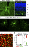
Opn5 expression is limited to RGCs. (A) Retinal localization of reporter+ cells (green) in en face retina from cre-dependent cAMPer (A) or in sections from the Ai14 (B) mouse lines crossed to Opn5cre. NFL, nerve fiber layer; GCL, ganglion cell layer; IPL, inner plexiform layer; INL, inner nuclear layer; OPL, outer plexiform layer; and ONL, outer nuclear layer. (C) Morphologies of Opn5 cells assessed through intravitreal injection of modified rabies virus (Rabies-∆G-tdTomato) in Opn5cre; RΦGT mice. (D and E) Detection and quantification of cells in the Opn5cre; Ai14 line (n = 6 mice) that express the RGC-specific marker, Rbpms (D, in red), represented as a proportion of all reporter+ cells (E). In E, an expanded set of axes is shown on the right.
Fig. 4.
Fig. 4.
The protective effects of VL in LIM requires OPN5. (AD) Measurements of refractive shift (A), AL (B), and choroidal thickness (C and D) in the eyes of mice subjected to the lens-induced myopia protocol. The data are displayed as box plots showing the minimum and maximum (error bars), the interquartile range (box), and the median value (bold horizontal line within the box). In A, B, and D, cohorts of Opn5fl/fl control (white and violet solid shaded bars) and Chx10-Cre; Opn5fl/fl experimental mice (black and violet cross-hatched bars) were assessed. These assays were performed either in standard WL (white bars and black cross-hatched bars) or in WL + evening VL (violet bars and violet cross-hatched bars). In C, all mice were wild type of the C57BL/6J background. The data shown is the difference between the treated and contralateral eyes normalized to baseline. *P < 0.05, **P < 0.01, ***P < 0.001, ****P < 0.0001. The values and statics for AD are shown in SI Appendix, Tables S5–S8, respectively.

Comment in

  • Seeing the violet light.
    Neff EP. Neff EP. Lab Anim (NY). 2021 Jul;50(7):167. doi: 10.1038/s41684-021-00807-x. Lab Anim (NY). 2021. PMID: 34188232 No abstract available.

References

    1. Ikuno Y., Overview of the complications of high myopia. Retina 37, 2347–2351 (2017). - PubMed
    1. Ohno-Matsui K., Lai T. Y., Lai C. C., Cheung C. M., Updates of pathologic myopia. Prog. Retin. Eye Res. 52, 156–187 (2016). - PubMed
    1. Ohno-Matsui K., Pathologic myopia. Asia Pac. J. Ophthalmol. (Phila.) 5, 415–423 (2016). - PubMed
    1. Dolgin E., The myopia boom. Nature 519, 276–278 (2015). - PubMed
    1. Holden B. A., et al. ., Global prevalence of myopia and high myopia and temporal trends from 2000 through 2050. Ophthalmology 123, 1036–1042 (2016). - PubMed

Publication types