. 2021 Jun 1;118(22):e2101713118.
doi: 10.1073/pnas.2101713118.

Zika virus employs the host antiviral RNase L protein to support replication factory assembly

Affiliations

Zika virus employs the host antiviral RNase L protein to support replication factory assembly

Jillian N Whelan et al. Proc Natl Acad Sci U S A. .

Abstract

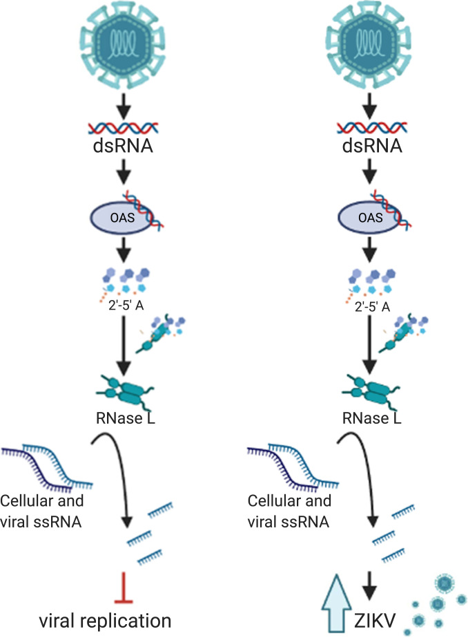
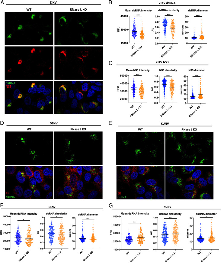
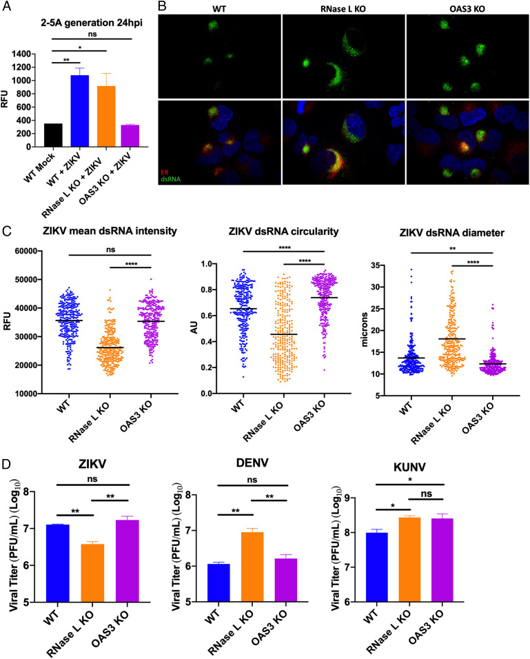
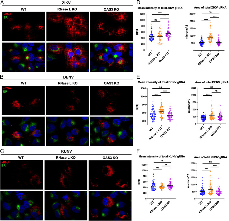
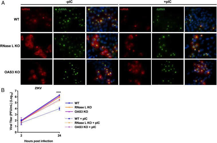
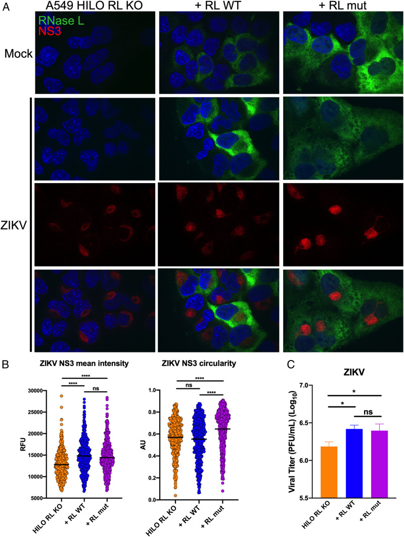
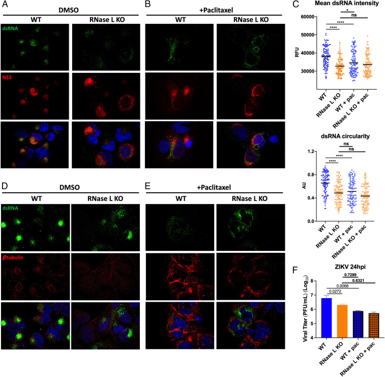
Infection with the flavivirus Zika virus (ZIKV) can result in tissue tropism, disease outcome, and route of transmission distinct from those of other flaviviruses; therefore, we aimed to identify host machinery that exclusively promotes the ZIKV replication cycle, which can inform on differences at the organismal level. We previously reported that deletion of the host antiviral ribonuclease L (RNase L) protein decreases ZIKV production. Canonical RNase L catalytic activity typically restricts viral infection, including that of the flavivirus dengue virus (DENV), suggesting an unconventional, proviral RNase L function during ZIKV infection. In this study, we reveal that an inactive form of RNase L supports assembly of ZIKV replication factories (RFs) to enhance infectious virus production. Compared with the densely concentrated ZIKV RFs generated with RNase L present, deletion of RNase L induced broader subcellular distribution of ZIKV replication intermediate double-stranded RNA (dsRNA) and NS3 protease, two constituents of ZIKV RFs. An inactive form of RNase L was sufficient to contain ZIKV genome and dsRNA within a smaller RF area, which subsequently increased infectious ZIKV release from the cell. Inactive RNase L can interact with cytoskeleton, and flaviviruses remodel cytoskeleton to construct RFs. Thus, we used the microtubule-stabilization drug paclitaxel to demonstrate that ZIKV repurposes RNase L to facilitate the cytoskeleton rearrangements required for proper generation of RFs. During infection with flaviviruses DENV or West Nile Kunjin virus, inactive RNase L did not improve virus production, suggesting that a proviral RNase L role is not a general feature of all flavivirus infections.

Keywords: OAS3; RNase L; Zika virus; flavivirus; replication factories.


Conflict of interest statement

Competing interest statement: S.R.W. is on the scientific advisory board of Immunome, Inc. and Ocugen, Inc. R.H.S. is a consultant to Inception Therapeutics, Inc.

Figures

Fig. 1.
Fig. 1.
Noncanonical RNase L function promotes infectious ZIKV production. (Left) Canonical RNase L antiviral activity. Viral dsRNA is detected by OAS3, which produces the small molecule 2-5A that binds inactive RNase L, inducing its homodimerization and catalytic activation, resulting in cleavage of host and viral ssRNA, leading to inhibition of viral infection. (Right) RNase L activity during ZIKV infection. ZIKV dsRNA is recognized by OAS3, which activates RNase L resulting in ssRNA cleavage; however, ZIKV production is improved with RNase L expression.
Fig. 2.
Fig. 2.
RNase L improves RF function to increase ZIKV RNA and protein expression at RFs. A549 WT and RNase L cells were infected at a multiplicity of infection of 1, cells were fixed at 20 hpi for IFA. (A) ZIKV-infected cells were stained for dsRNA (green) and ZIKV NS3 (red) with 4′,6-diamidino-2-phenylindole (DAPI, blue) staining of nuclei. Quantification of ZIKV (B) dsRNA and (C) NS3 mean intensity, circularity, and diameter of staining shown in A. (D) DENV- or (E) KUNV-infected cells were stained for dsRNA (green) and ER (PDI, red) with DAPI (blue) staining of nuclei. Quantification of (F) DENV or (G) KUNV dsRNA mean intensity, circularity, and diameter of staining shown in D and E, respectively. The data are representative of at least two independent experiments. Statistical significance was determined by Student’s t test. The black bars represent the mean. ns = not significant, *P < 0.05, and ****P < 0.0001. (Imaged at 100× magnification.) RFU = relative fluorescence units, AU = arbitrary units. See also SI Appendix, Figs. S1 and S2.
Fig. 3.
Fig. 3.
Effects of OAS3 KO on ZIKV RF assembly and virus output. (A) A549 WT, RNase L KO, or OAS3 KO cells were mock infected or infected with ZIKV at a multiplicity of infection (MOI) of 5, 24 hpi lysates were harvested for 2-5A fluorescence resonance energy transfer assay. (B) A549 WT, RNase L KO, or OAS3 KO cells were infected with ZIKV at an MOI of 1, fixed at 20 hpi, and stained for dsRNA (green) and ER (PDI, red) with 4′,6-diamidino-2-phenylindole (blue) staining of nuclei. (C) Quantification of ZIKV mean intensity, circularity, and diameter of staining shown in B. (D) A549 WT, RNase L KO, or OAS3 KO cells were infected with ZIKV, DENV, or KUNV at an MOI of 0.1, supernatants were harvested at 48 hpi for measurement of viral titers by plaque assay, shown as plaque forming units (PFU)/mL virus. The data are representative of at least two independent experiments. Statistical significance was determined by one-way ANOVA. Displayed is the mean of three replicates ± SD for replication assays. ns = not significant, *P < 0.05, **P < 0.01, and ****P < 0.0001. (Imaged at 100× magnification.) RFU = relative fluorescence units, AU = arbitrary units. See also SI Appendix, Figs. S3–S5.
Fig. 4.
Fig. 4.
RNase L maintains ZIKV RF structure in the absence of OAS3 to enhance virus replication. A549 WT, RNase L KO, or OAS3 KO cells were infected at a multiplicity of infection of 1 with (A) ZIKV, (B) DENV, or (C) KUNV. Cells were fixed at 20 hpi and stained with a virus genome–specific probe (red) using FISH before staining ER (PDI, green) and nuclei with 4′,6-diamidino-2-phenylindole (blue) by IFA. Quantification of the mean intensity and area of (D) ZIKV, (E) DENV, or (F) KUNV genome staining. The data are representative of at least two independent experiments. Statistical significance was determined by one-way ANOVA. ns = not significant, **P < 0.01, ***P < 0.001, and ****P < 0.0001. (Imaged at 100× magnification.) RFU = relative fluorescence units. See SI Appendix, Fig. S4B.
Fig. 5.
Fig. 5.
ZIKV RFs are unaffected by pIC treatment in the absence of OAS3. A549 WT, RNase L KO, or OAS3 KO cells were transfected with lipofectamine only or with pIC for 2 h before infection with ZIKV at a multiplicity of infection of 1. (A) Cells were fixed at 24 hpi and stained with a ZIKV positive–strand genome probe (red) using FISH before staining dsRNA (green) and nuclei with 4′,6-diamidino-2-phenylindole (blue) by IFA. (B) Supernatants from infected cells were harvested at 2 or 24 hpi for measurement of viral titers over time by plaque assay, shown as plaque forming units (PFU)/mL virus. The data are representative of at least two independent experiments. Statistical significance was determined by one-way ANOVA. Displayed is comparison of WT ± pIC (****) and the mean of three replicates ± SD; ****P < 0.0001. (Imaged at 60× magnification.).
Fig. 6.
Fig. 6.
Expression of RNase L R667A catalytic mutant in A549 HILO cells enhances ZIKV RF function and virus production. (A) A549 HILO Rnase L KO (RL KO) cells, or RL KO cells expressing doxycycline-inducible RNase L WT (+RL WT), or RNase L R667A nuclease dead mutant (+RL mut) were mock infected or infected with ZIKV at a multiplicity of infection (MOI) of 10. At 20 hpi, cells were fixed and stained for ZIKV NS3 (red) and flag-RNase L (green) with 4′,6-diamidino-2-phenylindole (blue) staining of nuclei. (B) Quantification of ZIKV NS3 mean intensity and circularity shown in A. (C) A549 HILO RL KO cells, +RL WT, and +RL mut were infected with ZIKV at an MOI of 0.1, supernatants were harvested at 48 hpi for measurement of viral titers by plaque assay, shown as plaque forming units (PFU)/mL virus. The data are representative of at least two independent experiments. Statistical significance was determined by one-way ANOVA. Displayed is the mean of three replicates ± SD for replication assays. ns = not significant, *P < 0.05, and ****P < 0.0001. (Imaged at 100× magnification.) RFU = relative fluorescence units, AU = arbitrary units. See also SI Appendix, Figs. S6 and S7.
Fig. 7.
Fig. 7.
Effects of RNase L deletion on ZIKV RFs resemble antiviral effects of microtubule stabilization on ZIKV RFs. A549 WT or RNase L KO cells were infected with ZIKV at a multiplicity of infection of 1, treated with 12.5 μM paclitaxel or DMSO 3 h after infection. For IFA staining, cells were fixed at 20 hpi. Cells treated with (A) DMSO or (B) paclitaxel were stained for dsRNA (green) and ZIKV NS3 (red) with 4′,6-diamidino-2-phenylindole (DAPI, blue) nuclei staining, mean dsRNA intensity and circularity was quantified in C. Cells treated with (D) DMSO or (E) paclitaxel were stained for dsRNA (green) and β-tubulin (red) with DAPI (blue) nuclei staining. (F) For infectious virus production, supernatants were harvested at 24 hpi for measurement of viral titers by plaque assay, shown as plaque forming units (PFU)/mL virus, displayed are P values. The data are representative of at least two independent experiments. Statistical significance was determined by one-way ANOVA. Displayed is the mean of three replicates ± SD for replication assays. ns = not significant, *P < 0.05, and ****P < 0.0001. (Imaged at 100× magnification.) RFU = relative fluorescence units, AU = arbitrary units. See also SI Appendix, Fig. S8.

References

    1. Wikan N., Smith D. R., Zika virus: History of a newly emerging arbovirus. Lancet Infect. Dis. 16, e119–e126 (2016). - PubMed
    1. Guzman M. G., Harris E., Dengue. Lancet 385, 453–465 (2015). - PubMed
    1. Kaiser J. A., Barrett A. D. T., Twenty years of progress toward West Nile virus vaccine development. Viruses 11, 823 (2019). - PMC - PubMed
    1. Wu K. Y., et al. ., Vertical transmission of Zika virus targeting the radial glial cells affects cortex development of offspring mice. Cell Res. 26, 645–654 (2016). - PMC - PubMed
    1. Cugola F. R., et al. ., The Brazilian Zika virus strain causes birth defects in experimental models. Nature 534, 267–271 (2016). - PMC - PubMed

Publication types

MeSH terms