. 2021 May 24;12(1):3065.
doi: 10.1038/s41467-021-23351-1.

Cryo-EM structure of cortical microtubules from human parasite Toxoplasma gondii identifies their microtubule inner proteins

Affiliations

Cryo-EM structure of cortical microtubules from human parasite Toxoplasma gondii identifies their microtubule inner proteins

Xiangli Wang et al. Nat Commun. .

Erratum in

Abstract

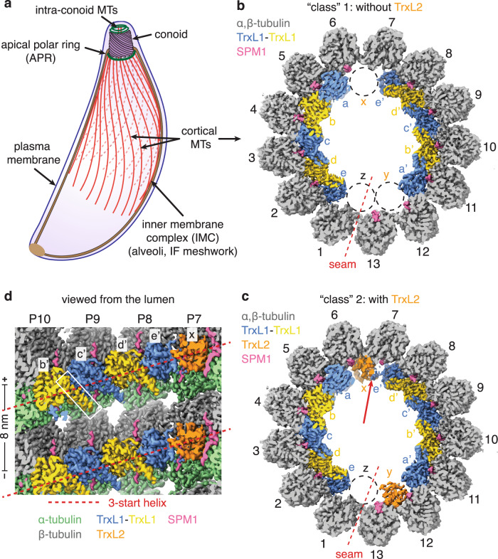
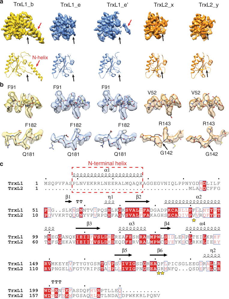
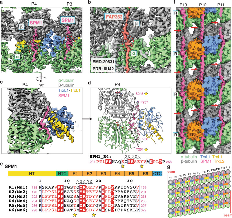
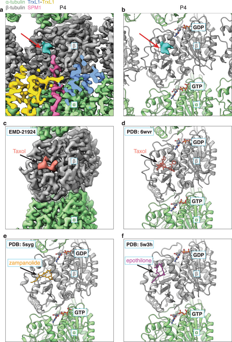
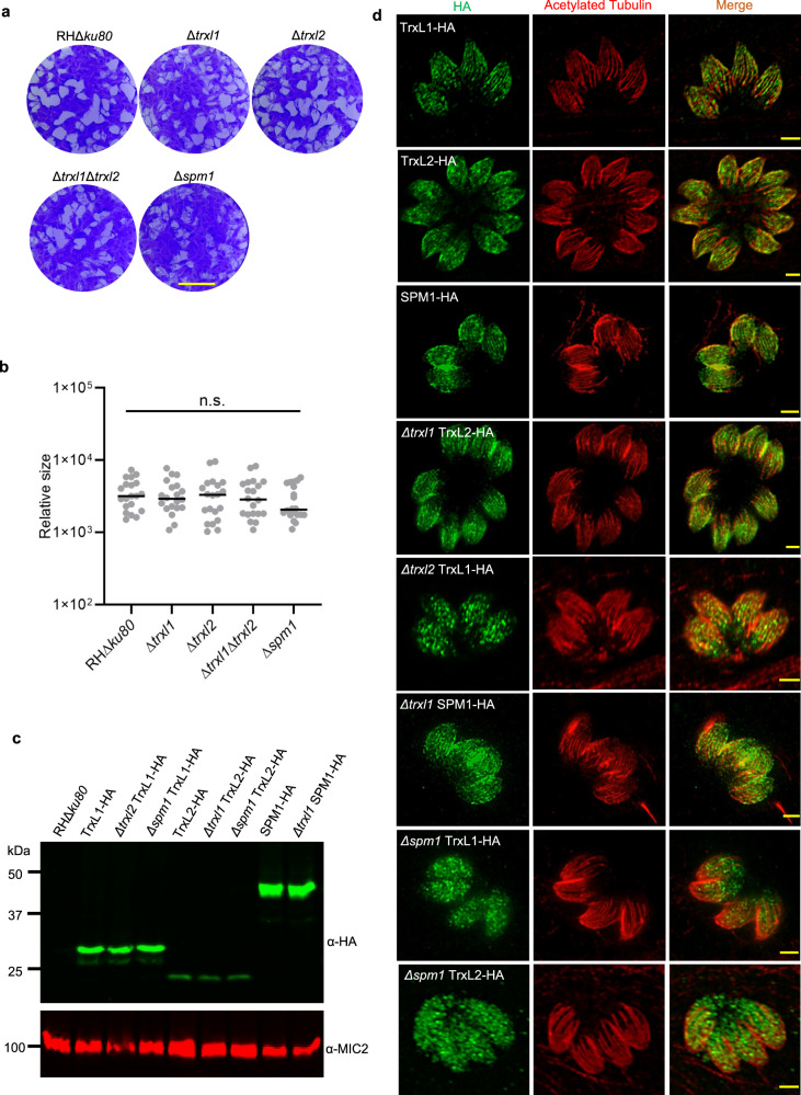
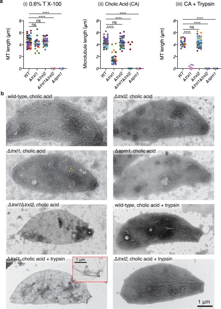
In living cells, microtubules (MTs) play pleiotropic roles, which require very different mechanical properties. Unlike the dynamic MTs found in the cytoplasm of metazoan cells, the specialized cortical MTs from Toxoplasma gondii, a prevalent human pathogen, are extraordinarily stable and resistant to detergent and cold treatments. Using single-particle cryo-EM, we determine their ex vivo structure and identify three proteins (TrxL1, TrxL2 and SPM1) as bona fide microtubule inner proteins (MIPs). These three MIPs form a mesh on the luminal surface and simultaneously stabilize the tubulin lattice in both longitudinal and lateral directions. Consistent with previous observations, deletion of the identified MIPs compromises MT stability and integrity under challenges by chemical treatments. We also visualize a small molecule like density at the Taxol-binding site of β-tubulin. Our results provide the structural basis to understand the stability of cortical MTs and suggest an evolutionarily conserved mechanism of MT stabilization from the inside.


Conflict of interest statement

The authors declare no competing interests.

Figures

Fig. 1
Fig. 1. Overview of the cryo-EM structures of cortical MT from T. gondii.
a Cartoon diagram of T. gondii showing plasma membrane, inner membrane complex (IMC) (including the alveoli and an intermediate filament (IF)-like protein meshwork), the apical complex (a conoid and two intra-conoid MTs), the apical polar ring (APR) and 22 cortical MTs. bc Cross section view of the cryo-EM structure of T. gondii cortical MT with (b) and without (c) TrxL2 at sites x and y. The structures show an asymmetric arrangement of the microtubule inner proteins (MIPs) including TrxL1 (gold or blue), TrxL2 (orange), and SPM1 (pink). An unoccupied site at the MT seam is labeled as z. Composite DeepEMhancer-sharpened maps (see “Methods”) are used for visualization. The quotation marks on “class” 1 and “class” 2 indicate that the two composite maps only differ in sub-regions containing sites x and y. d The same as (c), but showing the luminal surface of the tubulin and MIPs. The N-terminal helix of TrxL1 (marked by white rectangle) inserts into a binding pocket of the neighboring TrxL1 molecule. The helical paths for the 3-start helix of the MT are indicated by dashed red lines.
Fig. 2
Fig. 2. TrxL1 and TrxL2.
a Atomic models and segmented cryo-EM densities for TrxL1 (gold and blue) and TrxL2 (orange) at different locations as indicated in Fig. 1c. The black arrows highlight the distinct appearances of the N-termini of TrxL1 and TrxL2. b Side-chain densities of three residues (TrxL1: F91, Q181, and F182) that distinguish TrxL1 from TrxL2. DeepEMhancer-sharpened maps (see “Methods”) are used for visualization. c Sequence alignment of TrxL1 and TrxL2. Secondary structure elements of TrxL1 are shown above the sequences. The three residues shown in (b) are marked by yellow stars.
Fig. 3
Fig. 3. The interactions between SPM1, TrxL1/2, and tubulin.
a Cutaway view of the cryo-EM density showing a short α-helix (red dashed circle) of SPM1 (pink) bound at the intra-dimer interface between α- and β-tubulin (green and gray, respectively). b Cutaway view of the cryo-EM density of FAP363, a Mn-motif containing protein, bound to the Chlamydomonas ciliary doublet MT. c 90-degree rotation of panel a, showing SPM1 is sandwiched between tubulin and TrxL1. d Same view as panel c, but only displaying the atomic models. The two conserved residues (characteristic of the Mn motif) of SPM1 are marked by yellow stars (same in panel e). We arbitrarily assigned the protein sequence of the SPM1 density to be R4 for the purpose of model building. e Above: schematic of the domain organization of SPM1. NT: N-terminal domain, NTC: N-terminal conserved domain, CTC: C-terminal conserved domain. Below: sequence alignment of the six internal repeats (R1-R6) of SPM1. f Luminal view of the MIP densities at protofilaments 11–13 (P11–13). Unsharpened cryo-EM density map is used to better visualize the connectivity of SPM1. The red arrows point to a region of SPM1 with weaker densities (except for the SPM1 on P12), presumably due to incoherent averaging of divergent residues between R1-R6. g Schematic of the MT lumen that is cut open and unfurled at the seam.
Fig. 4
Fig. 4. Density at the Taxol-binding site of β-tubulin.
a Close-up view of the cryo-EM density of one α,β-tubulin dimer at protofilament P4. Unexplained density (cyan, pointed by a red arrow) can be observed at the Taxol-binding site of β-tubulin (gray), but its size and shape are inconsistent with Taxol (c, d). b Same view as a, but showing the atomic models together with the unexplained density. Atomic models of TrxL1 and SPM1 are hidden for clarity. c Close-up view of the cryo-EM density of one α,β-tubulin dimer of taxol-stabilized bovine MT (EMD-21924). d Atomic model of the cryo-EM density shown in (c) (PDB 6WVR). e Atomic model of zampanolide-stabilized porcine MT (PDB 5SYG). f Atomic model of epothilone-stabilized yeast MT (PDB 5W3H).
Fig. 5
Fig. 5. Knockout strains and HA-tagged strains of individual MIPs.
a Plaques formed by RHΔku80 (wild type), Δtrxl1, Δtrxl2, Δtrxl1Δtrxl2 and Δspm1 on human foreskin fibroblasts cells (HFF) monolayers infected with 200 parasites per monolayer. Scale bar = 5 mm. b Plaque area was measured by the pixels of each plaque and indicated as relative size. Plaque data represented 20 replicates for each parasite strain and was analyzed for statistical significance using one-way ANOVA. c Western blot analysis of different T. gondii strains expressing endogenously 3xHA (α-HA) tagged TrxL1, TrxL2, and SPM1. Tagged proteins were detected using anti-HA antibody (α-HA, green). MIC2 (α-MIC2, red) was used as a loading control. d Indirect immunofluorescence of different HA tagging strains . Tagged proteins were detected using anti-HA antibody (green) 24 h post-infection. Anti-acetylated tubulin antibody (red) was used to stain MTs for co-localization. Images were taken by laser scanning confocal microscope with Airyscan. Scale bar = 2 μm. Experiments in (a) and (c) were repeated twice with similar results. Each IFA image in (d) was selected from one of the three independent experiments with similar outcomes.
Fig. 6
Fig. 6. Cortical microtubule lengths in wild-type and knockout strains upon chemical treatments.
a Plot of MT lengths of wild-type and four knockout strains (Δtrxl1, Δtrxl2, Δspm1, and Δtrxl1Δtrxl2 double knockout) upon three different chemical treatments: (i) glycerol extraction followed by detergent treatment with 0.6% Triton X-100 (58, 32 and 37 MTs were measured from 10 WT, 5 Δtrxl1, and 10 Δtrxl2 parasites, respectively); (ii) detergent extraction with 1.5% cholic acid (68, 42, 59, and 4 MTs were measured from 10 WT, 10 Δtrxl1, 10 Δtrxl2, and 1 Δtrxl1Δtrxl2 parasites, respectively); (iii) detergent treatment with 1.5% cholic acid followed by trypsin digestion (27, 4, and 54 MTs were measured from 10 WT, 1 Δtrxl1 and 10 Δtrxl2 strains, respectively). Only visible cortical MTs within the cell bodies were measured. Each data point represents the measured length of a cortical MT, and each color represents one parasite. The numerical data were expressed as mean ± SD (the mean values are indicated in the plot as horizontal lines), ****P < 0.0001, two-sided Kruskal–Wallis test with Dunn’s Multiple Comparison Correction Test. b Representative negative-stain EM images of the whole parasite. The samples were stained with 1% aqueous phosphotungstic acid (PTA). In the image of Δtrxl1 with cholic acid treatment alone, the posterior ends of visible MTs were marked by yellow asterisks. For Δtrxl1 with cholic acid treatment plus trypsin digestion, no cortical MTs were observed in most of the cell bodies, while some ‘bare’ MTs can be occasionally seen in the background, as shown in the red inset. Those bare MTs were not included in the length measurement.

Similar articles

Cited by

  • Analysis of CDPK1 targets identifies a trafficking adaptor complex that regulates microneme exocytosis in Toxoplasma.
    Chan AW, Broncel M, Yifrach E, Haseley NR, Chakladar S, Andree E, Herneisen AL, Shortt E, Treeck M, Lourido S. Chan AW, et al. Elife. 2023 Nov 7;12:RP85654. doi: 10.7554/eLife.85654. Elife. 2023. PMID: 37933960 Free PMC article.
  • Ciliopathy patient variants reveal organelle-specific functions for TUBB4B in axonemal microtubules.
    Dodd DO, Mechaussier S, Yeyati PL, McPhie F, Anderson JR, Khoo CJ, Shoemark A, Gupta DK, Attard T, Zariwala MA, Legendre M, Bracht D, Wallmeier J, Gui M, Fassad MR, Parry DA, Tennant PA, Meynert A, Wheway G, Fares-Taie L, Black HA, Mitri-Frangieh R, Faucon C, Kaplan J, Patel M, McKie L, Megaw R, Gatsogiannis C, Mohamed MA, Aitken S, Gautier P, Reinholt FR, Hirst RA, O'Callaghan C, Heimdal K, Bottier M, Escudier E, Crowley S, Descartes M, Jabs EW, Kenia P, Amiel J, Bacci GM, Calogero C, Palazzo V, Tiberi L, Blümlein U, Rogers A, Wambach JA, Wegner DJ, Fulton AB, Kenna M, Rosenfeld M, Holm IA, Quigley A, Hall EA, Murphy LC, Cassidy DM, von Kriegsheim A; Scottish Genomes Partnership16; Genomics England Research Consortium45; Undiagnosed Diseases Network46; Papon JF, Pasquier L, Murris MS, Chalmers JD, Hogg C, Macleod KA, Urquhart DS, Unger S, Aitman TJ, Amselem S, Leigh MW, Knowles MR, Omran H, Mitchison HM, Brown A, Marsh JA, Welburn JPI, Ti SC, Horani A, Rozet JM, Perrault I, Mill P. Dodd DO, et al. Science. 2024 Apr 26;384(6694):eadf5489. doi: 10.1126/science.adf5489. Epub 2024 Apr 26. Science. 2024. PMID: 38662826 Free PMC article.
  • Microtubules in Martini: Parameterizing a heterogeneous elastic-network towards a mechanically accurate microtubule.
    Sahoo A, Hanson SM. Sahoo A, et al. PNAS Nexus. 2025 Jun 21;4(7):pgaf202. doi: 10.1093/pnasnexus/pgaf202. eCollection 2025 Jul. PNAS Nexus. 2025. PMID: 40599826 Free PMC article.
  • Microtubule inner proteins of Plasmodium are essential for transmission of malaria parasites.
    Hentzschel F, Binder AM, Dorner LP, Herzel L, Nuglisch F, Sema M, Röver K, He B, Aguirre-Botero MC, Cyrklaff M, Funaya C, Frischknecht F. Hentzschel F, et al. Proc Natl Acad Sci U S A. 2025 Feb 11;122(6):e2421737122. doi: 10.1073/pnas.2421737122. Epub 2025 Feb 5. Proc Natl Acad Sci U S A. 2025. PMID: 39908102 Free PMC article.
  • Ciliary central apparatus structure reveals mechanisms of microtubule patterning.
    Gui M, Wang X, Dutcher SK, Brown A, Zhang R. Gui M, et al. Nat Struct Mol Biol. 2022 May;29(5):483-492. doi: 10.1038/s41594-022-00770-2. Epub 2022 May 16. Nat Struct Mol Biol. 2022. PMID: 35578023 Free PMC article.

References

    1. Torgerson PR, Mastroiacovo P. The global burden of congenital toxoplasmosis: a systematic review. Bull. World Health Organ. 2013;91:501–508. - PMC - PubMed
    1. Dunay IR, Gajurel K, Dhakal R, Liesenfeld O, Montoya JG. Treatment of toxoplasmosis: historical perspective, animal models, and current clinical practice. Clin. Microbiol. Rev. 2018;31:1965. - PMC - PubMed
    1. Parlog A, Schlüter D, Dunay IR. Toxoplasma gondii-induced neuronal alterations. Parasite Immunol. 2015;37:159–170. - PubMed
    1. Morrissette N. Targeting Toxoplasma tubules: tubulin, microtubules, and associated proteins in a human pathogen. Eukaryot. Cell. 2015;14:2–12. - PMC - PubMed
    1. Russell DG, Burns RG. The polar ring of coccidian sporozoites: a unique microtubule-organizing centre. J. Cell Sci. 1984;65:193–207. - PubMed

Publication types

MeSH terms