. 2021 May 24;7(1):46.
doi: 10.1038/s41522-021-00217-4.

Metabolic plasticity enables lifestyle transitions of Porphyromonas gingivalis

Affiliations

Metabolic plasticity enables lifestyle transitions of Porphyromonas gingivalis

M Fata Moradali et al. NPJ Biofilms Microbiomes. .

Abstract

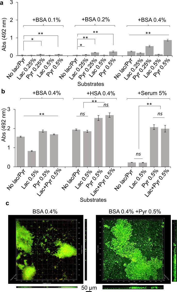
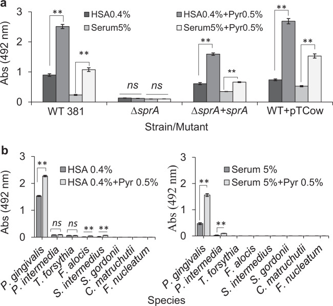
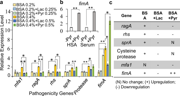
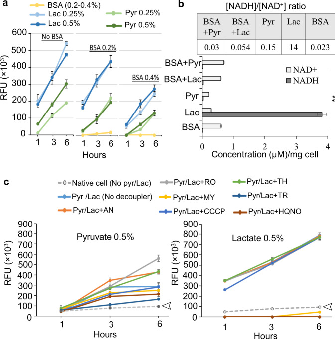
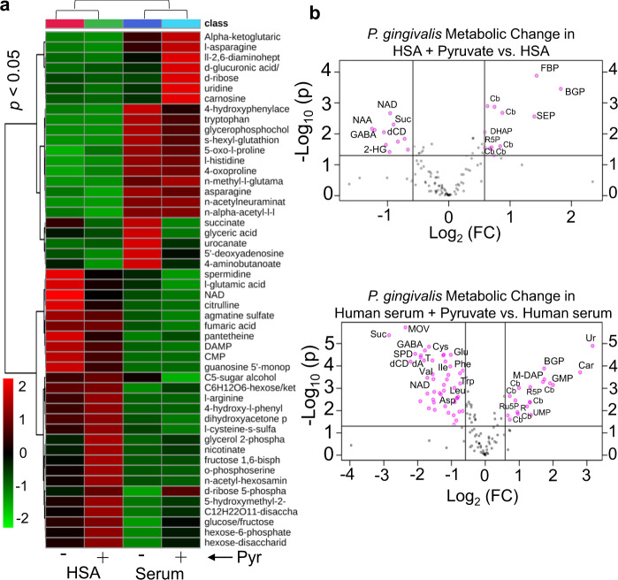
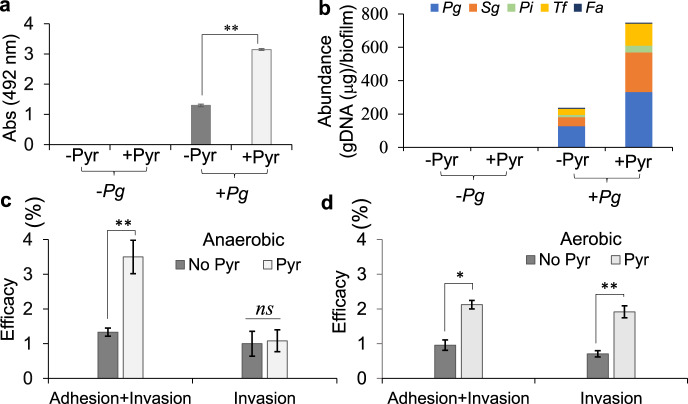
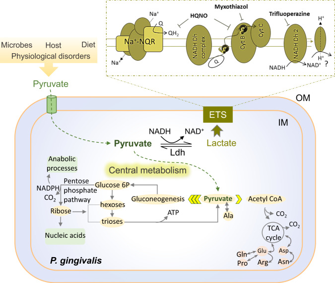
Our understanding of how the oral anaerobe Porphyromonas gingivalis can persist below the gum line, induce ecological changes, and promote polymicrobial infections remains limited. P. gingivalis has long been described as a highly proteolytic and asaccharolytic pathogen that utilizes protein substrates as the main source for energy production and proliferation. Here, we report that P. gingivalis displays a metabolic plasticity that enables the exploitation of non-proteinaceous substrates, specifically the monocarboxylates pyruvate and lactate, as well as human serum components, for colonization and biofilm formation. We show that anabolism of carbohydrates from pyruvate is powered by catabolism of amino acids. Concomitantly, the expression of fimbrial adhesion is upregulated, leading to the enhancement of biofilm formation, stimulation of multispecies biofilm development, and increase of colonization and invasion of the primary gingival epithelial cells by P. gingivalis. These studies provide the first glimpse into the metabolic plasticity of P. gingivalis and its adaptation to the nutritional condition of the host niche. Our findings support the model that in response to specific nutritional parameters, P. gingivalis has the potential to promote host colonization and development of a pathogenic community.


Conflict of interest statement

The authors declare no competing interests.

Figures

Fig. 1
Fig. 1. Synergistic effects of the metabolism of pyruvate, lactate, and serum components on the growth and biofilm formation of P. gingivalis.
a Biofilm assessments show that the utilization of BSA enhances P. gingivalis biofilm in a concentration-dependent manner. Pyruvate and lactate as sole carbon and energy sources cannot support P. gingivalis growth and biofilm formation, while the combination of BSA-pyruvate and BSA-lactate, respectively, promotes and inhibits biofilm formation. b Pyruvate enhances biofilm development in combination with human serum albumin (HSA) or human serum, but lactate is not inhibitive to biofilm growth neither alone nor in combination with pyruvate. c Images of 24-h biofilms were acquired by CLSM. Top and side views show that pyruvate significantly promotes surface attachment and biofilm development by P. gingivalis. Graphs represent mean biomass of biofilms ± SE (two biological replicates, in each n = 3) at 48 h, as determined by safranin staining. Biofilm biomasses were normally distributed (Shapiro–Wilk test: p > 0.05), and thus analyzed using ANOVA test. Asterisks indicate pairs of significantly different values (post hoc Tukey’s HSD test: *p < 0.05; **p < 0.01; ns not significant).
Fig. 2
Fig. 2. Synergistic effects of the metabolism of pyruvate, lactate, and serum components on the biofilm formation of ∆sprA mutant, that lacks the essential translocon of the T9SS, and other oral pathogens.
a Biofilm assay shows P. gingivalissprA mutant cannot efficiently utilize serum components for biofilm development; this mutant is not responsive to pyruvate addition. But, upon in trans complementation with sprA, biofilm development was restored by the mutant, indicating there must be a metabolic coupling system to utilize protein components and pyruvate whose synergistic effect enhances metabolic flux towards biofilm development. b Mono-species biofilm assays show that among tested oral pathogens, only P. gingivalis effectively benefits from the synergistic effects of concomitant utilization of pyruvate and serum components for enhancing biofilm. Statistically, P. intermedia, S. intermedius, and F. alocis positively responds to pyruvate addition but at very low levels. Graphs represent mean biomass of biofilms ± SE (two biological replicates, in each n = 3) at 48 h, as determined by safranin staining. Biofilm biomasses were normally distributed (Shapiro–Wilk test: p > 0.05), and thus analyzed using ANOVA test. Asterisks indicate pairs of significantly different values (post hoc Tukey’s HSD test: *p < 0.05; **p < 0.01; ns not significant).
Fig. 3
Fig. 3. Various pathogenicity genes are differentially expressed in response to the availability of specific energy substrates, i.e., pyruvate or lactate and/or serum components.
a RT-qPCR analysis and relative expression levels shows that, consistent with biofilm results, BSA utilization increases the expression of fimA in a concentration-dependent manner; the addition of pyruvate in the BSA-based medium remarkably increases the expression of fimA. In addition, the provision of higher concentrations of BSA correlates positively with the expression of those pathogenicity genes involved in the degradation of protein substrates and oligopeptide uptake. Exogenous lactate significantly decreases the expression of fimA and other pathogenicity genes when compared with untreated controls, leading to decreased biofilm formation. b The addition of pyruvate in HSA or human serum also significantly increases the expression of fimA. c Table summarizes RT-qPCR results and shows the correlation of utilized energy substrates and expression of pathogenicity genes. Graphs represent mean expression ± SE (two biological replicates, in each n = 3). Asterisks indicate pairs of significantly different values (post hoc Tukey’s HSD test: *p < 0.05; **p < 0.01).
Fig. 4
Fig. 4. The impact of proteinaceous substrates, pyruvate, and lactate on the redox status of P. gingivalis.
a Resazurin fluorometric (RF) assay indicates that exogenous lactate and pyruvate greatly change cellular redox status, but BSA utilization does not; concomitant utilization of BSA and pyruvate or lactate effectively controls redox changes. b Quantification of [NADH]/[NAD+] cofactors shows that the metabolism of BSA enhances cellular accumulation of [NAD+] when compared with the utilization of exogenous pyruvate that reduces it. Lactate metabolism remarkably favored cellular accumulation of [NADH] and the increase of [NADH]/[NAD+] ratio, while it is significantly lowered upon BSA addition. c The use of a series of chemical uncouplers in RF assay to shed light on the bioenergetics system of P. gingivalis and the effects of pyruvate and lactate on it. RF assays over 6-h incubation period indicated that the uncouplers HQNO (a potent inhibitor of quinone oxidation at the Na+–NQR complex in bacteria), TR (a calmodulin antagonist and type II NADH oxidoreductase (NDH-2) inhibitor in M. tuberculosis), and MY (a Qo site inhibitor at cytochrome b) have strong inhibitory effects on lactate-induced redox changes upon decoupling electron transport system from lactate metabolism while others, including AN, RO, CCCP, and TH did not show inhibitory effects. The uncouplers HQNO, TR, and MY were less inhibitive to pyruvate-induced redox change. Arrows indicate the basal redox status of the cells in the absence of any treatment. The data in graphs represent the means ± SD error for biological replicates (n = 4), and asterisk in the bar graph indicates significantly different values (post hoc Tukey’s HSD test: **p < 0.01). RFU relative fluorescence units, BSA bovine serum albumin, Pyr pyruvate, Lac lactate, HQNO 2-heptyl-4-hydroxyquinoline N-oxide, TR trifluoperazine, MY myxothiazol, AN antimycin A, RO rotenone, CCCP carbonyl cyanide m-chlorophenyl hydrazine, TH thenoyltrifluoroaceton.
Fig. 5
Fig. 5. Statistical analysis of metabolic changes among all groups of P. gingivalis biofilm cells grown in HSA or human serum with and without adding pyruvate.
a Heatmap represents multigroup analysis for the metabolites of all groups that are found as differentially produced upon one-way ANOVA followed by post hoc analysis (p value <0.05, n = 3) and variable importance in projection (VIP) values >1.0. b Two-group volcano plot analysis of metabolic changes in HSA or human serum with and without pyruvate. The points in each plot that satisfy the condition p value <0.05 (t test, n = 3) and fold change ≥ 1.5 appear in pink color, whereas the others that appear in gray are not significant. OAA oxaloacetate, KG α-ketoglutarate, Lac lactate, PEP phosphoenolpyruvate, ETC electron transport chain, BGP glycerol 2-phosphate, FBP fructose-1,6-bisphosphate, SEP O-phosphoserine, Cb carbohydrate derivative, NAD nicotinamide adenine dinucleotide, Suc succinate, GABA 4-aminobutyric acid or 4-aminobutanoate, NAA N-acetylaspartic acid, 2-HG 2-hydroxyglutarate, DHAP dihydroxyacetone phosphate, Ur uridine, MOV 3-methyl-2-oxovaleric acid, dCD deoxycytidine, SPD spermidine, dA deoxyadenosine, T thymine, G6P glucose-6-phosphate, M-DAP LL-2,6-diaminopimelic acid, R ribose, R5P ribose-5-phosphate, Ru5P ribulose-5-phosphate, UMP uridine monophosphate, Cys cysteine, Glu glutamic acid, Ile isoleucine, Val valine, Trp tryptophan, Leu leucine, Asp aspartic acid.
Fig. 6
Fig. 6. The impact of the co-utilization of pyruvate and human serum by P. gingivalis on development of multispecies biofilms, host colonization, and invasion.
a Multispecies biofilm assay shows that P. gingivalis is necessary for the formation of a heterotypic community composed of S. gordonii, T. forsythia, P. intermedia, and F. alocis when human serum (5%) is used; pyruvate addition (0.5%) augments the biomass of the heterotypic biofilm. b Graph shows relative abundance of each species in each mixed biofilm that is calculated on the basis of the quantification of the genomic DNA of individual population using qPCR and 16S ribosomal RNA primers. c, d Graphs show the efficacy of colonization and invasion of the primary HGEP cells by P. gingivalis (MOI of 100 and incubated for 1 h) when grown in human serum with and without adding pyruvate, incubated under anaerobic (c) or aerobic (d) conditions. Pyruvate availability significantly increases the adherence of P. gingivalis to the primary HGEP under both aerobic and anaerobic conditions; but invasion was augmented when the interaction was aerobically incubated. Biofilm graph represents the mean ± SE (two biological replicates, in each n = 3) of biomass incubated for 48 h. Obtained data were normally distributed (Shapiro–Wilk test: p > 0.05), and thus analyzed using ANOVA test. Asterisks indicate pairs of significantly different values (post hoc Tukey’s HSD test: **p < 0.01). Values in graphs are shown as the mean ± SE of CFU obtained from two independent experiments, each with four replicates, and were analyzed with a Student’s t test (*p < 0.05; **p < 0.01; ns nonsignificant). Pg P. gingivalis 381, Sg S. gordonii DL-1, Pi P. intermedia strain 17, Fa F. alocis ATCC 35896, Tf T. forsythia ATCC 43037.
Fig. 7
Fig. 7. Proposed model of possible bioenergetics system and metabolism of the monocarboxylates pyruvate and lactate in P. gingivalis that are deduced from the effectiveness of applied uncouplers in RF assay and metabolomic analysis.
Pyruvate availability in the intracellular and extracellular milieu of host cells is impacted by various external factors. P. gingivalis efficiently transports exogenous pyruvate inside to couple with the metabolism of serum components toward the enhancement of the PPP intermediates that are required for biosynthetic processes, resulting in the increase of cell proliferation and biomass in the biofilm community. ETS electron transport system, Ldh lactate dehydrogenase, Cyt cytochrome, NADH Dh NADH dehydrogenase, Na+–NQR the sodium pumping NADH quinone oxidoreductase, Q quinone, e electron, NAD nicotinamide adenine dinucleotide, NADPH nicotinamide adenine dinucleotide phosphate, Glucose 6P glucose-6-phosphate, Glu glutamic acid, Asp aspartic acid, Gln glutamine, Pro proline, Arg arginine, Asn asparagine, Ala alanine, ATP adenosine triphosphate; HQNO 2-heptyl-4-hydroxyquinoline N-oxide, IM inner membrane, OM outer membrane.

Similar articles

Cited by

References

    1. Socransky SS, Haffajee AD, Cugini MA, Smith C, Kent RL., Jr. Microbial complexes in subgingival plaque. J. Clin. Periodontol. 1998;25:134–144. doi: 10.1111/j.1600-051X.1998.tb02419.x. - DOI - PubMed
    1. Petersen PE, Ogawa H. The global burden of periodontal disease: towards integration with chronic disease prevention and control. Periodontol. 2000. 2012;60:15–39. doi: 10.1111/j.1600-0757.2011.00425.x. - DOI - PubMed
    1. Kumar PS. Oral microbiota and systemic disease. Anaerobe. 2013;24:90–93. doi: 10.1016/j.anaerobe.2013.09.010. - DOI - PubMed
    1. Maddi A, Scannapieco FA. Oral biofilms, oral and periodontal infections, and systemic disease. Am. J. Dent. 2013;26:249–254. - PubMed
    1. Lamont RJ, Koo H, Hajishengallis G. The oral microbiota: dynamic communities and host interactions. Nat. Rev. Microbiol. 2018;16:745–759. doi: 10.1038/s41579-018-0089-x. - DOI - PMC - PubMed

Publication types

LinkOut - more resources