

IGF1R and Src inhibition induce synergistic cytotoxicity in HNSCC through inhibition of FAK
- PMID: 34031486
- PMCID: PMC8144381
- DOI: 10.1038/s41598-021-90289-1
IGF1R and Src inhibition induce synergistic cytotoxicity in HNSCC through inhibition of FAK
Abstract
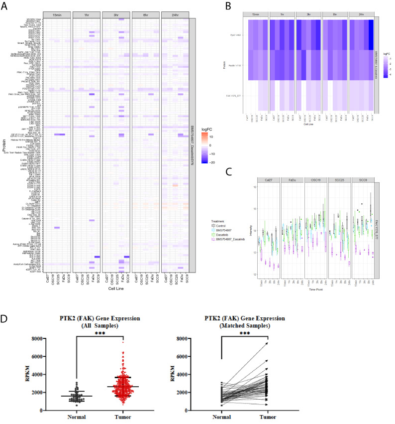
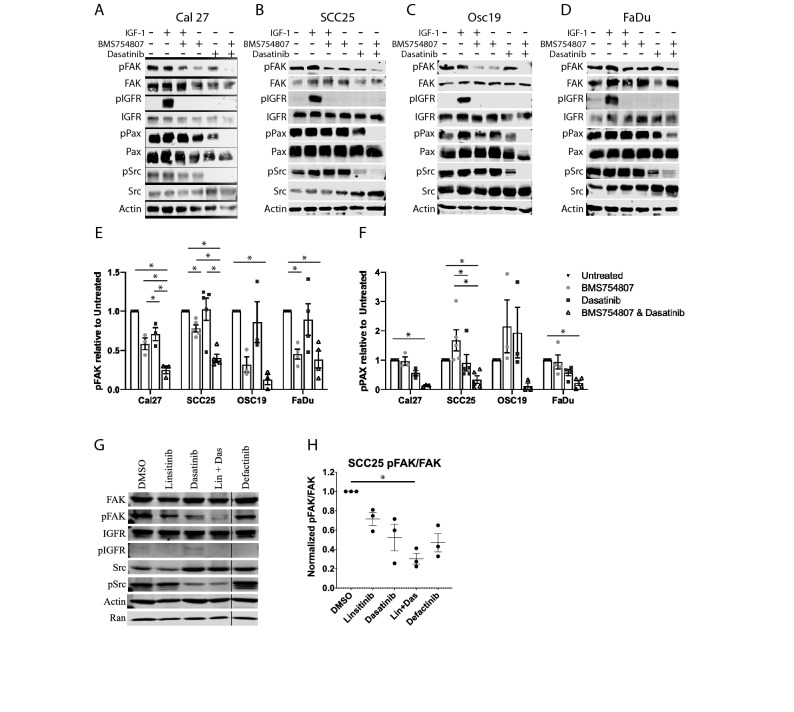
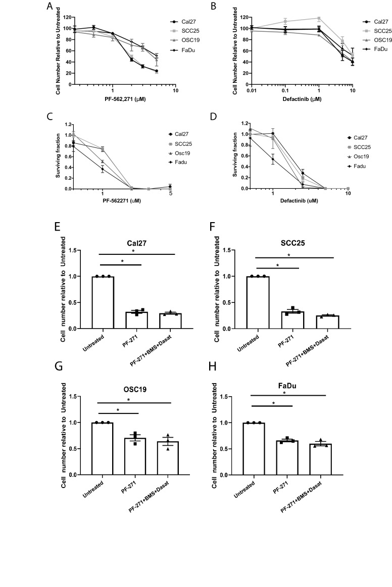
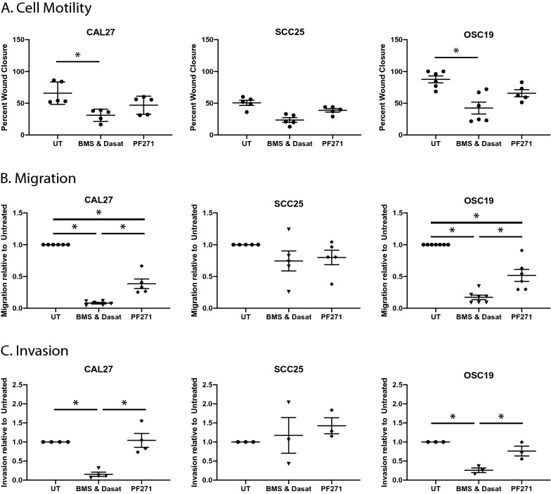
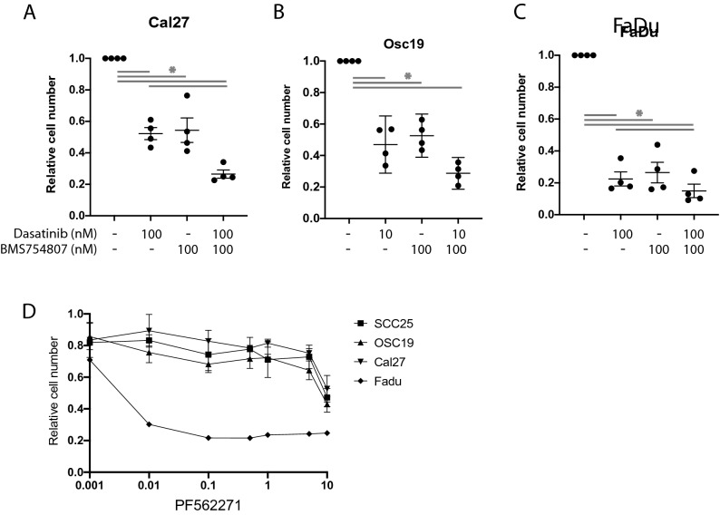
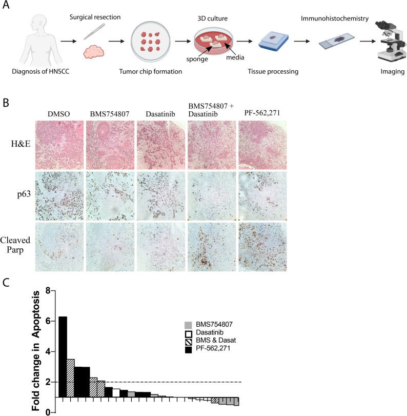
Head and neck cancer is the sixth most common cancer worldwide with a 5-year survival of only 65%. Targeting compensatory signaling pathways may improve therapeutic responses and combat resistance. Utilizing reverse phase protein arrays (RPPA) to assess the proteome and explore mechanisms of synergistic growth inhibition in HNSCC cell lines treated with IGF1R and Src inhibitors, BMS754807 and dasatinib, respectively, we identified focal adhesion signaling as a critical node. Focal Adhesion Kinase (FAK) and Paxillin phosphorylation were decreased as early as 15 min after treatment, and treatment with a FAK inhibitor, PF-562,271, was sufficient to decrease viability in vitro. Treatment of 3D spheroids demonstrated robust cytotoxicity suggesting that the combination of BMS754807 and dasatinib is effective in multiple experimental models. Furthermore, treatment with BMS754807 and dasatinib significantly decreased cell motility, migration, and invasion in multiple HNSCC cell lines. Most strikingly, treatment with BMS754807 and dasatinib, or a FAK inhibitor alone, significantly increased cleaved-PARP in human ex-vivo HNSCC patient tissues demonstrating a potential clinical utility for targeting FAK or the combined targeting of the IGF1R with Src. This ex-vivo result further confirms FAK as a vital signaling node of this combinatorial treatment and demonstrates therapeutic potential for targeting FAK in HNSCC patients.
Conflict of interest statement
The authors declare no competing interests.
Figures
Similar articles
-
Regulation of cisplatin-resistant head and neck squamous cell carcinoma by the SRC/ETS-1 signaling pathway.BMC Cancer. 2019 May 22;19(1):485. doi: 10.1186/s12885-019-5664-7. BMC Cancer. 2019. PMID: 31118072 Free PMC article.
-
Antitumor effect of insulin-like growth factor-1 receptor inhibition in head and neck squamous cell carcinoma.Laryngoscope. 2020 Jun;130(6):1470-1478. doi: 10.1002/lary.28236. Epub 2019 Aug 21. Laryngoscope. 2020. PMID: 31433065
-
Synergistic apoptosis in head and neck squamous cell carcinoma cells by co-inhibition of insulin-like growth factor-1 receptor signaling and compensatory signaling pathways.Head Neck. 2015 Dec;37(12):1722-32. doi: 10.1002/hed.23822. Epub 2015 Apr 6. Head Neck. 2015. PMID: 24986420 Free PMC article.
-
Dasatinib (BMS-354825) tyrosine kinase inhibitor suppresses invasion and induces cell cycle arrest and apoptosis of head and neck squamous cell carcinoma and non-small cell lung cancer cells.Clin Cancer Res. 2005 Oct 1;11(19 Pt 1):6924-32. doi: 10.1158/1078-0432.CCR-05-0757. Clin Cancer Res. 2005. PMID: 16203784
-
Targeting FAK radiosensitizes 3-dimensional grown human HNSCC cells through reduced Akt1 and MEK1/2 signaling.Int J Radiat Oncol Biol Phys. 2012 Aug 1;83(5):e669-76. doi: 10.1016/j.ijrobp.2012.01.065. Epub 2012 Apr 6. Int J Radiat Oncol Biol Phys. 2012. PMID: 22483702
Cited by
-
Emerging tyrosine kinase inhibitors for head and neck cancer.Expert Opin Emerg Drugs. 2022 Sep;27(3):333-344. doi: 10.1080/14728214.2022.2125954. Epub 2022 Sep 21. Expert Opin Emerg Drugs. 2022. PMID: 36131561 Free PMC article. Review.
-
Inhibiting IGF1R-mediated Survival Signaling in Head and Neck Cancer with the Peptidomimetic SSTNIGF1R.Cancer Res Commun. 2023 Jan 19;3(1):97-108. doi: 10.1158/2767-9764.CRC-22-0274. eCollection 2023 Jan. Cancer Res Commun. 2023. PMID: 36968227 Free PMC article.
-
Functional and clinical characteristics of focal adhesion kinases in cancer progression.Front Cell Dev Biol. 2022 Nov 2;10:1040311. doi: 10.3389/fcell.2022.1040311. eCollection 2022. Front Cell Dev Biol. 2022. PMID: 36407100 Free PMC article. Review.
-
Combination Therapy as a Promising Way to Fight Oral Cancer.Pharmaceutics. 2023 Jun 4;15(6):1653. doi: 10.3390/pharmaceutics15061653. Pharmaceutics. 2023. PMID: 37376101 Free PMC article. Review.
-
Does Bentonite Cause Cytotoxic and Whole-Transcriptomic Adverse Effects in Enterocytes When Used to Reduce Aflatoxin B1 Exposure?Toxins (Basel). 2022 Jun 26;14(7):435. doi: 10.3390/toxins14070435. Toxins (Basel). 2022. PMID: 35878173 Free PMC article.
References
-
- Axelrod MJ, Mendez RE, Khalil A, Leimgruber SS, Sharlow ER, Capaldo B, et al. Synergistic apoptosis in head and neck squamous cell carcinoma cells by co-inhibition of insulin-like growth factor-1 receptor signaling and compensatory signaling pathways. Head Neck. 2015;37(12):1722–1732. doi: 10.1002/hed.23822. - DOI - PMC - PubMed
Publication types
MeSH terms
Substances
Grants and funding
LinkOut - more resources
Full Text Sources
Other Literature Sources
Medical
Miscellaneous
