[Preprint]. 2021 May 21:rs.3.rs-513461.
doi: 10.21203/rs.3.rs-513461/v1.

SARS-CoV-2 Infection Causes Dopaminergic Neuron Senescence

Affiliations

SARS-CoV-2 Infection Causes Dopaminergic Neuron Senescence

Yuling Han et al. Res Sq. .

Update in

  • SARS-CoV-2 infection causes dopaminergic neuron senescence.
    Yang L, Kim TW, Han Y, Nair MS, Harschnitz O, Zhu J, Wang P, Koo SY, Lacko LA, Chandar V, Bram Y, Zhang T, Zhang W, He F, Pan C, Wu J, Huang Y, Evans T, van der Valk P, Titulaer MJ, Spoor JKH, Furler O'Brien RL, Bugiani M, D J Van de Berg W, Schwartz RE, Ho DD, Studer L, Chen S. Yang L, et al. Cell Stem Cell. 2024 Feb 1;31(2):196-211.e6. doi: 10.1016/j.stem.2023.12.012. Epub 2024 Jan 17. Cell Stem Cell. 2024. PMID: 38237586 Free PMC article.

Abstract

COVID-19 patients commonly present with neurological signs of central nervous system (CNS)1-3 and/or peripheral nervous system dysfunction4. However, which neural cells are permissive to infection by severe acute respiratory syndrome coronavirus 2 (SARS-CoV-2) has been controversial. Here, we show that midbrain dopamine (DA) neurons derived from human pluripotent stem cells (hPSCs) are selectively permissive to SARS-CoV-2 infection both in vitro and upon transplantation in vivo, and that SARS-CoV-2 infection triggers a DA neuron inflammatory and cellular senescence response. A high-throughput screen in hPSC-derived DA neurons identified several FDA approved drugs, including riluzole, metformin, and imatinib, that can rescue the cellular senescence phenotype and prevent SARS-CoV-2 infection. RNA-seq analysis of human ventral midbrain tissue from COVID-19 patients, using formalin-fixed paraffin-embedded autopsy samples, confirmed the induction of an inflammatory and cellular senescence signature and identified low levels of SARS-CoV-2 transcripts. Our findings demonstrate that hPSC-derived DA neurons can serve as a disease model to study neuronal susceptibility to SARS-CoV-2 and to identify candidate neuroprotective drugs for COVID-19 patients. The susceptibility of hPSC-derived DA neurons to SARS-CoV-2 and the observed inflammatory and senescence transcriptional responses suggest the need for careful, long-term monitoring of neurological problems in COVID-19 patients.

Keywords: SARS-CoV-2; midbrain dopamine; neuron senescence.


Conflict of interest statement

Competing Interests. R.E.S. is on the scientific advisory board of Miromatrix Inc and is a paid consultant and speaker for Alnylam Inc. L.S. is a scientific cofounder and paid consultant of BlueRock Therapeutics Inc. The other authors declare no competing interests.

Figures

Extended Data Figure 1.
Extended Data Figure 1.. hPSC-derived dopaminergic cells can be infected by SARS-CoV-2 pseudo-entry virus.
a, Representative confocal images of hPSC-derived DA neurons stained with antibodies recognizing Nurr1-GFP, TH or FOXA2. Scale bar=50μm. b, Representative confocal images of hPSC-derived DA neurons stained with ACE2 antibody. Scale bar=50μm. c, Luciferase activity in lysates from hPSC-derived DA neurons at 24 hpi following exposure to SARS-CoV-2-entry virus at MOI=0.01. d, Representative confocal images of hPSC-derived DA neurons infected with SARS-CoV-2-entry virus (MOI=0.01) at 24 hpi using antibodies against luciferase and DA markers. Scale bar=50μm. Data was presented as mean ± STDEV. P values were calculated by unpaired two-tailed Student’s t test. ***P < 0.001.
Figure 1.
Figure 1.. hPSC-derived DA neurons, and in particular A9 DA neurons, are permissive to SARS-CoV-2 infection.
a, Schematic for in vivo infection. b, Representative confocal image of DA neuron xenograft stained with antibodies against ACE2 and Nurr1-GFP. Scale bar=50μm. c, Representative confocal image of DA neuron xenograft at 24 hpi stained with antibodies against Luc and Nurr1-GFP. Scale bar=60μm. d. qRT-PCR analysis of total RNA extracted from hPSC-derived DA neurons at 48 hpi of SARS-CoV-2 infection (MOI=0.2) for viral N sgRNA. The graph depicts the mean sgRNA level normalized to ACTB. e, Representative confocal images of hPSC-DA neurons infected with SARS-CoV-2 (MOI=0.1) at 72 hpi using antibodies against SARS-CoV-2 Nucleocapsid protein (SARS-N) and markers for DA neurons. Scale bar=50μm. f, Transmission electron microscope (TEM) images of DA neurons at 72 hpi of SARS-CoV-2 (MOI=1.0). Arrows point to SARS-CoV-2 viral particles. Right panel: Zoom in images. Scale bar=1μm. g, RNA-seq read coverage of the viral genome in infected hPSC-derived DA neurons at 48 hpi (MOI=0.2). The schematic below depicts the SARS-CoV-2 genome and was created using BioRender. h, PCA plot of gene expression profiles from mock infected and SARS-CoV-2 infected hPSC-derived DA neurons at 48 hpi (MOI=0.2). i, Clustering analysis of mock or SARS-CoV-2 infected hPSC-derived DA neurons at 48 hpi (MOI=0.2). j, Heatmap of DA neurons and A9 DA neuron marker genes expression levels in mock or SARS-CoV-2 infected hPSC-derived DA neurons at 48 hpi (MOI=0.2). k, l, Fluorescence in situ hybridization (k) and quantification (l) of A9 DA marker, LMO3, in mock or SARS-CoV-2 infected hPSC-derived DA neurons at 48 hpi (MOI=0.2) Scale bar=10μm. N=3 independent biological replicates. Data was presented as mean ± STDEV. P values were calculated by unpaired two-tailed Student’s t test. **P < 0.01.
Figure 2.
Figure 2.. SARS-CoV-2 infection induces senescence of DA neurons.
a, Volcano plot indicating differentially expressed genes in mock or SARS-CoV-2 infected hPSC-derived DA neurons at 48 hpi (MOI=0.2). Differentially expressed genes (p-adjusted value < 0.05) are indicated in red. Non-significant differentially expressed genes with a log2 (Fold Change) > 0.5 are indicated in black. b, c, Heatmap of chemokine/cytokines (b) and inflammation associated genes (c) in mock or SARS-CoV-2 infected hPSC-derived DA neurons at 48 hpi (MOI=0.2). d, IPA analysis of differentially expressed genes in a. e, Heatmap of senescence associated genes in mock or SARS-CoV-2 infected hPSC-derived DA neurons at 48 hpi (MOI=0.2). f, g, Beta-Gal staining (f) and quantification (g) of mock or SARS-CoV-2 infected hPSC-derived DA neurons at 72 hpi (MOI=0.1). Scale bar=75μm. h. qRT-PCR analysis of senescence related genes of mock or SARS-CoV-2 infected hPSC-derived DA neurons at 48 hpi (MOI=0.2). i. TEM images of mock or SARS-CoV-2 infected hPSC-derived DA neurons at 72 hpi (MOI=1.0). Scale bar=2μm. N=3 independent biological replicates. Data was presented as mean ± STDEV. P values were calculated by unpaired two-tailed Student’s t test. *P < 0.05, **P < 0.01, and ***P < 0.001.
Figure 3.
Figure 3.. Riluzole, metformin, and imatinib rescue SARS-CoV-2 induced senescence of DA neurons.
a, Primary screening results. X-axis is the compound number. Y axis is the Z-score. Red line is Z-score <−2, which means the luminescent signal is lower than average-2xSTDEV. b-d, Chemical structures of riluzole (b), metformin (c), and imatinib (d). e-g, Efficacy and cytotoxicity curves of riluzole (e), metformin (f), and imatinib (g). h, i, Beta-Gal staining (h) and the quantification (i) of DMSO or drug candidate-treated hPSC-derived DA neurons at 72 hpi upon SARS-CoV-2 infection (MOI=0.1). Scale bar=100μm. j, qRT-PCR analysis of senescence related genes of DMSO or drug candidate-treated hPSC-derived DA neurons at 48 hpi upon SARS-CoV-2 infection (MOI=0.1). k, qRT-PCR analysis of total RNA extracted from DMSO or drug candidate-treated hPSC-derived DA neurons at 48 hpi upon SARS-CoV-2 infection (MOI=0.1) for viral N sgRNA. The graph depicts the mean sgRNA level normalized to ACTB. l, m, Representative confocal images (l) and quantification (m) of DMSO or drug candidate-treated hPSC-derived DA neurons at 72 hpi upon SARS-CoV-2 infection (MOI=0.1) using antibodies against SARS-CoV-2 Nucleocapsid protein (SARS-N) and markers for DA neurons. Scale bar=100μm. n, o, PCA plot of gene expression profiles (n) and clustering analysis (o) of DMSO or drug candidate-treated hPSC-derived DA neurons at 48 hpi upon SARS-CoV-2 infection (MOI=0.1). p, Heatmap of senescence associated genes of DMSO or drug candidate-treated hPSC-derived DA neurons at 48 hpi upon SARS-CoV-2 infection (MOI=0.1). N=3 independent biological replicates. Data was presented as mean ± STDEV. P values were calculated by unpaired two-tailed Student’s t test. *P < 0.05, **P < 0.01, and ***P < 0.001.
Figure 4.
Figure 4.. SARS-CoV-2 is detected in autopsy substantia nigra samples of COVID-19 patients.
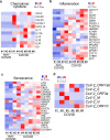
a-c, Heatmap of chemokine/cytokine (a), inflammation associated genes (b) and senescence associated genes (c) in the autopsy substantia nigra sections of COVID-19 patients versus non-COVID-19 patients. (N=6 COVID-19 patients; N=3 non-COVID-19 patients). d, Heatmap of viral transcripts in autopsy substantia nigra sections of COVID-19 patients.

References

    1. Mao L. et al. Neurologic Manifestations of Hospitalized Patients With Coronavirus Disease 2019 in Wuhan, China. JAMA Neurol (2020). - PMC - PubMed
    1. Huang C. et al. Clinical features of patients infected with 2019 novel coronavirus in Wuhan, China. Lancet 395, 497–506 (2020). - PMC - PubMed
    1. Helms J. et al. Neurologic Features in Severe SARS-CoV-2 Infection. N Engl J Med 382, 2268–2270 (2020). - PMC - PubMed
    1. Scheidl E., Canseco D.D., Hadji-Naumov A. & Bereznai B. Guillain-Barre syndrome during SARS-CoV-2 pandemic: A case report and review of recent literature. J Peripher Nerv Syst 25, 204–207 (2020). - PMC - PubMed
    1. Pezzini A. & Padovani A. Lifting the mask on neurological manifestations of COVID-19. Nat Rev Neurol 16, 636–644 (2020). - PMC - PubMed

Publication types

LinkOut - more resources