[Preprint]. 2021 May 19:rs.3.rs-515215.
doi: 10.21203/rs.3.rs-515215/v1.

CryoEM and AI reveal a structure of SARS-CoV-2 Nsp2, a multifunctional protein involved in key host processes

Meghna Gupta #  1 Caleigh M Azumaya #  1 Michelle Moritz #  1 Sergei Pourmal #  1 Amy Diallo #  1 Gregory E Merz #  1 Gwendolyn Jang #  2   3   4   5 Mehdi Bouhaddou #  2   3   4   5 Andrea Fossati #  2   3   4   5 Axel F Brilot  1 Devan Diwanji  1 Evelyn Hernandez  1 Nadia Herrera  1 Huong T Kratochvil  1 Victor L Lam  1 Fei Li  1 Yang Li  1 Henry C Nguyen  1 Carlos Nowotny  1 Tristan W Owens  1 Jessica K Peters  1 Alexandrea N Rizo  1 Ursula Schulze-Gahmen  1 Amber M Smith  1 Iris D Young  1 Zanlin Yu  1 Daniel Asarnow  1 Christian Billesbølle  1 Melody G Campbell  1   6 Jen Chen  1 Kuei-Ho Chen  2   3   4   5 Un Seng Chio  1 Miles Sasha Dickinson  1 Loan Doan  1 Mingliang Jin  1 Kate Kim  1 Junrui Li  1 Yen-Li Li  1 Edmond Linossi  1 Yanxin Liu  1 Megan Lo  1 Jocelyne Lopez  1 Kyle E Lopez  1 Adamo Mancino  1 Frank R Moss 3rd  1 Michael D Paul  1 Komal Ishwar Pawar  1 Adrian Pelin  2   3   4   5 Thomas H Pospiech Jr  1 Cristina Puchades  1 Soumya Govinda Remesh  1 Maliheh Safari  1 Kaitlin Schaefer  1 Ming Sun  1   7 Mariano C Tabios  1 Aye C Thwin  1 Erron W Titus  1 Raphael Trenker  1 Eric Tse  1 Tsz Kin Martin Tsui  1 Feng Wang  1 Kaihua Zhang  1 Yang Zhang  1 Jianhua Zhao  1 Fengbo Zhou  1 Yuan Zhou  2   3   4   5 Lorena Zuliani-Alvarez  1   2   3   4   5 QCRG Structural Biology ConsortiumDavid A Agard  1   2   3   8 Yifan Cheng  1   2   3   8   9 James S Fraser  1   2   3   8 Natalia Jura  1   2   3   10 Tanja Kortemme  1   2   3   11   12 Aashish Manglik  1   2   3   13 Daniel R Southworth  1   2   3   8 Robert M Stroud  1   2   3   8 Danielle L Swaney  2   3   4   5 Nevan J Krogan  2   3   4   5   14 Adam Frost  1   2   3   8   15 Oren S Rosenberg  1   2   3   8   15   16 Kliment A Verba  1   2   3   13
Affiliations

CryoEM and AI reveal a structure of SARS-CoV-2 Nsp2, a multifunctional protein involved in key host processes

Meghna Gupta et al. Res Sq. .

Abstract

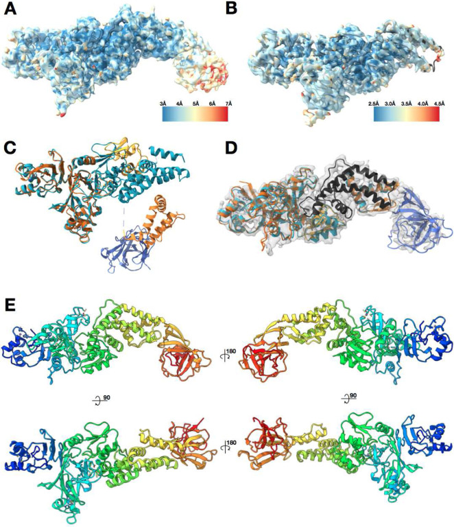
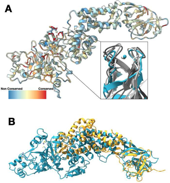
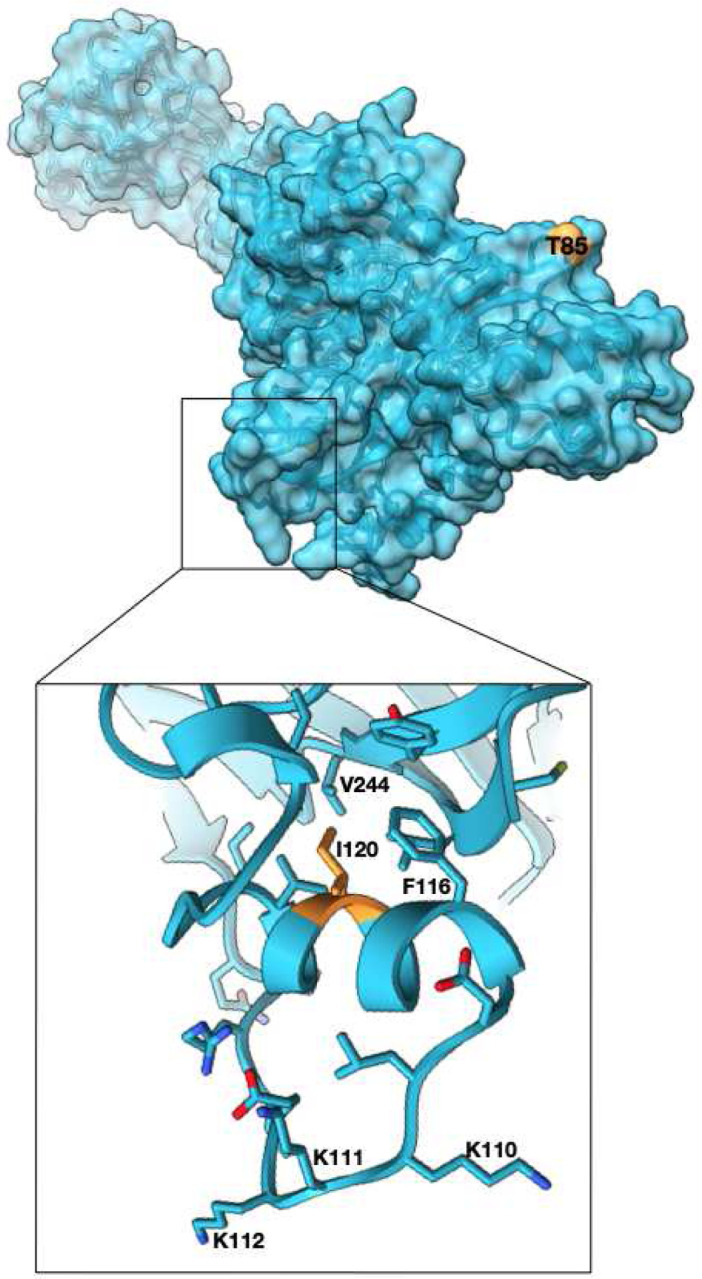
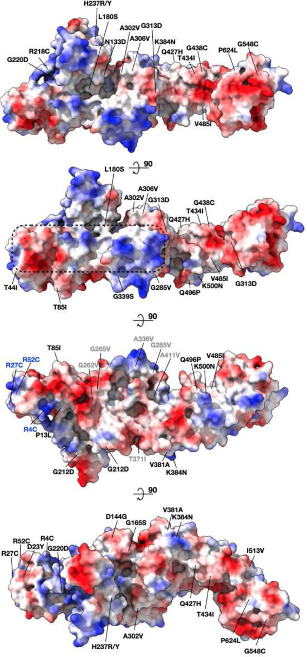
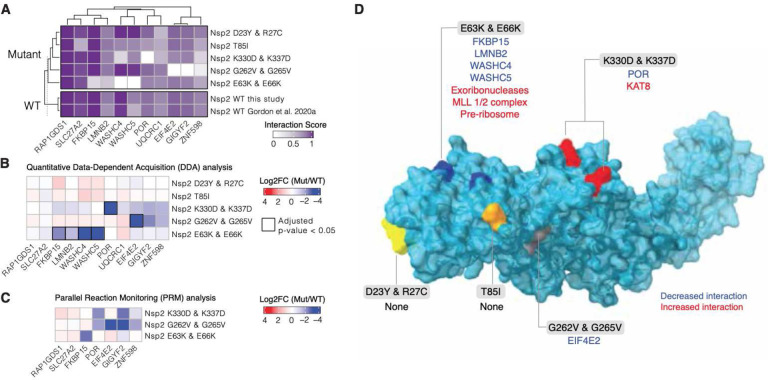
The SARS-CoV-2 protein Nsp2 has been implicated in a wide range of viral processes, but its exact functions, and the structural basis of those functions, remain unknown. Here, we report an atomic model for full-length Nsp2 obtained by combining cryo-electron microscopy with deep learning-based structure prediction from AlphaFold2. The resulting structure reveals a highly-conserved zinc ion-binding site, suggesting a role for Nsp2 in RNA binding. Mapping emerging mutations from variants of SARS-CoV-2 on the resulting structure shows potential host-Nsp2 interaction regions. Using structural analysis together with affinity tagged purification mass spectrometry experiments, we identify Nsp2 mutants that are unable to interact with the actin-nucleation-promoting WASH protein complex or with GIGYF2, an inhibitor of translation initiation and modulator of ribosome-associated quality control. Our work suggests a potential role of Nsp2 in linking viral transcription within the viral replication-transcription complexes (RTC) to the translation initiation of the viral message. Collectively, the structure reported here, combined with mutant interaction mapping, provides a foundation for functional studies of this evolutionary conserved coronavirus protein and may assist future drug design.

Keywords: SARS-CoV-2; drug design; proteins.


Figures

Figure 1.
Figure 1.. Nsp2 cryo-EM density and model overview.
(A) 3.8 Å cryo-EM map of Nsp2 colored by local resolution showing the extra density at the C-terminus (B) 3.2 Å cryo-EM map of Nsp2 colored by local resolution with the resulting model in ribbon (C) Most up to date AlphaFold2 Nsp2 model (multicolored) was aligned to the experimentally built Nsp2 model shown in cyan ribbon. The missing 93 amino acids from the latest AlphaFold2 prediction are indicated by a dashed line. (D) AlphaFold2 Nsp2 predicted model (same as C) was broken into 4 regions and then individually aligned to the experimentally built model (domains segmented from the AlphaFold2 prediction are in shades of orange, experimental model is in cyan, in black is the region missing from the AlphaFold2 prediction but built into the experimental model, in blue is the C-terminal domain as predicted by AlphaFold2 and fit into the 3.8 Å cryo-EM map) (E) The resulting full length Nsp2 structure depicted as ribbon and colored as rainbow, blue for N terminus to red for C-terminus.
Figure 2.
Figure 2.. Nsp2 has a conserved zinc binding motif but otherwise shows low conservation.
(A) Nsp2 structure depicted as ribbon and colored by conservation (see methods for details). The four cysteines show the highest conservation and are indicated in red. The magnified insert shows the zinc ribbon motif of Nsp2 in cyan aligned to zinc ribbon motifs from structurally similar structures in the PDB in shades of gray (PDBs: 1JJ2, 5XON, 1QUP, 4C2M). (B) Structure of IBV Nsp2 (PDB:3LD1, yellow ribbon) aligns well to the C-terminal region of SARS-CoV-2 Nsp2 (cyan) even though it has less than 10% sequence identity.
Figure 3.
Figure 3.. High frequency mutations in Nsp2 may provide host specific advantages.
T85I mutation which is present in 13% of all the SARS-CoV-2 sequences is at the surface and may mediate host specific protein-protein interactions (Nsp2 surface in cyan, T85 in orange). Another mutated site, I120 points into a hydrophobic core stabilizing a small helix which is attached to a highly positively charged surface loop. Phe substitution at the site may further stabilize the helix. (zoomed panel, I120 in orange).
Figure 4.
Figure 4.. Mapping surface mutations in SARS-CoV-2 Nsp2 shows both potentially constrained surfaces and rapidly changing regions.
All the positively selected mutations on Nsp2 mapped to the protein surface are colored by the surface charge. The region that is less susceptible to mutations is inscribed in a dashed rectangle. Residues that became less charged at the N-terminus are marked in blue (cluster 1). Residues that became more hydrophobic are marked in gray (cluster 2).
Figure 5.
Figure 5.. Nsp2 possesses multiple interaction surfaces for host proteins.
(A) Interaction scores (average between MiST and Saint Scores) for human proteins (“preys”) deemed high-confidence interactions in at least one affinity purification (“bait”) mass spectrometry assay and detected to interact with both the wild-type Nsp2 in this study and in Gordon et al (2020a). Interaction scores range from zero to one, one being the most high-confidence. (B) Quantitative statistical analysis of data-dependent acquisition (DDA) mass spectrometry data using MSstats for interactions selected and depicted in A. Prey intensities were normalized by bait expression abundance. Log2 fold changes and BH-adjusted p-values were calculated by comparing each mutant to the wild-type from this study. Square black outlines depict adjusted p-values < 0.05. (C) Parallel reaction monitoring (PRM) analysis of select preys from B for mutants found to possess significantly-changed interactions (adjusted p-value < 0.05). (D) Nsp2 structure depicted as surface (light blue) with the mutations considered in this study depicted on the surface: E63K/E66K (dark blue), K330D/K337D (red), D23Y/R27C (yellow), T85I (orange), and G262V/G265V (grey). Lost interactions (adjusted p-value < 0.05) from data-dependent acquisition global proteomics analysis (DDA) from B depicted in blue and gained protein complexes depicted in red (see Sup Fig 4)

References

    1. V’kovski P., Kratzel A., Steiner S., Stalder H. & Thiel V. Coronavirus biology and replication: implications for SARS-CoV-2. Nat. Rev. Microbiol. (2020) doi: 10.1038/s41579-020-00468-6. - DOI - PMC - PubMed
    1. Gordon D. E. et al. A SARS-CoV-2 protein interaction map reveals targets for drug repurposing. Nature 583, 459–468 (2020). - PMC - PubMed
    1. Thoms M. et al. Structural basis for translational shutdown and immune evasion by the Nsp1 protein of SARS-CoV-2. Science 369, 1249–1255 (2020). - PMC - PubMed
    1. Miorin L. et al. SARS-CoV-2 Orf6 hijacks Nup98 to block STAT nuclear import and antagonize interferon signaling. Proc. Natl. Acad. Sci. U. S. A. 117, 28344–28354 (2020). - PMC - PubMed
    1. García-Sastre A. Ten Strategies of Interferon Evasion by Viruses. Cell Host Microbe 22, 176–184 (2017). - PMC - PubMed

Publication types

LinkOut - more resources