

Cu-Ferrocene-Functionalized CaO2 Nanoparticles to Enable Tumor-Specific Synergistic Therapy with GSH Depletion and Calcium Overload
- PMID: 34032026
- PMCID: PMC8292872
- DOI: 10.1002/advs.202100241
Cu-Ferrocene-Functionalized CaO2 Nanoparticles to Enable Tumor-Specific Synergistic Therapy with GSH Depletion and Calcium Overload
Abstract
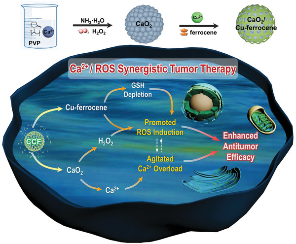
The conversion of endogenous H2 O2 into toxic hydroxyl radical (• OH) via catalytic nanoparticles is explored for tumor therapy and received considerable success. The intrinsic characteristics of microenvironment in tumor cells, such as limited H2 O2 and overexpressed glutathione (GSH), hinder the intracellular • OH accumulation and thus weaken therapeutic efficacy considerably. In this study, fine CaO2 nanoparticles with Cu-ferrocene molecules at the surface (CaO2 /Cu-ferrocene) are successfully designed and synthesized. Under an acidic condition, the particles release Ca2+ ions and H2 O2 in a rapid fashion, while they can remain stable in neutral. In addition, agitated production of • OH occurs following the Fenton reaction of H2 O2 and ferrocene molecules, and GSH is consumed by Cu2+ ions to avoid the potential • OH consumption. More interestingly, in addition to the exogenous Ca2+ released by the particles, the enhanced • OH production facilitates intracellular calcium accumulation by regulating Ca2+ channels and pumps of tumor cells. It turns out that promoted • OH induction and intracellular calcium overload enable significant in vitro and in vivo antitumor phenomena.
Keywords: CaO2; Cu-ferrocene; GSH depletion; calcium overload; synergistic tumor therapy.
© 2021 The Authors. Advanced Science published by Wiley-VCH GmbH.
Conflict of interest statement
The authors declare no conflict of interest.
Figures


Similar articles
-
A ROS storm generating nanocomposite for enhanced chemodynamic therapy through H2O2 self-supply, GSH depletion and calcium overload.Nanoscale. 2024 May 2;16(17):8479-8494. doi: 10.1039/d3nr06422k. Nanoscale. 2024. PMID: 38590261
-
A metal-organic framework functionalized CaO2-based cascade nanoreactor induces synergistic cuproptosis/ferroptosis and Ca2+ overload-mediated mitochondrial damage for enhanced sono-chemodynamic immunotherapy.Acta Biomater. 2025 Jan 24;193:455-473. doi: 10.1016/j.actbio.2024.12.010. Epub 2024 Dec 21. Acta Biomater. 2025. PMID: 39637958
-
Copper(II) Oxide Spindle-like Nanomotors Decorated with Calcium Peroxide Nanoshell as a New Nanozyme with Photothermal and Chemodynamic Functions Providing ROS Self-Amplification, Glutathione Depletion, and Cu(I)/Cu(II) Recycling.ACS Appl Mater Interfaces. 2025 Jan 8;17(1):632-649. doi: 10.1021/acsami.4c17852. Epub 2024 Dec 25. ACS Appl Mater Interfaces. 2025. PMID: 39720911 Free PMC article.
-
Advances in nanotherapeutics for tumor treatment by targeting calcium overload.Colloids Surf B Biointerfaces. 2025 Jan;245:114190. doi: 10.1016/j.colsurfb.2024.114190. Epub 2024 Sep 2. Colloids Surf B Biointerfaces. 2025. PMID: 39232477 Review.
-
Application of calcium overload-based ion interference therapy in tumor treatment: strategies, outcomes, and prospects.Front Pharmacol. 2024 Feb 15;15:1352377. doi: 10.3389/fphar.2024.1352377. eCollection 2024. Front Pharmacol. 2024. PMID: 38425645 Free PMC article. Review.
Cited by
-
Programmable Release of Chemotherapeutics from Ferrocene-Based Injectable Hydrogels Slows Melanoma Growth.Adv Healthc Mater. 2024 Oct;13(27):e2400265. doi: 10.1002/adhm.202400265. Epub 2024 Jul 15. Adv Healthc Mater. 2024. PMID: 39007274 Free PMC article.
-
Metal Peroxide Nanoparticles for Modulating the Tumor Microenvironment: Current Status and Recent Prospects.Cancers (Basel). 2024 Oct 24;16(21):3581. doi: 10.3390/cancers16213581. Cancers (Basel). 2024. PMID: 39518022 Free PMC article. Review.
-
Recent progress of metal-based nanomaterials with anti-tumor biological effects for enhanced cancer therapy.Exploration (Beijing). 2023 Jun 30;3(5):20220001. doi: 10.1002/EXP.20220001. eCollection 2023 Oct. Exploration (Beijing). 2023. PMID: 37933288 Free PMC article. Review.
-
Phototherapy techniques for the management of musculoskeletal disorders: strategies and recent advances.Biomater Res. 2023 Nov 28;27(1):123. doi: 10.1186/s40824-023-00458-8. Biomater Res. 2023. PMID: 38017585 Free PMC article. Review.
-
Multifunctional Fe-based coordination polymer nano-bomb modified with β-lapachone and CaO2 for targeted tumor dual chemodynamic therapy with enhanced ferroptosis and H2O2 self-supply.J Nanobiotechnology. 2024 Jan 3;22(1):3. doi: 10.1186/s12951-023-02287-2. J Nanobiotechnology. 2024. PMID: 38166978 Free PMC article.
References
-
- Nosaka Y., Nosaka A. Y., Chem. Rev. 2017, 11, 11302. - PubMed
-
- Gorrini C., Harris I. S., Mak T. W., Nat. Rev. Drug Discovery 2013, 12, 931. - PubMed
-
- Felsher D. W., Nat. Rev. Cancer 2003, 3, 375. - PubMed
-
- a) Lu Z., Gao J., Fang C., Zhou Y., Li X., Han G., Adv. Sci. 2020, 7, 2001223; - PMC - PubMed
- b) Chen T., Gu T., Cheng L., Li X., Han G., Liu Z., Biomaterials 2020, 255, 120202; - PubMed
- c) Gu T., Wang Y., Lu Y., Cheng L., Feng L., Zhang H., Li X., Han G., Liu Z., Adv. Mater. 2019, 31, 1806803. - PubMed
Publication types
MeSH terms
Substances
Grants and funding
LinkOut - more resources
Full Text Sources
Other Literature Sources
Medical
Miscellaneous