

Dynamic SWIR imaging near the 1950-nm water absorption band for caries lesion diagnosis
- PMID: 34032036
- PMCID: PMC8144227
- DOI: 10.1117/1.JBO.26.5.056006
Dynamic SWIR imaging near the 1950-nm water absorption band for caries lesion diagnosis
Abstract
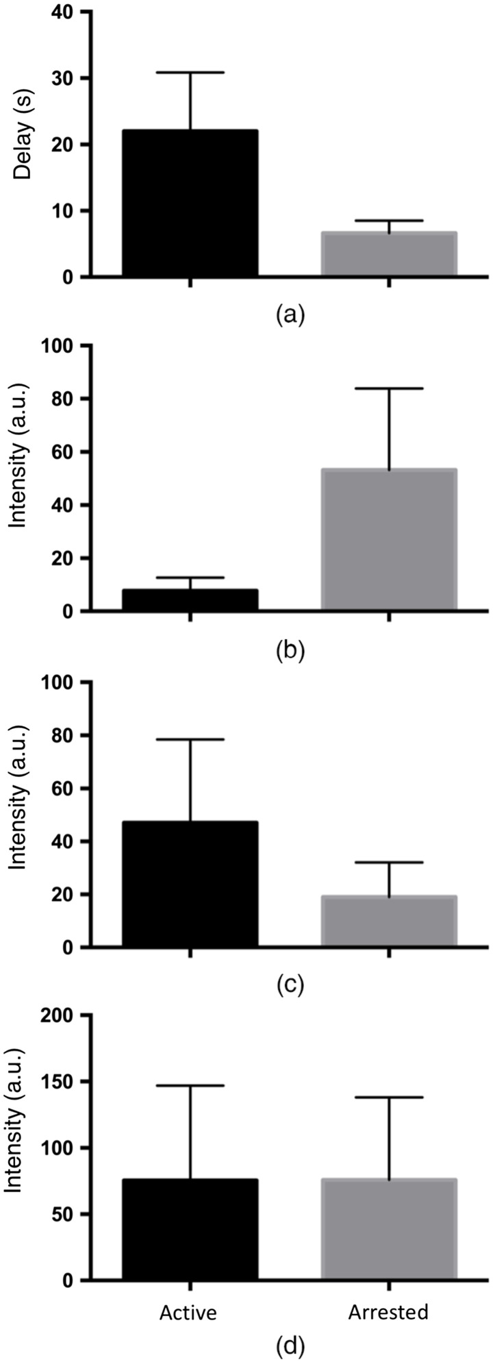
Significance: It is not sufficient to detect caries lesions on tooth surfaces; it is also necessary to measure the activity of the lesions to determine if intervention is needed. Changes in the reflectivity of lesion areas during dehydration with forced air at short wavelength infrared (SWIR) wavelengths can be used to assess lesion activity since these changes represent the evaporation dynamics of water from the lesion.
Aim: The aim of this study is to develop new optical methods for assessing lesion activity on tooth surfaces utilizing the strong water absorption band near 1950-nm.
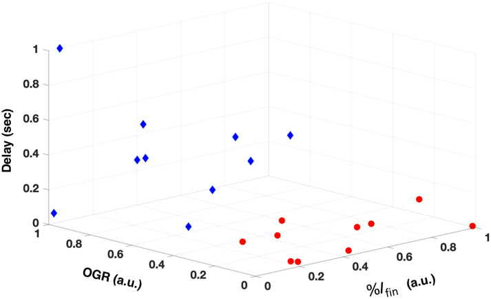
Approach: The time-resolved reflectivity of 20 active and arrested caries lesions on the surfaces of human extracted teeth was monitored at 1300 to 2000 nm using broadband light sources and an extended range InGaAs camera during drying with air.
Results: Multiple parameters representing the rate of change of the lesion reflectivity correlated with the presence of a highly mineralized outer surface zone indicative of lesion arrest measured with x-ray microtomography (microCT). Performance at 1950-nm was higher than for other wavelengths.
Conclusions: This study demonstrates that SWIR imaging near 1950-nm has great potential for the assessment of lesion activity.
Keywords: dental caries; lesion activity; short wavelength infrared imaging.
Figures
Similar articles
-
Assessment of the activity of secondary caries lesions with short-wavelength infrared, thermal, and optical coherence tomographic imaging.J Biomed Opt. 2023 Sep;28(9):094801. doi: 10.1117/1.JBO.28.9.094801. Epub 2023 Jan 10. J Biomed Opt. 2023. PMID: 36761935 Free PMC article.
-
Time-resolved SWIR imaging for the assessment of the activity of occlusal caries lesions.J Biophotonics. 2023 Oct;16(10):e202300165. doi: 10.1002/jbio.202300165. Epub 2023 Jun 22. J Biophotonics. 2023. PMID: 37316468 Free PMC article.
-
High Contrast Reflectance Imaging of Enamel Demineralization and Remineralization at 1950-nm for the Assessment of Lesion Activity.Lasers Surg Med. 2021 Sep;53(7):968-977. doi: 10.1002/lsm.23371. Epub 2021 Jan 13. Lasers Surg Med. 2021. PMID: 33442896 Free PMC article.
-
Review of short-wave infrared spectroscopy and imaging methods for biological tissue characterization.J Biomed Opt. 2015 Mar;20(3):030901. doi: 10.1117/1.JBO.20.3.030901. J Biomed Opt. 2015. PMID: 25803186 Free PMC article. Review.
-
Early caries imaging and monitoring with near-infrared light.Dent Clin North Am. 2005 Oct;49(4):771-93, vi. doi: 10.1016/j.cden.2005.05.008. Dent Clin North Am. 2005. PMID: 16150316 Review.
Cited by
-
Atlas of Dental Near-Infrared Transillumination Images.Diagnostics (Basel). 2024 May 30;14(11):1154. doi: 10.3390/diagnostics14111154. Diagnostics (Basel). 2024. PMID: 38893679 Free PMC article.
-
In vitro Assessment of lesion activity using simultaneous time-resolved reflectance imaging at 1300 and 1950 nm.Lasers Med Sci. 2024 Aug 27;39(1):223. doi: 10.1007/s10103-024-04168-y. Lasers Med Sci. 2024. PMID: 39191998
-
Caries inhibition of simulated active caries lesions with CO2 laser irradiation and fluoride.Proc SPIE Int Soc Opt Eng. 2022 Jan-Feb;11942:119420B. doi: 10.1117/12.2608308. Epub 2022 Mar 4. Proc SPIE Int Soc Opt Eng. 2022. PMID: 35450400 Free PMC article.
-
Mouthwash as a non-invasive method of indocyanine green delivery for near-infrared fluorescence dental imaging.J Biomed Opt. 2022 Jun;27(6):066001. doi: 10.1117/1.JBO.27.6.066001. J Biomed Opt. 2022. PMID: 35689334 Free PMC article.
-
Use of SWIR dehydration and OCT to assess the complete arrest of simulated incipient caries lesions.Proc SPIE Int Soc Opt Eng. 2022 Jan-Feb;11942:119420A. doi: 10.1117/12.2608297. Epub 2022 Mar 4. Proc SPIE Int Soc Opt Eng. 2022. PMID: 35444361 Free PMC article.
References
-
- Fejerskov O., Nyvad B., Kidd E., Dental Caries: The Disease and its Clinical Management, Wiley Blackwell, Hoboken, New Jersey: (2015).
-
- Ando M., Stookey G. K., Zero D. T., “Ability of quantitative light-induced fluorescence (QLF) to assess the activity of white spot lesions during dehydration,” Am. J. Dent. 19(1), 15–8 (2006). - PubMed
Publication types
MeSH terms
Substances
Grants and funding
LinkOut - more resources
Full Text Sources
Other Literature Sources
Medical
