. 2021 Sep;239(3):545-556.
doi: 10.1111/joa.13451. Epub 2021 May 25.

High-resolution magnetic resonance and mass spectrometry imaging of the human larynx

Affiliations

High-resolution magnetic resonance and mass spectrometry imaging of the human larynx

Ayami Ohno Kishimoto et al. J Anat. 2021 Sep.

Abstract

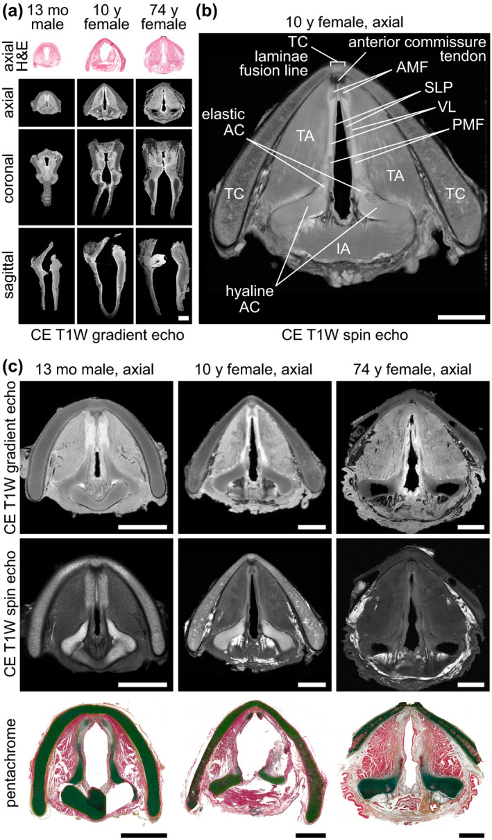
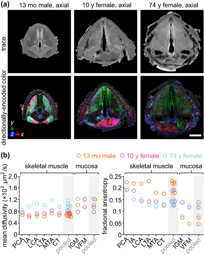
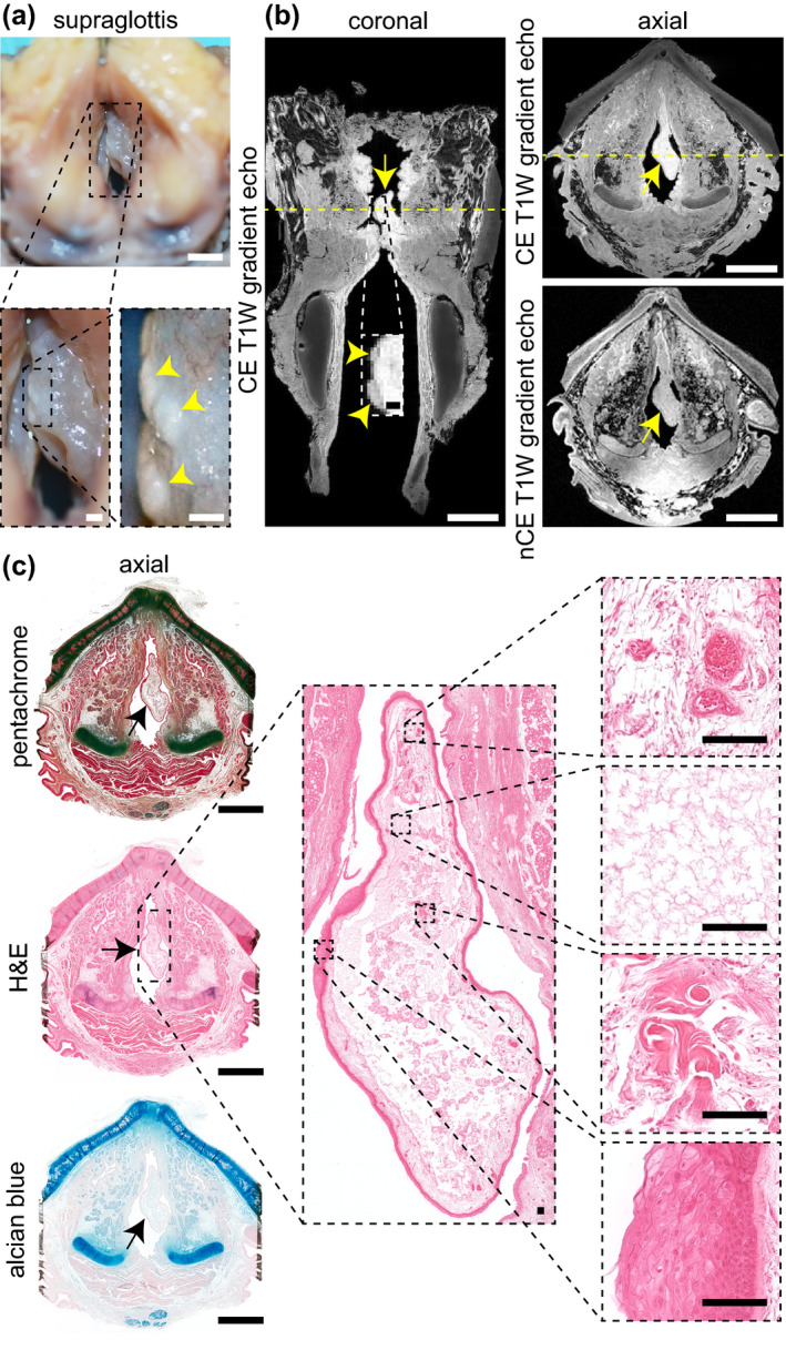
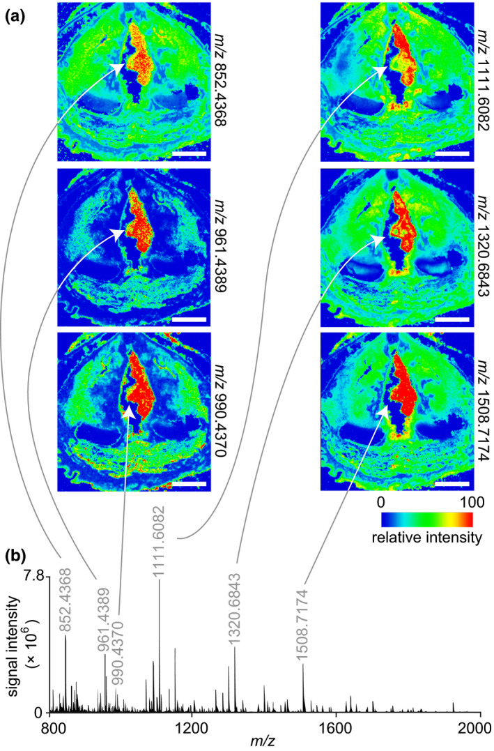
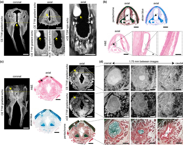
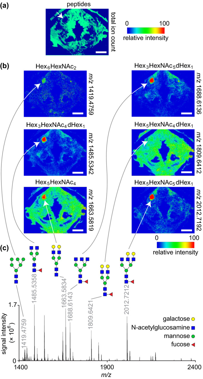
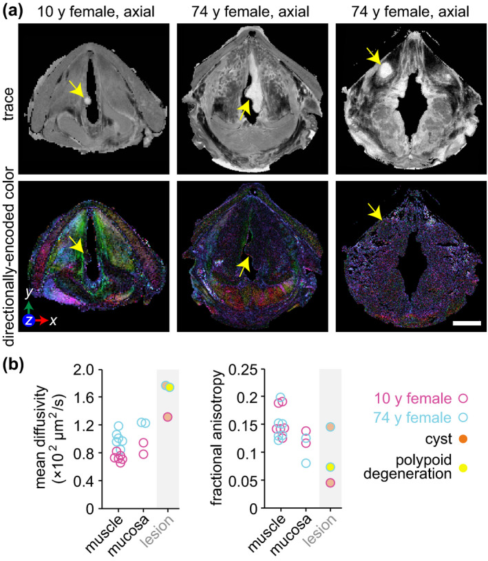
High-resolution, noninvasive and nondestructive imaging of the subepithelial structures of the larynx would enhance microanatomic tissue assessment and clinical decision making; similarly, in situ molecular profiling of laryngeal tissue would enhance biomarker discovery and pathology readout. Towards these goals, we assessed the capabilities of high-resolution magnetic resonance imaging (MRI) and matrix-assisted laser desorption/ionisation-mass spectrometry (MALDI-MS) imaging of rarely reported paediatric and adult cadaveric larynges that contained pathologies. The donors were a 13-month-old male, a 10-year-old female with an infraglottic mucus retention cyst and a 74-year-old female with advanced polypoid degeneration and a mucus retention cyst. MR and molecular imaging data were corroborated using whole-organ histology. Our MR protocols imaged the larynges at 45-117 μm2 in-plane resolution and capably resolved microanatomic structures that have not been previously reported radiographically-such as the vocal fold superficial lamina propria, vocal ligament and macula flavae; age-related tissue features-such as intramuscular fat deposition and cartilage ossification; and the lesions. Diffusion tensor imaging characterised differences in water diffusivity, primary tissue fibre orientation, and fractional anisotropy between the intrinsic laryngeal muscles, mucosae and lesions. MALDI-MS imaging revealed peptide signatures and putative protein assignments for the polypoid degeneration lesion and the N-glycan constituents of one mucus retention cyst. These imaging approaches have immediate application in experimental research and, with ongoing technology development, potential for future clinical application.

Keywords: Reinke's oedema; diffusion tensor imaging; glycomics; molecular imaging; mucus retention cyst; polypoid degeneration; proteomics; vocal fold; whole-larynx histology.


Figures

FIGURE 1
FIGURE 1
High‐resolution MRI of paediatric and adult cadaveric human larynges at the glottis. (a) Orthogonal CE T1W gradient echo images showing orientation of the glottis in the axial, coronal and sagittal planes. The corresponding H&E‐stained histological images are in the axial plane. Images are shown at a uniform scale to reflect relative specimen size. (b) CE T1W spin echo axial image showing resolution of vocal fold substructures in a 10‐year‐old female larynx. (c) CE T1W gradient echo and spin echo and corresponding pentachrome‐stained histological images of a 13‐month‐old male, 10‐year‐old female and 74‐year‐old female larynx at the glottis in the axial plane. Note: The 13‐month‐old specimen exhibits a hyperintense signal artefact at the anterior glottis due to fluid trapped between the vocal folds during MRI; the 74‐year‐old specimen had the lateral TC laminae resected prior to imaging; the histological images exhibit moderate connective tissue shrinkage and tearing artefacts. AC, arytenoid cartilage; AMF, anterior macula flava; CE, contrast‐enhanced; H&E, haematoxylin and eosin; IA, interarytenoid muscle; mo, month; MRI, magnetic resonance imaging; PMF, posterior macula flava; SLP, superficial lamina propria; T1W, T1‐weighted; TA, thyroarytenoid muscle; TC, thyroid cartilage; VL, vocal ligament; y, year. Scale bars, 5 mm
FIGURE 2
FIGURE 2
DTI of paediatric and adult cadaveric human larynges. (a) Trace and directionally encoded colour maps of a 13‐month‐old male, 10‐year‐old female and 74‐year‐old female larynx at the glottis in the axial plane. The trace maps show the diffusivity of water within the specimens. The directionally encoded colour maps are weighted by anisotropy and show the primary orientation of tissue fibres: green, anterior‐posterior; red, medial‐lateral; blue, cranial‐caudal. Note: The 74‐year‐old specimen had the lateral thyroid cartilage laminae resected prior to imaging. (b) Mean diffusivity and fractional anisotropy of six skeletal muscle and two mucosal regions within each larynx. Measurements were obtained via manual tracing of each region of interest in the relevant DTI images from each full larynx scan. DTI, diffusion tensor imaging; IA, interarytenoid muscle; IGM, infraglottic mucosa; LCA, lateral cricoarytenoid muscle; LTA, lateral thyroarytenoid muscle; mo, month; MTA, medial thyroarytenoid muscle; PCA, posterior cricoarytenoid muscle; VFM; vocal fold mucosa; y, year. Scale bar, 5 mm
FIGURE 3
FIGURE 3
MRI and histological characterisation of polypoid degeneration. (a) Superior view of the supraglottis and vocal fold mucosae at the time of procurement from a 74‐year‐old female donor. Yellow arrowheads denote leukoplakia on the epithelial surface. (b) CE (coronal and axial) and nCE (axial) T1W gradient echo images showing the lesion boundary in both planes. Yellow arrows denote the lesion; yellow arrowheads denote leukoplakia. Dashed yellow lines denote the position of orthogonal views in the respective CE coronal and axial images. Note: The lateral thyroid cartilage laminae were resected prior to imaging. (c) Pentachome‐, H&E‐ and alcian blue‐stained histological images of the lesion in the same axial view as shown on MRI. Black arrows denote the lesion. The high‐magnification H&E images show (top‐to‐bottom): capillary wall thickening and extravascular leakage of red blood cells; an edematous lake; dense fibrin deposits; epithelial hyperparakeratosis and basement membrane thickening. CE, contrast‐enhanced; H&E, haematoxylin and eosin; nCE, non‐contrast‐enhanced; MRI, magnetic resonance imaging; T1W, T1‐weighted. Scale bars, 5 mm (whole‐larynx photography, MRI and histology); 500 μm (high‐magnification photography and MRI); 100 μm (high‐magnification histology)
FIGURE 4
FIGURE 4
MALDI‐MS imaging and proteomic analysis of polypoid degeneration. (a) Ion maps from a peptide acquisition run showing the spatial distribution of six peptides with elevated relative intensity throughout the lesion. White arrows denote the lesion. The donor was a 74‐year‐old female. (b) Representative mass spectrum acquired from the lesion region. The annotated peaks correspond to the ion maps in panel (a). MALDI‐MS, matrix‐assisted laser desorption/ionisation‐mass spectrometry; m/z, mass‐to‐charge ratio. Scale bars, 5 mm
FIGURE 5
FIGURE 5
MRI and histological characterisation of mucus retention cysts. (a) CE T1W gradient echo coronal and axial images, alongside high‐magnification CE T1W gradient echo, nCE T1W gradient echo, and nCE T2W spin echo axial images, showing an infraglottic lesion in a 10‐year‐old female. Yellow arrows denote the lesion; white arrows denote adjacent mucous glands within the posterior infraglottis. Dashed yellow lines denote the position of orthogonal views in the respective CE coronal and axial images. (b) Pentachome‐, alcian blue‐ and H&E‐stained histological images of the lesion in the same axial view as shown on MRI. Black arrows denote the lesion. The high‐magnification H&E images show a cyst wall comprised of stroma and both internal and external epithelia; the cyst contents are absent. Note: The images exhibit moderate connective tissue shrinkage and tearing artefacts. (c) CE (coronal and axial) and nCE (axial) T1W gradient echo images showing a supraglottic lesion in a 74‐year‐old female. The corresponding H&E‐, alcian blue, and pentachrome‐ stained images show the same axial view as seen on MRI. Yellow, black and white arrows denote the lesion. The dashed yellow lines denote the position of orthogonal views in the respective CE coronal and axial images; additionally, the dashed lines in the coronal image denote the positions of the high‐magnification axial images in panel (d). (d) Serial CE and nCE T1W gradient echo and corresponding pentachrome‐stained histological images of the lesion at high magnification in the axial plane. The images are 1.75 mm apart and show a decrease in lesion size followed by entry to the LV in the cranial‐to‐caudal direction. The histology shows a circumferential epithelial lining and mucin‐dense lesion core (blue‐green signal). Note: The 74‐year‐old specimen had the lateral thyroid cartilage laminae resected prior to imaging; the histologic images exhibit moderate connective tissue shrinkage and tearing artifacts. CE, contrast‐enhanced; H&E, haematoxylin and eosin; LV, laryngeal ventricle; MRI, magnetic resonance imaging; nCE, non‐contrast‐enhanced; T1W, T1‐weighted; T2W, T2‐weighted. Scale bars, 5 mm (whole‐larynx MRI and histology); 1 mm (high‐magnification MRI and histology in panel [d]); 100 μm (high‐magnification histology in panel [b])
FIGURE 6
FIGURE 6
MALDI‐MS imaging and glycomic analysis of a mucus retention cyst. (a) Total ion count map from a peptide acquisition run showing the spatial distribution of summed peptide intensities. The white arrow denotes the lesion and shows no peptide signal in this region. The donor was a 74‐year‐old female. (b) Ion maps from a glycan acquisition run showing the spatial distribution of six N‐glycans identified throughout the lesion. White arrows denote the lesion. (c) Representative mass spectrum acquired from the lesion region. The annotated peaks correspond to the ion maps in panel (b). N‐glycan structures are depicted using the Symbol Nomenclature for Glycans (SNFG) format; note that the identified glycans are all in the sodiated adduct form as [M + Na]+. MALDI‐MS, matrix‐assisted laser desorption/ionisation‐mass spectrometry; m/z, mass‐to‐charge ratio. Scale bars, 5 mm
FIGURE 7
FIGURE 7
DTI of mucus retention cysts and polypoid degeneration. (a) Trace and directionally encoded colour maps of a infraglottic mucus rentention cyst in a 10‐year‐old female, polypoid degeneration in a 74‐year‐old female and supraglottic mucus retention cyst in the same 74‐year‐old female. The trace maps show the diffusivity of water within the specimens. The directionally encoded colour maps are weighted by anisotropy and show the primary orientation of tissue fibres: green, anterior‐posterior; red, medial‐lateral; blue, cranial‐caudal. Note: The 74‐year‐old specimen had the lateral thyroid cartilage laminae resected prior to imaging. (b) Mean diffusivity and fractional anisotropy of the three lesions compared to skeletal muscle and mucosa in the same larynges. The muscle and mucosal regions are those shown in Figure 2b. Measurements were obtained via manual tracing of each region of interest in the relevant DTI images from each full larynx scan. DTI, diffusion tensor imaging; y, year. Scale bar, 5 mm

Similar articles

Cited by

References

    1. Agnello, F. , Cupido, F. , Sparacia, G. , Midiri, F. , Miroddi, M. , Grassedonio, E. et al. (2017) Computerised tomography and magnetic resonance imaging of laryngeal squamous cell carcinoma: A practical approach. The Neuroradiology Journal, 30, 197–204. - PMC - PubMed
    1. Armstrong, W.B. , Ridgway, J.M. , Vokes, D.E. , Guo, S. , Perez, J. , Jackson, R.P. et al. (2006) Optical coherence tomography of laryngeal cancer. Laryngoscope, 116, 1107–1113. - PubMed
    1. Burns, J.A. , Kim, K.H. , deBoer, J.F. , Anderson, R.R. & Zeitels, S.M. (2011) Polarization‐sensitive optical coherence tomography imaging of benign and malignant laryngeal lesions: An in vivo study. Otolaryngology – Head and Neck Surgery, 145, 91–99. - PubMed
    1. Burns, J.A. , Zeitels, S.M. , Anderson, R.R. , Kobler, J.B. , Pierce, M.C. & de Boer, J.F. (2005) Imaging the mucosa of the human vocal fold with optical coherence tomography. Annals of Otology, Rhinology, and Laryngology, 114, 671–676. - PubMed
    1. Calligaris, D. , Feldman, D.R. , Norton, I. , Brastianos, P.K. , Dunn, I.F. , Santagata, S. et al. (2015) Molecular typing of meningiomas by desorption electrospray ionization mass spectrometry imaging for surgical decision‐making. International Journal of Mass Spectrometry, 377, 690–698. - PMC - PubMed

Publication types

LinkOut - more resources