. 2021 May 25;13(16):20094-20115.
doi: 10.18632/aging.203016. Epub 2021 May 25.

Overexpressed DEPDC1B contributes to the progression of hepatocellular carcinoma by CDK1

Affiliations

Overexpressed DEPDC1B contributes to the progression of hepatocellular carcinoma by CDK1

Xiao-Wei Dang et al. Aging (Albany NY). .

Abstract

Background: Hepatocellular carcinoma (HCC) is the main type of primary liver cancer and shows a heavy burden worldwide. Its recurrence and mortality rate are still uncontrolled by the usage of present treatments. More attention has been focused on exploring specific genes that play important roles in HCC procession, and the function of DEP domain containing 1B (DEPDC1B) in HCC has not been researched.

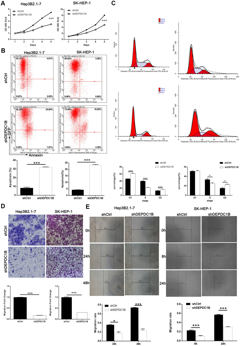
Methods: Immunohistochemical staining was used to detect the expression level of DEPDC1B in tumor tissues and adjacent normal tissues. After DEPDC1B and CDK1 knockdown in cell lines HEP3B2.1-7 and SK-HEP-1, MTT assay and colony formation assay was used to detect cell growth, flow cytometry assay was used to investigate cell apoptosis and cell cycle, wound-healing assay and Transwell assay were used to examine the tumor cell migration. Moreover, a xenograft model was constructed to research functions of DEPDC1B in tumor growth in vivo.

Results: The results show that DEPDC1B knockdown inhibit the progression of HCC, through inhibiting cell proliferation, migration, colony formation, leading to G2 phase arrest, and promoting cell apoptosis in vitro, and CDK1 was selected for further mechanic research according to the results of Human GeneChip prime view. The results of recovery experiment displayed that the functions of DEPDC1B on HCC progression were mediated by CDK1. DEPDC1B knockdown can also inhibit tumor growth in vivo.

Conclusions: The study confirmed that DEPDC1B knockdown restrains the tumor growth in vitro and vivo, and it can interact with CDK1 and rescued by CDK1. The study suggested that DEPDC1B was as a potential therapeutic target involved in HCC growth and progression.

Keywords: CDK1; DEPDC1B; hepatocellular carcinoma (HCC); migration; proliferation.


Conflict of interest statement

CONFLICTS OF INTEREST: The authors declare that they have no conflicts of interest.

Figures

Figure 1
Figure 1
Overexpressed DEPDC1B in tumor tissues and it was related to advanced stage. The expression of DEPDC1B was detected by immunohistochemistry and overexpressed in cancer tissues compared with normal tissues (Magnification ×200, scale bar = 50 μm).
Figure 2
Figure 2
Successful construction of DEPDC1B knockdown. (A) The expression level of DEPDC1B in SK-HEP-1, Hep3B2.1-7, huh-7 was significantly higher than in HCCLM3 (P<0.05). (B) The fluorescence of cells, which were infected with shCtrl, shDEPDC1B for 72 h, observed by microscope demonstrates a >80% efficiency of infection and the normal cell condition. The results of qRT-PCR show that, after the infection of lentivirus: (C) Compared with the shCtrl group, in HEP3B2.1-7 cells, the knockdown efficiency of DEPDC1B in shDEPDC1B group was 71.35% (P<0.001) and in SK-HEP-1 cells, the knockdown efficiency of DEPDC1B in shDEPDC1B group was 74.1% (P<0.001). The results of Western blot show that, after the infection of lentivirus: (D) compared with shCtrl group, in HEP3B2.1-7 and SK-HEP-1 cells: the protein level of DEPDC1B in shDEPDC1B group was down-regulated. *: P <0.05. **: P <0.01. ***: P <0.001.
Figure 3
Figure 3
DEPDC1B knockdown inhibited HCC cell progression in vivo. (A) The results of MTT assay show that, after the infection of lentivirus: compared with shCtrl group, the cells in shDEPDC1B group exhibited slower proliferation rate (P<0.001). (B) The results of flow cytometry demonstrate that, after the infection of lentivirus: compared with shCtrl group, apoptosis percentage was increased in shDEPDC1B group (P<0.001). (C) The results of flow cytometry show that, compared with shCtrl group, in shDEPDC1B group, the percentage of SK-HEP-1 cells in G2 phase increased (P<0.01), and the percentage of Hep3B2.1-7cells in G2 phase increased significantly in shDEPDC1B group (P<0.001). Transwell assay showed that, after the infection of lentivirus: compared with shCtrl group, the migration ability of cells in shDEPDC1B group was inhibited (P<0.001) (D). (E) The results of wound-healing assay showed that, in HEP3B2.1-7 and SK-HEP-1cells, compared with shCtrl group, the migration rate of cells in shDEPDC1B group was decreased by 68% (P<0.001) and 47% (P<0.001). **: P <0.01. ***: P <0.001.
Figure 4
Figure 4
Potential mechanisms of DEPDC1B functions on HCC. (AC) Bcl-2, Bcl-w, HSP27, HSP70, Livin, sTNF-R2, TNF-α, and TNF-β was significantly down-regulated in shDEPDC1B (P<0.05). (D) P-Akt, CDK6, Cyclin D1, and PIK3CA was down-regulated in shDEPDC1B group.
Figure 5
Figure 5
Obtained the DEGs by human GeneChip PrimeView and the expression at mRNA and protein level. (A) Genes were represented as red points or green points, when Fold Change ≤-1.5 and FDR<0.05 or Fold Change ≥1.5 and FDR<0.05. (B) Up-regulated genes and down-regulated genes were labeled as red and green, respectively. (C) Salvage Pathways of Pyrimidine Ribonucleotides, NER Pathway, Estrogen-mediated S-phase Entry, and Cyclins and Cell Cycle Regulation were significantly inhibited. (D) The enrichment of DEGS in disease and function. (E) The interaction network between DEPDC1B, Cyclins and Cell Cycle Regulation, and Wnt/β-catenin Signaling pathway. (F) The results of qRT-PCR showed that, compared to shCtrl group, the expression level of E2F1, CCNA1, TFDP1, CCNA2, FOS, UBA52, CCNB1, JUN, CCNB2, CCNE2, MYC, CDK1, CDK2, CDK6, RBL1, CDKN2C, CDC25A, SKP2, DKK1, and TCF3 was down-regulated (P<0.05). (G) The results of western blot showed that CCNE2, CDK1, CDK6, and E2F1was significantly down-regulated in shDEPDC1B group, compared to shCtrl group. (H) The results of IHC showed that the expression level of CDK1 in cancer tissues was higher than in normal tissues (scale bar = 50 μm).
Figure 6
Figure 6
Functions on cell progression. (A) The results of Celigo cell counting assay show that, compared with NC(OE+KD) group: the cells in DEPDC1B+NC (KD) group exhibited faster proliferation rate (P<0.001), and the cells in shCDK1+NC(OE) group exhibited slower proliferation rate (P<0.001). Compared to DEPDC1B+NC(KD) group, the cells in DEPDC1B+shCDK1 group exhibited slower proliferation rate (P<0.01). The cells in DEPDC1B+shCDK1 group exhibit faster proliferation rate, compared with shCDK1+NC(OE) group (P<0.01). (B) The results of colony formation assay show that, compared to NC(OE+KD) group: the cell colony number in DEPDC1B+NC(KD) group was significantly increased (P<0.001), while the cell colony number in shCDK1+NC(OE) group was significantly decreased (P<0.001). Compared with DEPDC1B+NC(KD) group, cell colony number was significantly decreased in DEPDC1B+shCDK1 group (P<0.001). Cell colony number in DEPDC1B+shCDK1 group was significantly increased, compared to shCDK1+NC(OE) group (P<0.01). (C) The results of flow cytometry demonstrate that: compared to NC(OE+KD) group: cell apoptosis was decreased in DEPDC1B+NC(KD) group (P<0.01), and in shCDK1+NC(OE) group, cell apoptosis was significantly increased (P<0.001). Compared with DEPDC1B+NC(KD) group, cell apoptosis was significantly increased in DEPDC1B+shCDK1 group (P<0.001). Compared to shCDK1+NC(OE) group, cell apoptosis was decreased in DEPDC1B+shCDK1 group (P<0.001). **: P <0.01. ***: P <0.001.
Figure 7
Figure 7
Detection of cell migration ability, and interaction between CDK1 and DEPDC1B. (A) The results of wound-healing assay and Transwell assay showed that, compared to NC(OE+KD) group: the migration rate of cells was significantly increased in DEPDC1B+NC(KD) group (48 h) (P<0.001), while in shCDK1+NC(OE) group (48 h), the migration rate of cells was significantly decreased (P<0.001). Compared to DEPDC1B+NC(KD) group, the migration rate of cells was significantly decreased in DEPDC1B+shCDK1 group (48 h) (P<0.001). Compared with shCDK1+NC(OE) group, the migration rate of cells was significantly increased in DEPDC1B+shCDK1 group (48 h) (P<0.05). (B) The transwell assay shows that, compared to NC(OE+KD) group: the migration ability of cells in DEPDC1B+NC(KD) group was significantly increased (P<0.001), and the migration ability of cells in shCDK1+NC(OE) group is significantly decreased (P<0.001). Compared to DEPDC1B+NC(KD) group, the migration ability of cells was significantly decreased in DEPDC1B+shCDK1 group (P<0.001). Compared with shCDK1+NC(OE) group, the migration ability of cells in DEPDC1B+shCDK1 group was significantly increased (P<0.001). (C, D) The results of co-immunoprecipitation. *: P <0.05. **: P <0.01. ***: P <0.001.
Figure 8
Figure 8
Influences of DEPDC1B knockdown on tumor in vitro. The volumes of tumors were obviously decreased in shDEPDC1B group (A). The weight (B) and fluorescence intensity (C) of tumors in shDEPDC1B were significantly lighter and weaker than that in shCtrl group (P<0.01). (D, E) Images of mice were taken under small animal living imaging system and digital camera. **: P <0.01.

References

    1. Bray F, Ferlay J, Soerjomataram I, Siegel RL, Torre LA, Jemal A. Global cancer statistics 2018: GLOBOCAN estimates of incidence and mortality worldwide for 36 cancers in 185 countries. CA Cancer J Clin. 2018; 68:394–424. 10.3322/caac.21492 - DOI - PubMed
    1. Anstee QM, Reeves HL, Kotsiliti E, Govaere O, Heikenwalder M. From NASH to HCC: current concepts and future challenges. Nat Rev Gastroenterol Hepatol. 2019; 16:411–28. 10.1038/s41575-019-0145-7 - DOI - PubMed
    1. The Cancer Genome Atlas Research Network. Comprehensive and Integrative Genomic Characterization of Hepatocellular Carcinoma. Cell. 2017; 169:1327–41.e23. 10.1016/j.cell.2017.05.046 - DOI - PMC - PubMed
    1. Yang JD, Hainaut P, Gores GJ, Amadou A, Plymoth A, Roberts LR. A global view of hepatocellular carcinoma: trends, risk, prevention and management. Nat Rev Gastroenterol Hepatol. 2019; 16:589–604. 10.1038/s41575-019-0186-y - DOI - PMC - PubMed
    1. Llovet JM, Montal R, Villanueva A. Randomized trials and endpoints in advanced HCC: Role of PFS as a surrogate of survival. J Hepatol. 2019; 70:1262–77. 10.1016/j.jhep.2019.01.028 - DOI - PubMed

Publication types