

Increased tibial tubercle-trochlear groove and patellar height indicate a higher risk of recurrent patellar dislocation following medial reefing
- PMID: 34032867
- PMCID: PMC9007812
- DOI: 10.1007/s00167-021-06581-0
Increased tibial tubercle-trochlear groove and patellar height indicate a higher risk of recurrent patellar dislocation following medial reefing
Abstract
Purpose: Identifying anatomical risk factors on recurrent dislocation after medial reefing is important for deciding surgical treatment. The present study aimed to retrospectively analyze the preoperative magnetic resonance imaging (MRI)-based parameters of patients treated with medial reefing and whether these parameters lead to a higher risk of recurrent dislocation.
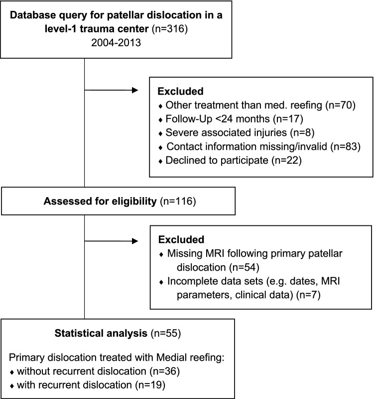
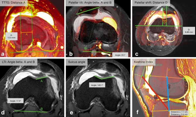
Methods: Fifty-five patients (18.6 ± 6.6 years) who underwent medial reefing after primary traumatic patellar dislocation (84% with medial patellofemoral ligament [MPFL] rupture) were included. Patients were followed up for at least 24 months postoperatively (3.8 ± 1.2 years) to assess the incidence of recurrent patellar dislocation. In patients without recurrent dislocation, the Kujala and subjective IKDC scores were assessed. Moreover, the tibial tubercle-trochlear groove (TT-TG), sulcus angle, patellar tilt, patellar shift, and lateral trochlea index (LTI) were measured. The patellar height was measured using the Caton-Dechamps (CDI), Blackburne-Peel (BPI), and Insall-Salvati index (ISI). The cohort was subclassified into two groups with and without recurrent dislocation. Differences between groups were analyzed with respect to the MRI parameters.
Results: Forty percent had a pathological sulcus angle of > 145°, 7.2% had an LTI of < 11°, 47.3% had a patellar tilt of > 20°, and 36.4% had a TT-TG of ≥ 16 mm. Increased patellar height was observed in 34.5, 65.5, and 34.5% of the patients as per CDI, BPI, and ISI, respectively. Nineteen (34.5%) patients suffered from recurrent dislocation. Compared with patients without recurrent dislocation, those with recurrent dislocation had a significantly lower LTI (p = 0.0467). All other parameters were not significantly different between the groups. Risk factor analysis showed higher odds ratios (OR > 2), although not statistically significant, for MPFL rupture (OR 2.05 [95% confidence interval 0.38-11.03], LTI (6.6 [0.6-68.1]), TT-TG (2.9 [0.9-9.2]), and patellar height according to ISI (2.3 [0.7-7.5]) and CDI (2.3 [0.7-7.5])). Patients without recurrent dislocation had a Kujala score of 93.7 ± 12.1 (42-100) points and an IKDC score of 90.6 ± 11.7 (55.2-100) points.
Conclusion: Anatomical, MRI-based parameters should be considered before indicating medial reefing. A ruptured MPFL, an LTI < 11°, a TT-TG ≥ 16 mm, a patellar tilt > 20 mm, and an increased patellar height according to ISI and CDI were found to be associated, although not significantly, with a higher risk (OR > 2) of recurrent patellar dislocation after medial reefing. Thorough preoperative analysis is crucial to reduce the risk of recurrent dislocation in young patient cohorts.
Level of evidence: Level IV.
Keywords: MRI; Medial reefing; Patellar dislocation; Recurrent dislocation.
© 2021. The Author(s).
Conflict of interest statement
SS is a member of the AO Joint Preservation and Osteotomy Expert Group. All other authors declare no potential conflict of interest.
Figures
References
-
- Arendt EA, Askenberger M, Agel J, Tompkins MA. Risk of redislocation after primary patellar dislocation: a clinical prediction model based on magnetic resonance imaging variables. Am J Sports Med. 2018;46:3385–3390. - PubMed
-
- Arendt EA, England K, Agel J, Tompkins MA. An analysis of knee anatomic imaging factors associated with primary lateral patellar dislocations. Knee Surg Sports Traumatol Arthrosc. 2017;25:3099–3107. - PubMed
-
- Balcarek P, Oberthür S, Hopfensitz S, Frosch S, Walde TA, Wachowski MM, et al. Which patellae are likely to redislocate? Knee Surg Sports Traumatol Arthrosc. 2014;22:2308–2314. - PubMed
-
- Biedert RM, Albrecht S. The patellotrochlear index: a new index for assessing patellar height. Knee Surg Sports Traumatol Arthrosc. 2006;14:707–712. - PubMed
-
- Biedert RM, Tscholl PM. Patella Alta: A Comprehensive Review of Current Knowledge. Am J Orthop (Belle Mead NJ) 2017;46:290–300. - PubMed
MeSH terms
LinkOut - more resources
Full Text Sources
Other Literature Sources
Medical
Miscellaneous
