Observational Study
. 2021 Sep 15;204(6):651-666.
doi: 10.1164/rccm.202012-4510OC.

Metformin: Experimental and Clinical Evidence for a Potential Role in Emphysema Treatment

Affiliations
Observational Study

Metformin: Experimental and Clinical Evidence for a Potential Role in Emphysema Treatment

Francesca Polverino et al. Am J Respir Crit Care Med. .

Abstract

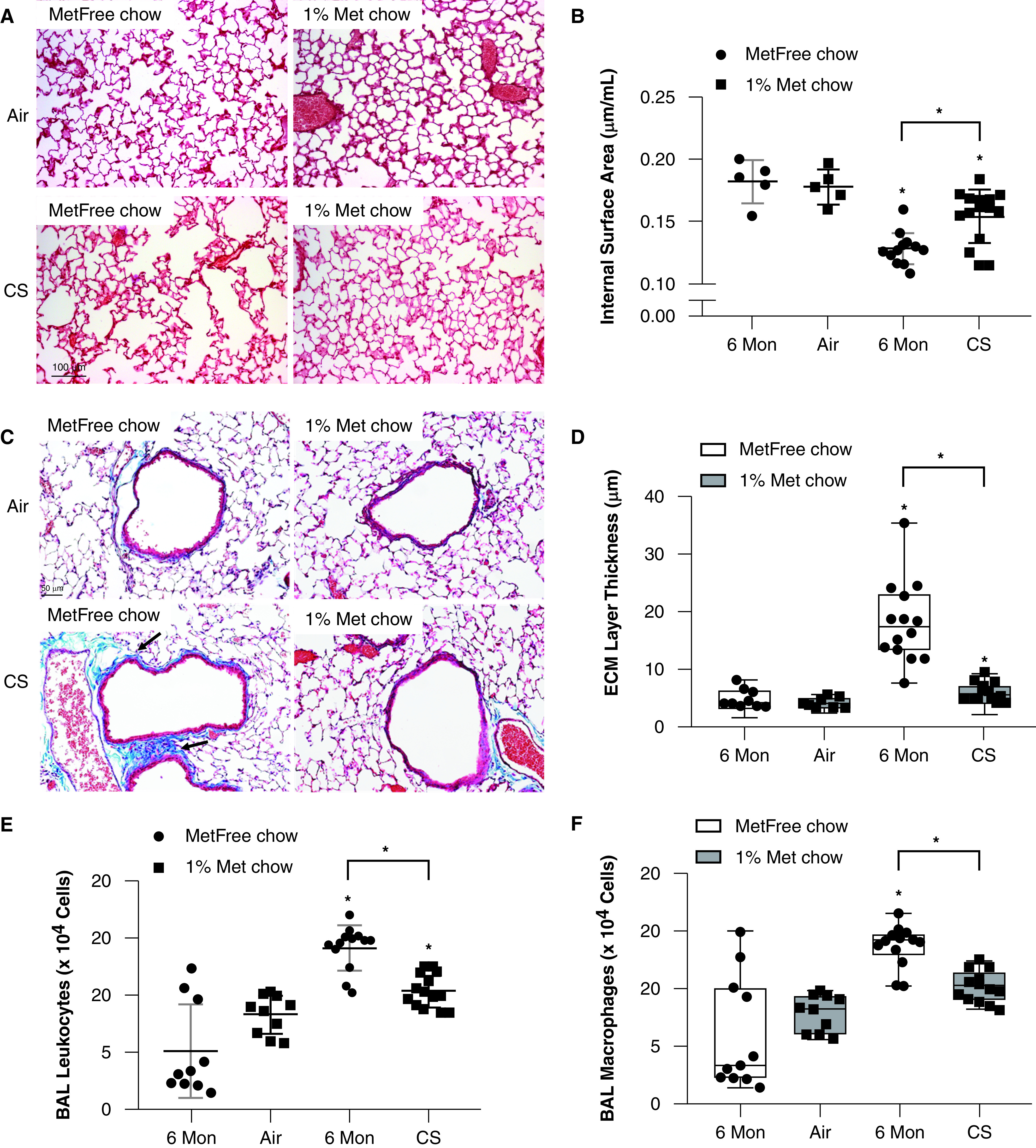
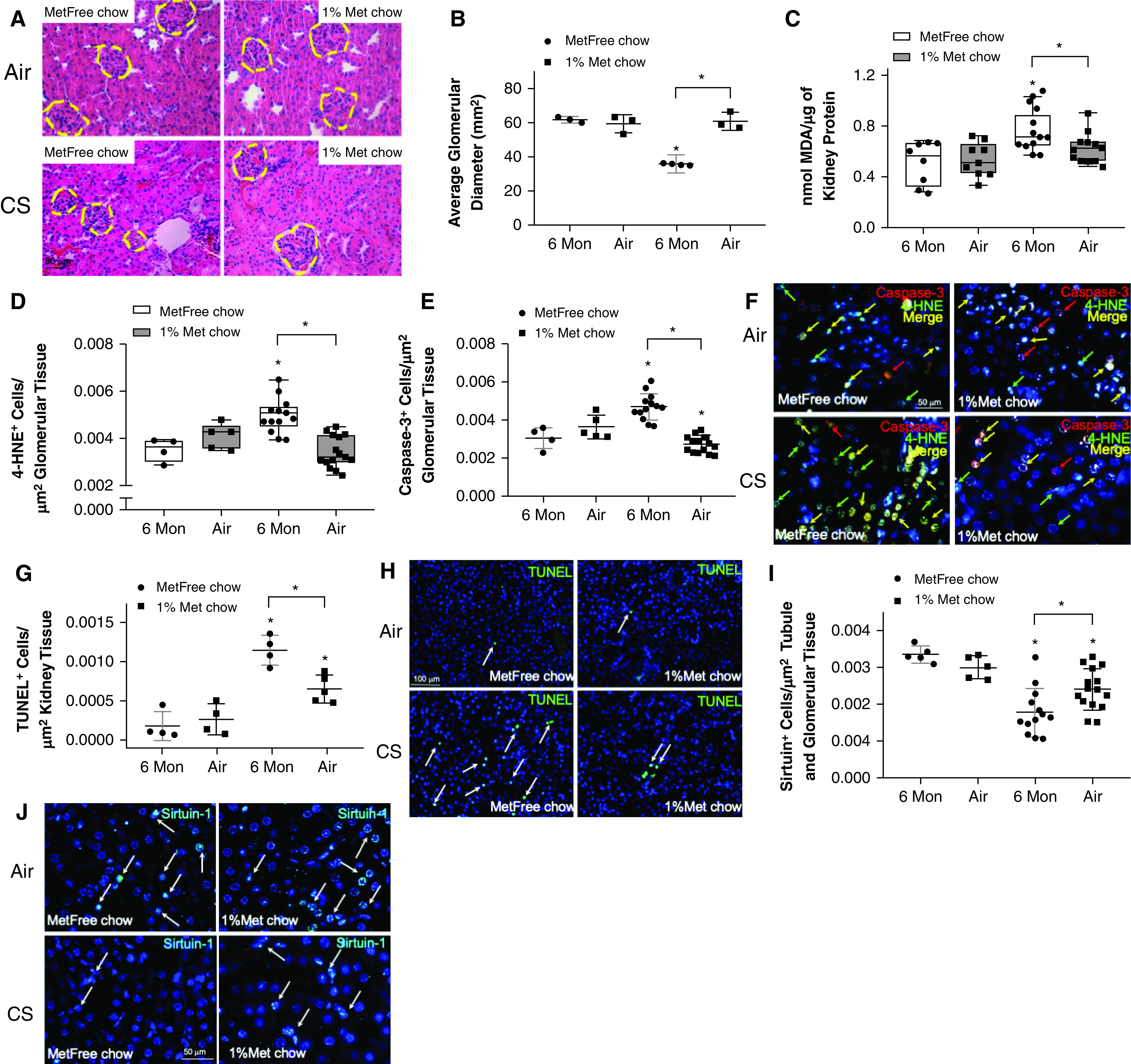
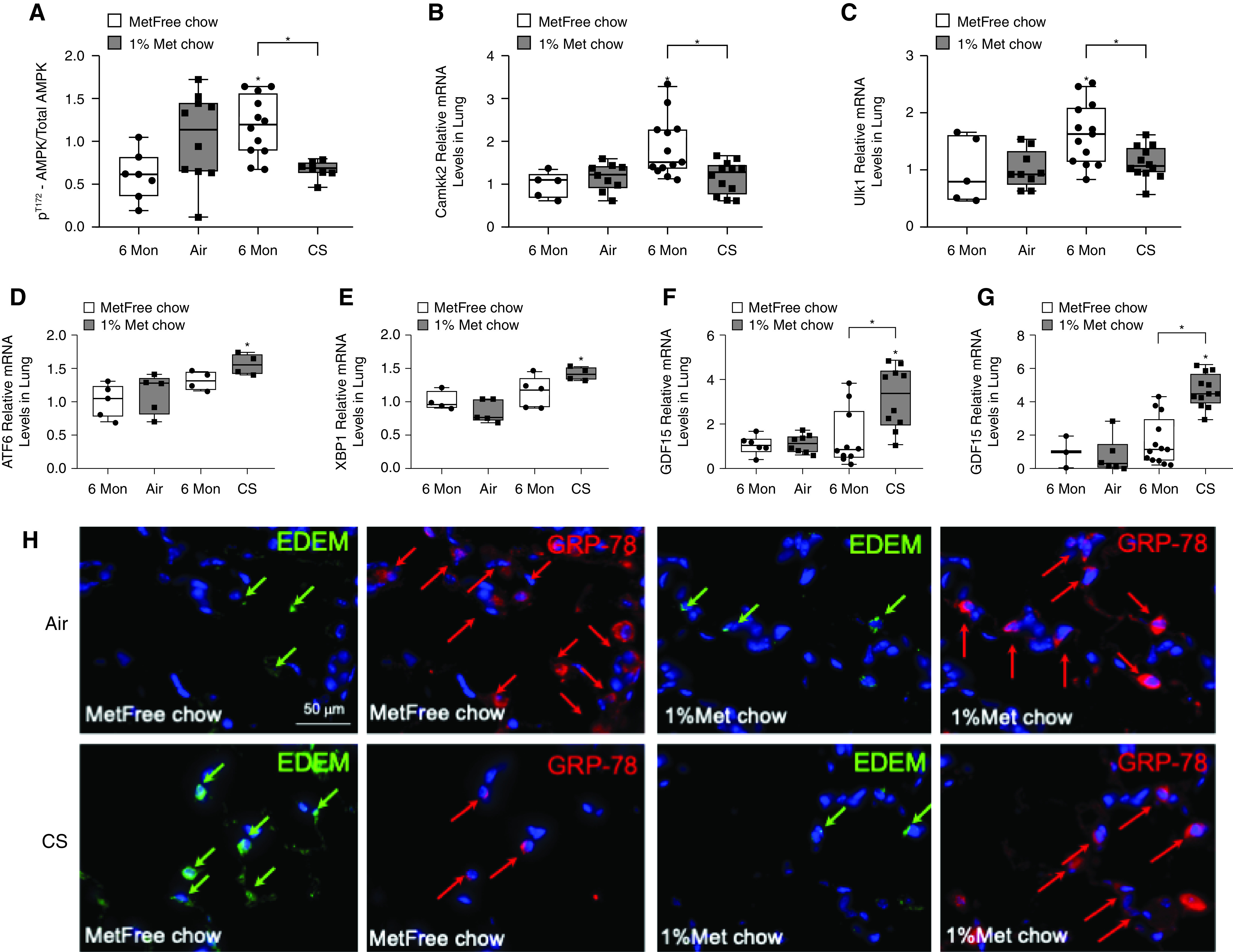
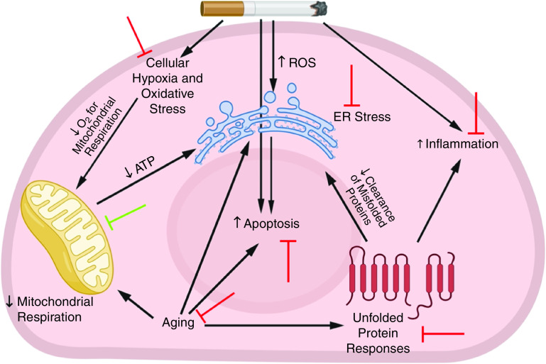
Rationale: Cigarette smoke (CS) inhalation triggers oxidative stress and inflammation, leading to accelerated lung aging, apoptosis, and emphysema, as well as systemic pathologies. Metformin is beneficial for protecting against aging-related diseases. Objectives: We sought to investigate whether metformin may ameliorate CS-induced pathologies of emphysematous chronic obstructive pulmonary disease (COPD). Methods: Mice were exposed chronically to CS and fed metformin-enriched chow for the second half of exposure. Lung, kidney, and muscle pathologies, lung proteostasis, endoplasmic reticulum (ER) stress, mitochondrial function, and mediators of metformin effects in vivo and/or in vitro were studied. We evaluated the association of metformin use with indices of emphysema progression over 5 years of follow-up among the COPDGene (Genetic Epidemiology of COPD) study participants. The association of metformin use with the percentage of emphysema and adjusted lung density was estimated by using a linear mixed model. Measurements and Main Results: Metformin protected against CS-induced pulmonary inflammation and airspace enlargement; small airway remodeling, glomerular shrinkage, oxidative stress, apoptosis, telomere damage, aging, dysmetabolism in vivo and in vitro; and ER stress. The AMPK (AMP-activated protein kinase) pathway was central to metformin's protective action. Within COPDGene, participants receiving metformin compared with those not receiving it had a slower progression of emphysema (-0.92%; 95% confidence interval [CI], -1.7% to -0.14%; P = 0.02) and a slower adjusted lung density decrease (2.2 g/L; 95% CI, 0.43 to 4.0 g/L; P = 0.01). Conclusions: Metformin protected against CS-induced lung, renal, and muscle injury; mitochondrial dysfunction; and unfolded protein responses and ER stress in mice. In humans, metformin use was associated with lesser emphysema progression over time. Our results provide a rationale for clinical trials testing the efficacy of metformin in limiting emphysema progression and its systemic consequences.

Keywords: aging; chronic obstructive pulmonary disease; cigarette smoke; comorbidities; metformin.


Figures

Figure 1.
Figure 1.
Metformin ameliorates the development of airspace enlargement, small airway remodeling, and inflammation in the lungs of mice exposed to cigarette smoke (CS). Wild-type mice were exposed to air or CS for 24 weeks and fed metformin-free (MetFree) or metformin-enriched (Met) chow during the last 12 weeks of exposure. (A) The lung parenchyma of Met chow–fed, CS-exposed mice showed milder airspace enlargement than that of the MetFree chow–fed, CS-exposed mice. Scale bars: CS, 50 μm; air, 100 μm. (B) Metformin treatment was protective against CS-induced decreases in the internal surface area quantified in formalin-fixed, paraffin-embedded Gill-stained lung sections and adjusted for the total lung volume (5 air-exposed mice/group and 13–15 CS-exposed mice/group). N = 5–15 mice/group. (C) Representative images of extracellular matrix (ECM) deposition around small airways (in bright blue color; see arrows) in Masson’s Trichrome–stained lung sections. Scale bar, 50 μm. (D) The thickness of the ECM protein layer deposited around small airways (300–799 μm in diameter) was significantly lower in Met chow–fed, CS-exposed mice than in MetFree chow–fed, CS-exposed mice. (E and F) The numbers of total leukocytes and macrophages quantified in BAL samples were lower in MetFree chow–fed, CS-exposed mice than in Met chow–fed, CS-exposed mice. *P < 0.05 for each time point indicated. Mon = months.
Figure 2.
Figure 2.
Metformin protects the lung against cigarette smoke (CS)-induced apoptosis and oxidative stress. Wild-type mice were exposed to air or CS for 24 weeks and were fed metformin-free (MetFree) or metformin-enriched (Met) chow during the last 12 weeks of exposure. (A and B) The levels of MDA and 4-hydroxy-2-nonenal (4-HNE) were lower in CS-exposed, Met chow–fed mice than in CS-exposed, MetFree chow–fed mice, respectively. (C) The number of cells positive for the apoptosis marker caspase-3 normalized for squared micrometers of lung tissue was lower in Met chow–fed, CS-exposed mice than in MetFree chow–fed, CS-exposed mice. (D) Representative double-immunofluorescence pictures of pulmonary alveoli positive for 4-HNE (green arrows), caspase-3 (red arrows), or both (yellow arrows). N = 5–15 mice/group. Scale bar, 50 μm. (E and F) The number of alveolar cells with DNA fragmentation (see arrows) as determined by using a TUNEL (terminal deoxynucleotidyl transferase dUTP nick end labeling) assay and normalized for cells/μm2 of lung tissue was higher in MetFree chow–fed, CS-exposed mice than in Met chow–fed, CS-exposed mice. Scale bar, 100 μm. *P < 0.05 versus the air group that received the same chow or versus the group indicated. MDA = malondialdehyde; Mon = months.
Figure 3.
Figure 3.
Metformin protects the lung against cigarette smoke (CS)-induced aging and telomere damage. Wild-type mice were exposed to air or CS for 24 weeks and were fed metformin-free (MetFree) or metformin-enriched (Met) chow during the last 12 weeks of exposure. (A and B) The numbers of alveolar cells/μm2 of alveolar tissue expressing the aging markers p16 and p21 were lower in CS-exposed, Met chow–fed mice than in CS-exposed, MetFree chow–fed mice, respectively. (C) Representative double-immunofluorescence pictures showing a lower number of cells positive for the aging markers p16 (green arrows), p21 (red arrows), or both (yellow arrows) in Met chow–fed, CS-exposed mice than in MetFree chow–fed, CS-exposed mice. Scale bar, 50 μm. (D) The number of alveolar cells/μm2 of alveolar tissue expressing the prosurvival marker sirtuin-1 was higher in CS-exposed, Met chow–fed mice than in CS-exposed, MetFree chow–fed mice. (E) Representative immunofluorescence pictures showing a higher number of cells positive for sirtuin-1 (arrows) in Met chow–fed, CS-exposed mice than in MetFree chow–fed, CS-exposed mice. Scale bar, 50 μm. (F) The mRNA mTERT (murine telomerase reverse transcriptase) levels quantified by using RT-PCR analysis were higher in Met chow–fed, CS-exposed mice than in MetFree chow–fed, CS-exposed mice. (G) The number of alveolar cells/μm2 of alveolar tissue expressing γ-H2AX (the phosphorylated form of H2AX [γ-H2A.X variant histone]) was higher in CS-exposed, MetFree chow–fed mice than in Met chow–fed mice. (H) Representative immunofluorescence pictures showing a higher number of cells positive for γ-H2AX (arrows) in MetFree chow–fed, CS-exposed mice than in Met chow–fed, CS-exposed mice. Scale bar, 50 μm. *P < 0.05 versus the air group that received the same chow or versus the group indicated. Mon = months.
Figure 4.
Figure 4.
Metformin protects against cigarette smoke (CS)-induced glomerular shrinkage by reducing oxidative stress, apoptosis, and aging in the kidneys. Wild-type mice were exposed to air or CS for 24 weeks and were fed metformin-enriched (Met) or metformin-free (MetFree) chow during the last 12 weeks of exposure. (A) Representative images of glomeruli in hematoxylin and eosin–stained kidney sections. Met chow–fed mice were protected against CS-induced glomerular shrinkage compared with MetFree chow–fed, CS-exposed mice. Scale bar, 50 μm. (B) The glomerular diameters were quantified in 3–4 mice/group. (C–E) The levels of MDA and the numbers of alveolar cells expressing 4-hydroxy-2-nonenal (4-HNE) and caspase-3 normalized for cells/μm2 of renal tissue were lower in CS-exposed, Met chow–fed mice than in CS-exposed, MetFree chow–fed mice, respectively. (F) Representative double-immunofluorescence pictures showing a lower number of cells positive for caspase-3 (red arrows), 4-HNE (green arrows), or both (yellow arrows) in Met chow–fed, CS-exposed mice than in MetFree chow–fed, CS-exposed mice. Scale bar, 50 μm. (G and H) The number of cells with DNA fragmentation (see arrows) as determined by using a TUNEL (terminal deoxynucleotidyl transferase dUTP nick end labeling) assay and normalized for cells/μm2 of renal tissue was higher in MetFree chow–fed, CS-exposed mice than in Met chow–fed, CS-exposed mice. Scale bar, 100 μm. (I) The number of cells/μm2 of renal tissue expressing sirtuin-1 was higher in CS-exposed, MetFree chow–fed mice than in CS-exposed, Met chow–fed mice. (J) Representative immunofluorescence pictures showing a higher number of cells positive for sirtuin-1 (arrows) in MetFree chow–fed, CS-exposed mice than in Met chow–fed, CS-exposed mice. Scale bar, 50 μm. *P < 0.05 versus the air group that received the same chow or versus the group indicated. MDA = malondialdehyde; Mon = months.
Figure 5.
Figure 5.
Metformin protects against cigarette smoke (CS)-induced skeletal muscle aging and waste by preserving quadriceps telomere length and type I fibers. (A) The number of cells/μm2 of peripheral muscle tissue expressing the prosurvival marker sirtuin-1 was higher in CS-exposed, metformin-enriched (Met) chow–fed mice than in metformin-free (MetFree) chow–fed mice. (B) Representative immunofluorescence pictures showing a higher number of cells positive for sirtuin-1 (arrows) in Met chow–fed, CS-exposed mice than in MetFree chow–fed, CS-exposed mice. Scale bar, 50 μm. (C) The quadriceps telomere length was higher in Met chow–fed, CS-exposed mice than in MetFree chow–fed, CS-exposed mice. (D) The number of cells/μm2 of peripheral muscle tissue expressing γ-H2AX (the phosphorylated form of H2AX [γ-H2A.X variant histone]) was higher in CS-exposed, MetFree chow–fed mice than in CS-exposed, Met chow–fed mice. (E) Representative immunofluorescence pictures showing a higher number of cells positive for γ-H2AX (arrows) in MetFree chow–fed, CS-exposed mice than in Met chow–fed, CS-exposed mice. Scale bar, 50 μm. (F) CS-exposed, Met chow–fed mice had a greater number of reduced nicotinamide adenine dinucleotide (NADH) muscle fibers (arrows in G) than CS-exposed, MetFree chow–fed mice. (G) The NADH staining of type I and type II muscle fibers in formalin-fixed, paraffin-embedded skeletal muscle sections. The type I fibers stain with dark blue, and the type II fibers stain with light blue. Met chow–fed mice were protected against CS-induced loss of type I fibers compared with MetFree chow–fed, CS-exposed mice. Scale bar, 50 μm. *P < 0.05 versus the air group that received the same chow or versus the group indicated. Mon = months.
Figure 6.
Figure 6.
Proteomic analyses of proteins associated with metformin-induced protection of the lung against cigarette smoke (CS)-induced mitochondrial damage and endoplasmic reticulum (ER) stress. (A) A volcano plot shows the log2 fold change plotted against log10-adjusted P values for CS-exposed versus CS-exposed and metformin-treated (CSM) samples for all significantly affected proteins, with mitochondria-associated proteins being highlighted in blue and ER proteins being shown in orange. The horizontal line represents the cutoff for a P value of <0.05, and the two vertical lines represent the cutoff values of 1.5-fold change in either the positive or negative direction. (B) Scatter plot of the GO biological processes category showing enrichment findings for the significantly affected proteins of the different pairwise comparisons. CS is used to indicate CS exposure alone. (C) Scatter plot of the mitochondrial proteins significantly affected by metformin treatment. The vertical dashed green line represents the cutoff P value of  <0.05. n = 4 per group. (D) Scatter plot of the mitochondrial proteins associated with COPD pathogenesis and CS-induced lung damage with proteins significantly affected by metformin treatment highlighted. The vertical dashed green line represents the P cutoff value of < 0.05. n = 4 per group. The entire list of proteins associated with CS exposure and metformin treatment is available in the online supplement. A = air-exposed, metformin-free chow–fed mice; AM = air-exposed, metformin-enriched chow–fed mice; COPD = chronic obstructive pulmonary disease; GO = Gene Ontology.
Figure 7.
Figure 7.
Metformin effects in cigarette smoke (CS)-exposed mice are associated with changes in components of the AMPK (AMP-activated protein kinase) pathway and in key stress sensors regulating unfolded protein responses (UPRs) and endoplasmic reticulum (ER) stress. (A) Western blot analyses of the lung homogenates were performed, and the levels of phosphoT172-AMPK/total AMPK were quantified and found to be decreased in CS-exposed, metformin-enriched (Met) chow–fed mice compared with CS-exposed, metformin-free (MetFree) chow–fed mice. Pulmonary (B) Camkk-2 (calcium/calmodulin-dependent protein kinase kinase 2) and (C) Ulk-1 levels were quantified by using RT-PCR analysis in the four experimental groups of mice and were found to be decreased in CS-exposed, Met chow–fed mice compared with CS-exposed, MetFree chow–fed mice. (D and E) The mRNA levels of the UPR sensors ATF-6 (activating transcription factor 6) and XBP-1 (X-box binding protein 1) were higher in CS-exposed, Met chow–fed mice than in CS-exposed, MetFree chow–fed mice and (F and G) were associated with higher gene expression levels of GDF15 (growth differentiation factor 15), a rescue cytokine that is expressed during cellular stress, in both the lungs and the kidneys, respectively. (H) Double-immunofluorescence staining for ER stress sensors, EDEMs (ER-associated degradation–enhancing α-mannosidase–like proteins) (green arrows) and BIP (binding immunoglobulin protein, also known as GRP-78) (red arrows) showing a decrease in EDEMs and an increase in GRP-78 in CS-exposed, Met chow–fed mice compared with CS-exposed, MetFree chow–fed mice. Scale bar, 50 μm. *P < 0.05 versus the air group that received the same chow or versus the group indicated. Mon = months.
Figure 8.
Figure 8.
Potential protective effects of metformin against cigarette smoke (CS)-induced pathologies. In response to irritants from CS exposure, a series of events occurs in the lung. These events include the recruitment of inflammatory cells, cellular hypoxia, and the generation of excessive ROS, which lead to lung structural-cell apoptosis, accelerated lung aging, endoplasmic reticulum (ER) stress, and mitochondrial damage and impaired respiration. In turn, accelerated lung aging is associated with unfolded protein responses that lead to the accumulation of misfolded proteins in the cytoplasm that further increase inflammation and ER stress. All of these factors contribute to the development of emphysema. Metformin protects the lung against CS-induced pathologies in several ways: the green blocking sign indicates a facilitative effect of metformin, and the red blocking signs indicate an inhibitory effect from metformin. ROS = reactive oxygen species.

Comment in

References

    1. Celli BR, Wedzicha JA. Update on clinical aspects of chronic obstructive pulmonary disease. N Engl J Med. 2019;381:1257–1266. - PubMed
    1. Regan EA, Lynch DA, Curran-Everett D, Curtis JL, Austin JH, Grenier PA, et al. Genetic Epidemiology of COPD (COPDGene) Investigators. Clinical and radiologic disease in smokers with normal spirometry. JAMA Intern Med. 2015;175:1539–1549. - PMC - PubMed
    1. Celli BR, Locantore N, Tal-Singer R, Riley J, Miller B, Vestbo J, et al. ECLIPSE Study Investigators. Emphysema and extrapulmonary tissue loss in COPD: a multi-organ loss of tissue phenotype. Eur Respir J. 2018;51:1702146. - PubMed
    1. Divo M, Cote C, de Torres JP, Casanova C, Marin JM, Pinto-Plata V, et al. BODE Collaborative Group. Comorbidities and risk of mortality in patients with chronic obstructive pulmonary disease. Am J Respir Crit Care Med. 2012;186:155–161. - PubMed
    1. Church DF, Pryor WA. Free-radical chemistry of cigarette smoke and its toxicological implications. Environ Health Perspect. 1985;64:111–126. - PMC - PubMed

Publication types

MeSH terms