Review
. 2021 May 25;26(1):49.
doi: 10.1186/s40001-021-00518-6.

An unusual case of chest wall glomus tumor presenting with axillary pain: a case report and literature review

Affiliations
Review

An unusual case of chest wall glomus tumor presenting with axillary pain: a case report and literature review

Leila Oryadi Zanjani et al. Eur J Med Res. .

Abstract

Background: Glomus tumor is an uncommon soft tissue tumor. However, as the tumor causes significant disability, its early diagnosis is essential. It involves subungual areas of fingers and toes in most cases, and its extra-digital involvement is rarely seen. To the best of the authors' knowledge, only a few chest wall involvement cases have been reported in the literature.

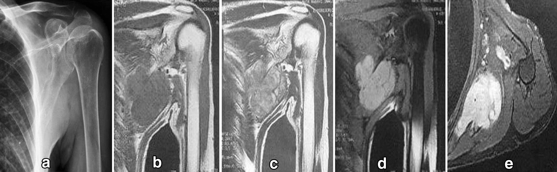
Case presentation: In this paper, we describe a 63-year-old patient with a chest wall glomus tumor presenting with axillary paroxysmal pain and limitation in his shoulder range of motion that had been missed for nearly 15 years. His symptoms were relieved immediately following surgical excision.

Conclusion: Glomus tumors may involve any part of the human body. It is curable with surgical excision in most cases. Therefore, a correct early diagnosis has paramount importance. A high index of suspicion is needed for early diagnosis, especially when the tumor involves uncommon anatomic areas.

Keywords: Case report; Chest wall; Glomus tumor.


Conflict of interest statement

The authors declare that they have no conflict of interest.

Figures

Fig. 1
Fig. 1
Diagnostic imaging. a Radiograph shows a soft tissue lesion affecting chest wall in the infra-axillary region. be, MRI shows a lobulated mass iso-signal in T1W and heterogeneous to high signal in T2W and high signal in fat-saturated sequences with strong post-contrast enhancement in the infra-axillary intermuscular fat plane. The tumor had close contact with adjacent vessels
Fig. 2
Fig. 2
Macroscopic appearance of tumor
Fig. 3
Fig. 3
Microscopic appearance. a Thin-walled blood vessels surrounded by a solid proliferation of round and epithelioid shape cells with round nuclei and lightly acidophilic cytoplasm (H&M,20×). b No significant nuclear atypia or mitosis was identified (H&M,40×). c, Cavernous hemangioma-like vascular structures surrounded by clusters of glomus cells (H&E 10 ×)
Fig. 4
Fig. 4
Immunohistochemical staining. a, b SMA, and CD34 positivity. c, d CK, and desmin negativity
Fig. 5
Fig. 5
A schematic view of the glomus body in the dermis layer of the skin

References

    1. Schiefer TK, Parker WL, Anakwenze OA, et al. Extradigital glomus tumors: a 20-year experience. Mayo Clin Proc. 2006;81(10):1337–1344. doi: 10.4065/81.10.1337. - DOI - PubMed
    1. Hartert M, Wolf M, Marko C, et al. Glomus tumor of the trachea–Synopsis of histology & methodology of treatment. Respir Med Case Rep. 2019;28:100905. - PMC - PubMed
    1. Abela M, Cole AS, Hill GA, et al. Glomus tumor of the scapular region. J Shoulder Elbow Surg. 2000;9(6):532–533. doi: 10.1067/mse.2000.108961. - DOI - PubMed
    1. Lee DW, Yang JH, Chang S, et al. Clinical and pathological characteristics of extradigital and digital glomus tumours: a retrospective comparative study. J Eur Acad Dermatol Venereol. 2011;25(12):1392–1397. doi: 10.1111/j.1468-3083.2011.03979.x. - DOI - PubMed
    1. Bali GS, Hartman DJ, Haight JB, et al. A rare case of malignant glomus tumor of the esophagus. Case Rep Oncol Med. 2013;2013:287078. - PMC - PubMed